. 1992;2(2):99-110.

Identification of three mammalian proteins that bind to the yeast TATA box protein TFIID

Affiliations

Identification of three mammalian proteins that bind to the yeast TATA box protein TFIID

B Coulombe et al. Gene Expr. 1992.

Abstract

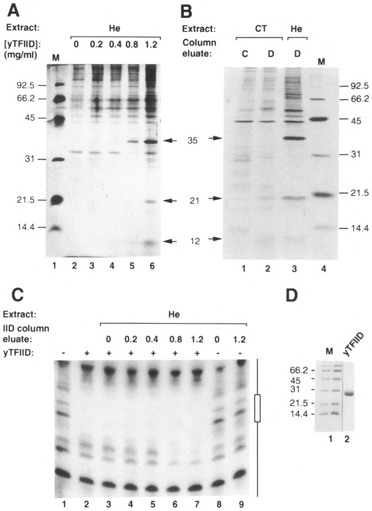
The TATA box binding transcription factor TFIID of S. cerevisiae was used as a ligand for affinity chromatography. Polypeptides that bind specifically to yeast TFIID (TFIID-associated proteins, DAPs) were purified from human HeLa (heDAPs) and calf thymus (ctDAPs) whole cell extracts. Both heDAP and ctDAP fractions altered the binding of TFIID to the TATA element, and substituted for the TFIIA transcription activity in a reconstituted in vitro system. The heDAP fraction also behaved like TFIIA in its ability to form a promoter-TFIID-TFIIA complex and to recruit TFIIB to such a complex. The interaction of DAPs with TFIID can confer heat-resistance (47 degrees C) on recombinant yeast or human TFIID. SDS-PAGE analysis revealed that three polypeptides from HeLa extracts specifically bound to yTFIID columns (heDAP35, heDAP21, and heDAP12). These data suggest that a multi-subunit transcription factor with the properties of TFIIA can bind to TFIID in the absence of DNA.


Figures

Figure 1
Figure 1
Gel mobility shift assays using yTFIID and DAPs. HeLa (He) and calf thymus (CT) eluates both from control (C) and yTFIID (D) columns were tested for their ability to bind DNA either in the presence (+) or absence (−) of yTFIID (25 ng). Lanes 1 and 14: no added proteins; lanes 2, 7, and 15: yTFIID alone; lanes 3–5: 0.5, 1.5, and 4.5 μl of He D eluate in the presence of yTFIID; lane 6: 4.5 μl of He C eluate in the presence of yTFIID; lanes 8–10: 0.5, 1.5, and 4.5 μl of CT D eluate in the presence of yTFIID; lane 11: 4.5 μl of CT C eluate in the presence of yTFIID; lane 12: 4.5 μl of He D eluate alone; lane 13: 4.5 μl of CT D eluate alone; lane 16: 4.5 μl of He D eluate in the presence of yTFIID; and lane 17: 4.5 μl of CT D eluate in the presence of yTFIID. Probes were either wild-type (TATAAAA; lanes 1-13) or mutated (TAGAGAA; lanes 14–17) oligonucleotides derived from the Ad2 ML promoter (from position −44 to −16). The positions of the yTFIID-specific complex and the complex of lower mobility are indicated by arrows.
Figure 2
Figure 2
Human DAPs but not calf thymus DAPs can form TFIIA-TFIID- and TFIIATFIID-TFIIB-type complexes. Proteins and the DNA probe (Ad2 MLP from position −50 to +33) were mixed together and incubated for 30 minutes at 28°C. Fifty ng of yTFIID were used per lane, and 30 ng of recombinant human TFIIB where indicated. Lane 1: free probe; lane 2: yTFIID plus 4 μl yTFIID-column eluate from HeLa cell extracts (He); lane 3: yTFIID plus 1 μl TFIIA fraction; lane 4: yTFIID plus 2 μl TFIIA fraction; lane 5: yTFIID plus 4 μl yTFIID-column eluate from calf thymus extracts (CT); lane 6: yTFIID plus 4 μl yTFIID-column eluate from HeLa cell extracts plus TFIIB; lane 7: yTFIID plus 1 μl TFIIA fraction plus TFIIB; lane 8: yTFIID plus 2 μl TFIIA fraction plus TFIIB; and lane 9: yTFIID plus 4 μl yTFIID-column eluate from calf thymus extract plus TFIIB. The positions of the complexes are indicated.
Figure 3
Figure 3
DNase I footprinting using yTFIID and DAPs. HeLa (He) and calf thymus (CT) eluates from both control (C) and yTFIID (D) columns were used in DNase I footprinting experiments in the presence of yTFIID (25 ng). Lanes 1 and 9: no added proteins; lanes 2 and 10: yTFIID alone; lanes 3 and 11: 2 μl of He D eluate in the presence of yTFIID; lanes 4 and 12: 4 μl of He D eluate in the presence of yTFIID; lanes 5 and 13: 2 μl of CT D eluate in the presence of yTFIID; lanes 6 and 14: 4 μl of CT D eluate in the presence of yTFIID; lanes 7 and 15: 4 μl of He C eluate in the presence of yTFIID; and lanes 8 and 16: 4 μl of CT C eluate in the presence of yTFIID Footprinting analyses were performed on both the coding (lanes 1-8) and the non-coding (lanes 9-16) strands of the Ad2 ML promoter (position −50 to +33). DNA coordinates, as well as the position of the TATA box (open boxes), are indicated; they were deduced from G + A sequencing reactions run on a separate gel. A schematic representation of the DNA probes and protection patterns is shown at the bottom. Arrows indicate sites hypersensitive to DNase I digestion.
Figure 4
Figure 4
In vitro transcription using a reconstituted system and DAPs. HeLa nuclear extract was loaded onto a P11 column, and fractions were generated by step-elution (flow-through 0.1 M = TFIIA fraction; 0.5 M = TFIIB/E/F fraction; and 1 M = TFIID fraction. Transcription reactions were performed using a mixture containing fraction TFIIB/E/F (2 μl; 1.8 mg/ml protein), purified RNA pol II (0.1 μl), and the fraction TFIID (2 μl; 0.9 mg/ml protein) (lanes 1–9) in the absence (lanes 2) or in the presence (lane 3) of fraction TFIIA (1 μl; 7.5 mg/ml protein), HeLa control-column eluate (+He C [2.5 μl]; lane 4), HeLa yTFIID column eluate (He D [2.5 and 5 μl]; lanes 5 and 6 respectively), calf thymus control-column eluate (CT C [2.5 μl]; lane 7), and calf thymus yTFIID-column eluate (CT D [2.5 and 5 μl]; lanes 8 and 9 respectively). The DNA template was plasmid pML(C2AT)Δ-50 linearized with restriction enzyme EcoR I. The formation of runoff transcripts (indicated by an arrow) was monitored on a denaturing-polyacrylamide gel. Positions of DNA molecular weight markers (M; lane 1) are indicated.
Figure 5
Figure 5
DAPs can confer heat-resistance to recombinant TFIIDs, but not to endogenous nor exogenous human TFIID present in a nuclear extract. A. Nuclear extract from HeLa cells was used in in vitro transcription assays either before (lane 1) or after (lanes 2–9) treatment for 20 minutes at 47°C. Increasing amounts of a yTFIID-column eluate (D eluate or DAPs; 0.5 [IX], 1 [2X], 2 [4X], and 4 μl [8X] in lanes 3-6 respectively), as well as a control-columneluate (C eluate; 2 μl [4X]; lane 7) were added to the nuclear extract prior to a heat-treatment. Recombinant TFIID (rTFIID), either human (h; lane 8) or yeast (y; lane 9), was added to the reaction after heat-treatment. Reactions were performed for 60 minutes at 30°C, using a template containing the Ad2 ML promoter. B. Recombinant TFIID (rTFIID), either human (h; lanes 1–6) or yeast (y; lanes 7–12), was added to the reaction (lanes 1 and 7) or heated at 47°C for 20 minutes in the absence (lanes 2 and 8) or in the presence of either increasing amounts of yTFIID-column eluate (0.5 [IX], 1 [2X], and 2 μl [4X]; lanes 3 and 9, 4 and 10, and 5 and 11 respectively) or control-columneluate (2 μl [4X]; lanes 6 and 12). Each reaction contained previously heat-treated nuclear extract. C. Recombinant TFIID (rTFIID), either human (h) or yeast (y), was added to heat-treated nuclear extract, incubated for 20 minutes at 30°C, and either used in transcription reactions (lanes 1 and 6) or heated again in the absence (lanes 2 and 7) or in the presence of increasing amounts of yTFIID-column eluates (1 [2X], 2 [4X] and 4 μl [8X]; lanes 3 and 8, lanes 4 and 9, and lanes 5 and 10 respectively). For heat-treatment, each reaction contained approximately 1 mg/ml BSA. The template and conditions were the same as in Figure 4, except that 2 μl of nuclear extract (15 mg/ml protein), 5 ng of ryTFIID, and 2ng of rhTFIID were used where indicated.
Figure 6
Figure 6
Affinity chromatography using yTFIID as a ligand. A. A volume of 400 μl of HeLa WCE (He) was chromatographed through 20 μl affinity columns containing different concentrations of immobilized yTFIID (0, 0.2, 0.4, 0.8, and 1.2 mgof yTFIID/ml of wet Affi-gel 10; lanes 2–6). Columns were washed with 200 μl loading buffer and eluted with loading buffer containing 0.5 M NaCl (Sopta et al., 1985). High salt eluates were analyzed using SDS-PAGE, followed by silver staining. Positions of DAP35, 21, and 12 (arrows), as well as molecular weight markers (M; lane 1), are indicated. B. Calf thymus WCE (CT) was chromatographed through control (C; lane 1) and yTFIID (D; lane 2) columns (0 and 2.5 mg of ligand/ml wet Affi-gel 10 respectively) and analyzed as described in A. A HeLa (He) eluate from a similar yTFIID column was included for comparison (lane 3). Positions of heDAP35, 21, and 12 (arrows), as well as positions of molecular weight standards (M; lane 4), are indicated. C. Eluates from yTFIID columns loaded with HeLa whole cell extract (He; see A) were analyzed using DNase I footprinting both in the absence (−) and presence (+) of yTFIID (20 ng). Lane 1: no added proteins; lane 2: yTFIID alone; lanes 3–7: yTFIID and eluates (1.2 μl) from 0, 0.2, 0.4, 0.8, or 1.2 mg/ml-yTFIID columns; lanes 8 and 9: eluates from the 0 and 1.2 mg/ml-yTFIID columns alone. The probe was the non-coding strand of the Ad2 ML promoter (positions −50 to +33) labeled at its 3′ end. The position of the TATA box is indicated as an open box. D. The yTFIID used as a ligand for affinity chromatography was analyzed by SDS-PAGE and stained with Coomassie blue. Positions of molecular weight markers are indicated (M).

Similar articles

Cited by

References

    1. Buratowski S., Hahn S., Sharp P. A., and Guarente L. (1988), Nature 334, 37–42. - PubMed
    1. Buratowski S., Hahn S., Guarente L., and Sharp P. A. (1989), Cell 56, 549–561. - PubMed
    1. Burton Z. F., Ortolan L. G., and Greenblatt J. (1986), EMBO J 5, 2923–2930. - PMC - PubMed
    1. Cavallini B., Huet J., Plassat J.-L., Sentenac A., Egly J.-M., and Chambon P. (1988), Nature 334, 77–80. - PubMed
    1. Conaway J. W., Reines D., and Conaway R. C. (1990), J Biol Chem 265, 7552–7558. - PMC - PubMed

Publication types

MeSH terms

LinkOut - more resources