. 2006 Mar 15;394(Pt 3):593-603.
doi: 10.1042/BJ20051659.

TNFalpha- and IKKbeta-mediated TANK/I-TRAF phosphorylation: implications for interaction with NEMO/IKKgamma and NF-kappaB activation

Affiliations

TNFalpha- and IKKbeta-mediated TANK/I-TRAF phosphorylation: implications for interaction with NEMO/IKKgamma and NF-kappaB activation

Marianne Bonif et al. Biochem J. .

Abstract

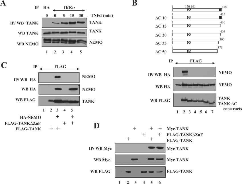
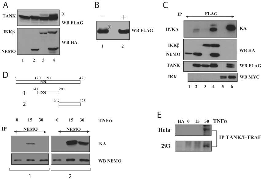
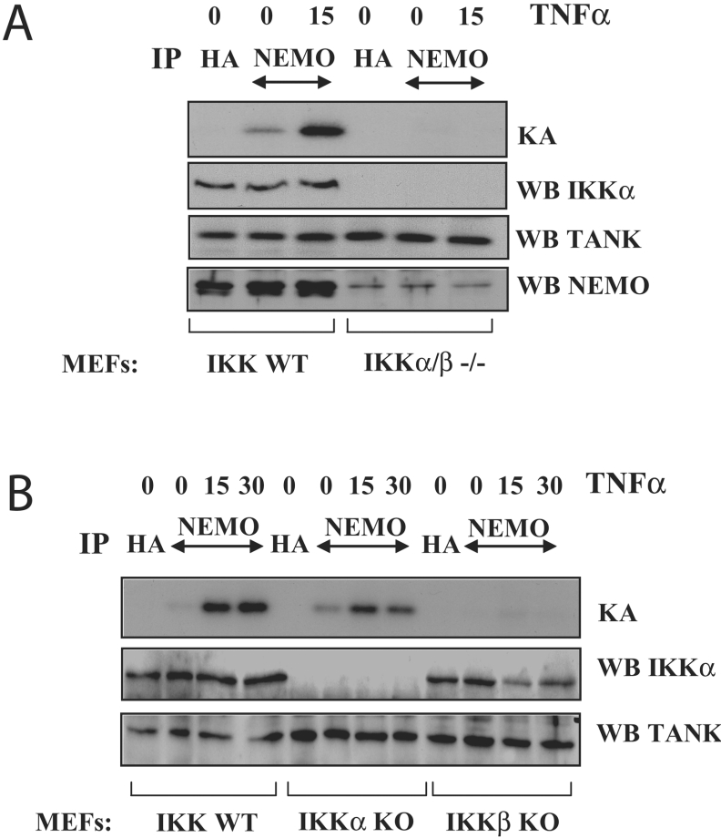
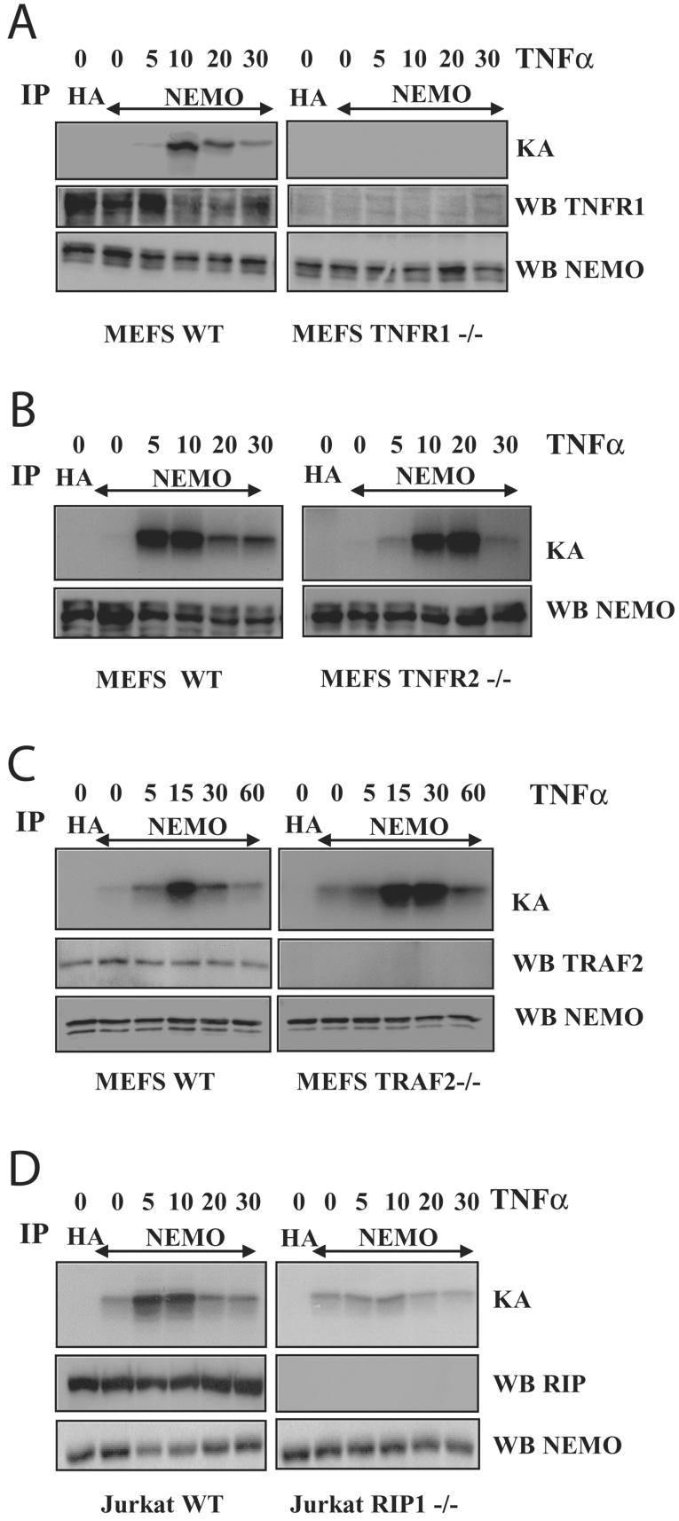
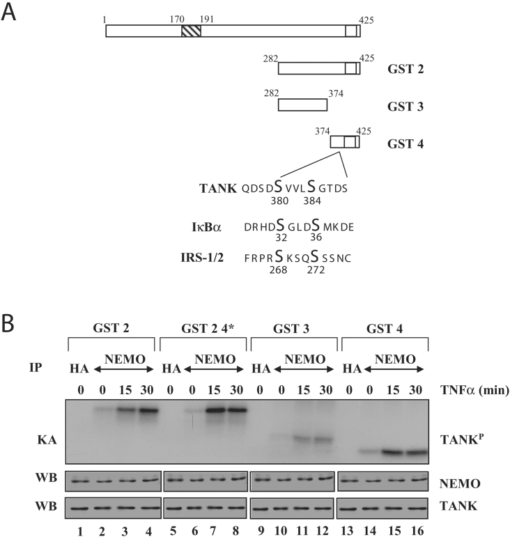
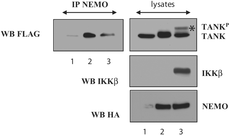
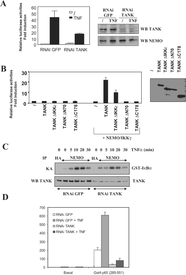
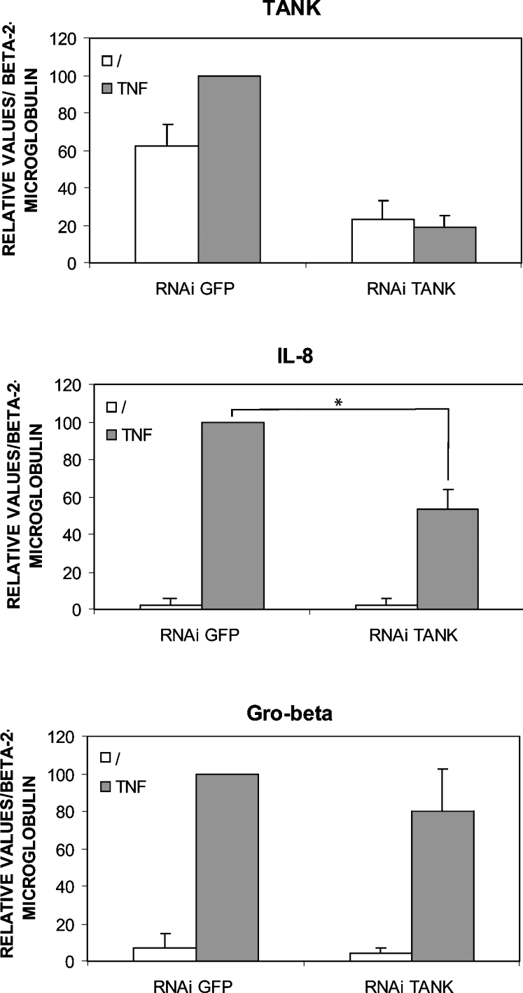
Pro-inflammatory cytokines trigger signalling cascades leading to NF-kappaB (nuclear factor-kappaB)-dependent gene expression through IKK [IkappaB (inhibitory kappaB) kinase]-dependent phosphorylation and subsequent degradation of the IkappaB proteins and via induced phosphorylation of p65. These signalling pathways rely on sequentially activated kinases which are assembled by essential and non-enzymatic scaffold proteins into functional complexes. Here, we show that the pro-inflammatory cytokine TNFalpha (tumour necrosis factor alpha) promotes TANK [TRAF (TNF receptor-associated factor) family member associated NF-kappaB activator] recruitment to the IKK complex via a newly characterized C-terminal zinc finger. Moreover, we show that TANK is phosphorylated by IKKbeta upon TNFalpha stimulation and that this modification negatively regulates TANK binding to NEMO (NF-kappaB essential modulator). Interestingly, reduced TANK expression by RNA interference attenuates TNFalpha-mediated induction of a subset of NF-kappaB target genes through decreased p65 transactivation potential. Therefore the scaffold protein TANK is required for the cellular response to TNFalpha by connecting upstream signalling molecules to the IKKs and p65, and its subsequent IKKbeta-mediated phosphorylation may be a mechanism to terminate the TANK-dependent wave of NF-kappaB activation.


Figures

Figure 1
Figure 1. TANK is recruited to the IKK complex upon TNFα stimulation and its zinc finger is required for interaction with NEMO
(A) TNFα triggers the recruitment of TANK to the IKK complex. HEK-293 cells were left untreated or stimulated with TNFα (100 units/ml) for 5–30 min and cell extracts were subjected to anti-IKKα immunoprecipitations followed by anti-TANK Western-blot analyses (top panel). The presence of TANK and NEMO in the cell extracts is illustrated by Western blots using the corresponding antibodies (second and third panels from the top respectively). (B, C) TANK interacts with NEMO through its C-terminal zinc finger. (B) The C-terminal domain of TANK is required for binding to NEMO. Upper panels: schematic representation of the TANK constructs used in the co-immunoprecipitation experiments. Lower panels: HEK-293 cells were transfected with HA–NEMO alone (lane 1) or in combination with FLAG–TANK or FLAG–TANKΔC10, -ΔC15, -ΔC20, -ΔC35 or -ΔC50 (lanes 2–7 respectively). Cell extracts were subjected to anti-FLAG immunoprecipitations followed by anti-HA Western-blot analyses (top panel). The presence of HA–NEMO and the FLAG-tagged TANK proteins in the cell extracts is illustrated in the second and third panels from the top respectively. (C) The TANK C-terminal zinc finger motif is required for interaction with NEMO. HEK-293 cells were transfected with pcDNA3 (lane 1) or with FLAG–TANK (lanes 2 and 3), FLAG–TANKΔZnF (lanes 4 and 5) and HA–NEMO (lanes 3 and 5), as indicated and cell extracts were subjected to an anti-FLAG immunoprecipitation followed by anti-HA Western-blot analyses (top panel). Cell extracts were subjected to anti-HA and -FLAG Western blots as well (middle and bottom panels respectively). (D) The TANK C-terminal zinc finger motif is dispensable for TANK dimerization. Cells were transfected with empty pcDNA3 (lane 1), FLAG–TANK or FLAG–TANKΔZnF alone (lanes 2 and 4 respectively), Myc–TANK alone (lane 3) or a combination of Myc–TANK with FLAG–TANK or FLAG–TANKΔZnF (lanes 5 and 6 respectively), as indicated. Anti-FLAG immunoprecipitations followed by anti-Myc Western-blot analyses were carried out (top panel), whereas anti-FLAG and -Myc Western blots were performed on the cell lysates (bottom panels). IP, immunoprecipitation; WB, Western blot.
Figure 2
Figure 2. TANK is phosphorylated by the IKK complex in response to TNFα
(A) Co-expression of TANK with NEMO and/or IKKβ leads to slower migrating forms of TANK, indicated by an asterisk. HEK-293 cells were transfected with FLAG–TANK alone (lane 1) or in combination with HA–NEMO (lane 2) or HA–IKKβ or both of them (lanes 3 and 4 respectively). Anti-FLAG and -HA Western blots were performed on cell lysates (upper and lower panels respectively). (B) Alkaline phosphatase treatment of the cell extracts derived from HEK-293 cells transfected with FLAG–TANK, HA–IKKβ and HA–NEMO inhibits the slower migrating forms of TANK. Cell extracts were left untreated (−) or incubated (+) with alkaline phosphatase for 1 h at 37 °C and anti-FLAG Western-blot analyses were performed. (C) IKK-mediated phosphorylation of TANK in vitro. HEK-293 cells were transfected with HA–NEMO alone (lane 1) or with FLAG–TANK with either HA–NEMO or HA–IKKβ (lanes 2 or 3 respectively) or with both of them (lane 4). As controls, HEK-293 cells were transfected with IKKϵ–Myc alone (negative control, lane 5) or with both IKKϵ–Myc and FLAG–TANK (positive control, lane 6). Anti-FLAG immunoprecipitates were subjected to an in vitro kinase assay (top panel). Anti-HA, anti-FLAG and anti-Myc Western-blot analyses were performed on cell extracts (second, third and bottom panels respectively). (D) TNFα-mediated phosphorylation of TANK on its C-terminal domain. Upper panel: schematic representation of the GST–TANK fusion proteins used as substrates in the kinase assays. Lower panels: HEK-293 cells were left untreated or stimulated with TNFα for the indicated times and anti-NEMO immunoprecipitations were performed. The GST–TANK fusion proteins 1 or 2 were used as substrates for the kinase assays (left and right panels respectively). An anti-NEMO Western blot performed on the cell extracts is illustrated (bottom panel). (E) TNFα-mediated phosphorylation of endogenous TANK in HeLa or HEK-293 cells (upper and lower panels respectively). Both cell lines were cultured in the presence of [32P]Pi and subsequently stimulated with TNFα for the indicated periods of time. Anti-TANK immunoprecipitations were carried out and the immunoprecipitates were migrated on an SDS/PAGE followed by autoradiography. IP, immunoprecipitation; KA, kinase assay; WB, Western blot.
Figure 3
Figure 3. TNFα-mediated phosphorylation of TANK requires IKKβ
(A) TNFα-mediated phosphorylation of TANK requires the IKK complex. WT (wild-type) or IKKα/IKKβ double KO MEFs were either untreated or stimulated for 15 min with TNFα. Anti-NEMO or -HA (negative control) immunoprecipitates (IP) were incubated with the GST–TANK used as substrate for a kinase assay (KA; top panel). Anti-IKKα, -TANK and -NEMO Western-blot (WB) analyses were performed on the cell lysates (second, third and bottom panels respectively). (B) IKKβ but not IKKα is required for TNFα-mediated TANK phosphorylation. WT, IKKα or IKKβ KO MEFs were used as cellular models to assess TNFα-mediated TANK phosphorylation as described above.
Figure 4
Figure 4. TNFα-mediated phosphorylation of TANK requires RIP1 and the TNFR1 but not the TNFR2, TRAF-2 and IKKα
(A, B) The TNFR1 but not the TNFR2 is required for the TNFα-mediated TANK phosphorylation. Wild-type (WT) (A, B), TNFR1 KO (A) or TNFR2 KO (B) MEF cells were left untreated or stimulated with TNFα for the indicated periods of time and in vitro kinase assays (KA) were carried out. Anti-NEMO (A, B) and -TNFR1 (A) Western-blot (WB) analyses were performed using the cell extracts (panels below the kinase assays). (C) IKK-mediated phosphorylation of TANK in TRAF-2 KO MEF cells. MEF cells lacking TRAF-2 were left untreated or stimulated for 5, 15, 30 or 60 min by TNFα and in vitro kinase assay using the GST–TANK as substrate was carried out. Anti-TRAF-2 and -NEMO Western blots performed on the cell lysates are illustrated (second and third panels from the top respectively). (D) IKK-mediated phosphorylation of TANK requires RIP1. Wild-type or RIP1-deficient Jurkat cells were left untreated or stimulated with TNFα for the indicated periods of time and in vitro kinase assays using the GST–TANK as substrate were carried out. Anti-RIP1 and -NEMO Western-blot analyses were performed with the cell extracts (second and third panels from the top respectively). IP, immunoprecipitation.
Figure 5
Figure 5. TNFα-mediated phosphorylation of TANK targets the C-terminal domain of this scaffold protein
(A) Schematic representation of the TANK protein with the potential C-terminal phosphorylated residues upon TNFα stimulation. (B) TANK is phosphorylated within its last 51 amino acids of the C-terminal domain upon TNFα stimulation. HEK-293 cells were left untreated or stimulated with TNFα for the indicated periods of time. Cell extracts were subjected to anti-HA (negative control) or anti-NEMO immunoprecipitations (IP) followed by kinase assays (KA) using these immunoprecipitates and a GST–TANK fusion protein as substrate. WB, Western blot.
Figure 6
Figure 6. IKKβ-mediated TANK phosphorylation attenuates its interaction with NEMO
HEK-293 cells were transfected with FLAG–TANK alone (lane 1) or with HA–NEMO only or in combination with HA–IKKβ (lanes 2 and 3 respectively) and anti-NEMO immunoprecipitations (IP) were performed on the cell extracts followed by anti-FLAG Western-blot (WB) analyses (left panel). Anti-FLAG, -IKKβ and -HA Western-blot analyses were also carried out on cell lysates (right panels). The asterisk illustrates the phosphorylated form of TANK (TANKP).
Figure 7
Figure 7. TANK positively regulates NF-κB activation in response to TNFα
(A) Decreased TNFα- and NF-κB-mediated gene activation in TANK RNAi cells. HEK-293 cells were transfected with 0.2 μg of RSV-βgal (where RSV is Rous sarcoma virus) and 1 μg of the κB luciferase reporter and were either left untreated or stimulated with TNFα (100 units/ml) for 5 h before lysis, as indicated. The cell extracts were collected for the measurement of both luciferase and β-galactosidase activities. The results of three independent experiments performed in triplicate, after normalization with β-Gal activities, are shown (means±S.D.). Anti-TANK and -NEMO Western blots (WB) performed on representative extracts are shown. RNAI, RNAi. (B) Wild-type TANK but not TANK mutants strongly activates NF-κB when co-expressed with NEMO. HEK-293 cells were transfected with the κB luciferase reporter, the RSV-βgal as well as the indicated expression vectors. Cell extracts were collected for the measurement of both luciferase and β-galactosidase activities and the results are shown as described above. An anti-FLAG Western blot performed to assess the levels of expression for each FLAG-tagged TANK expression vector is also illustrated. (C) IKK activation is unaltered in TANK RNAi cells. GFP or TANK RNAi cells were left unstimulated or treated with TNFα for the indicated periods of time and anti-NEMO immunoprecipitates (IP) were subjected to in vitro kinase assays (KA) using a purified GST–IκBα protein as substrate. Anti-TANK Western-blot analyses were also carried out on cell extracts. (D) TANK positively regulates p65 transactivation potential. Cells were transfected with a reporter plasmid harbouring a GAL4 DNA-responsive element (see the Materials and methods section) either alone (‘basal’) or in combination with the indicated GAL4–p65 fusion protein. p65 transactivation potential was assessed by measuring luciferase activities. The results of one representative experiment performed in triplicate, after normalization with Renilla luciferase activities, are shown (means±S.D.).
Figure 8
Figure 8. Identification of selected TNFα-induced genes that require TANK
Total RNAs from GFP or TANK RNAi HeLa cells either untreated or stimulated with TNFα were subjected to reverse transcription followed by real-time PCR analyses using the appropriate primers for amplification of the TANK, IL-8 or Gro-beta cDNAs. Amplification of the β2-microglobulin cDNA was used for normalization purposes. The results of four independent experiments performed in duplicate are shown (means±S.D.) and analysed by the Mann–Whitney test (*P<0.05).

References

    1. Tracey K. J., Cerami A. Tumor necrosis factor, other cytokines and disease. Annu. Rev. Cell Biol. 1993;9:317–343. - PubMed
    1. Karin M., Ben-Neriah Y. Phosphorylation meets ubiquitination: the control of NF-[kappa]B activity. Annu. Rev. Immunol. 2000;18:621–663. - PubMed
    1. Hayden M. S., Ghosh S. Signaling to NF-kappaB. Genes Dev. 2004;18:2195–2224. - PubMed
    1. Hsu H., Xiong J., Goeddel D. V. The TNF receptor 1-associated protein TRADD signals cell death and NF-kappa B activation. Cell (Cambridge, Mass.) 1995;81:495–504. - PubMed
    1. Devin A., Cook A., Lin Y., Rodriguez Y., Kelliher M., Liu Z. The distinct roles of TRAF2 and RIP in IKK activation by TNF-R1: TRAF2 recruits IKK to TNF-R1 while RIP mediates IKK activation. Immunity. 2000;12:419–429. - PubMed

Publication types

MeSH terms