. 2006 Mar 15;394(Pt 3):581-92.
doi: 10.1042/BJ20051471.

Class II phosphoinositide 3-kinase alpha-isoform regulates Rho, myosin phosphatase and contraction in vascular smooth muscle

Affiliations

Class II phosphoinositide 3-kinase alpha-isoform regulates Rho, myosin phosphatase and contraction in vascular smooth muscle

Yu Wang et al. Biochem J. .

Abstract

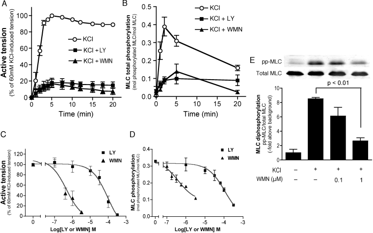
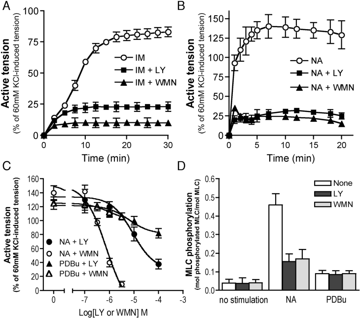
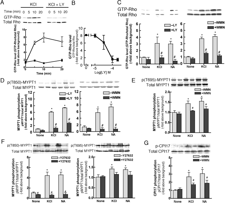
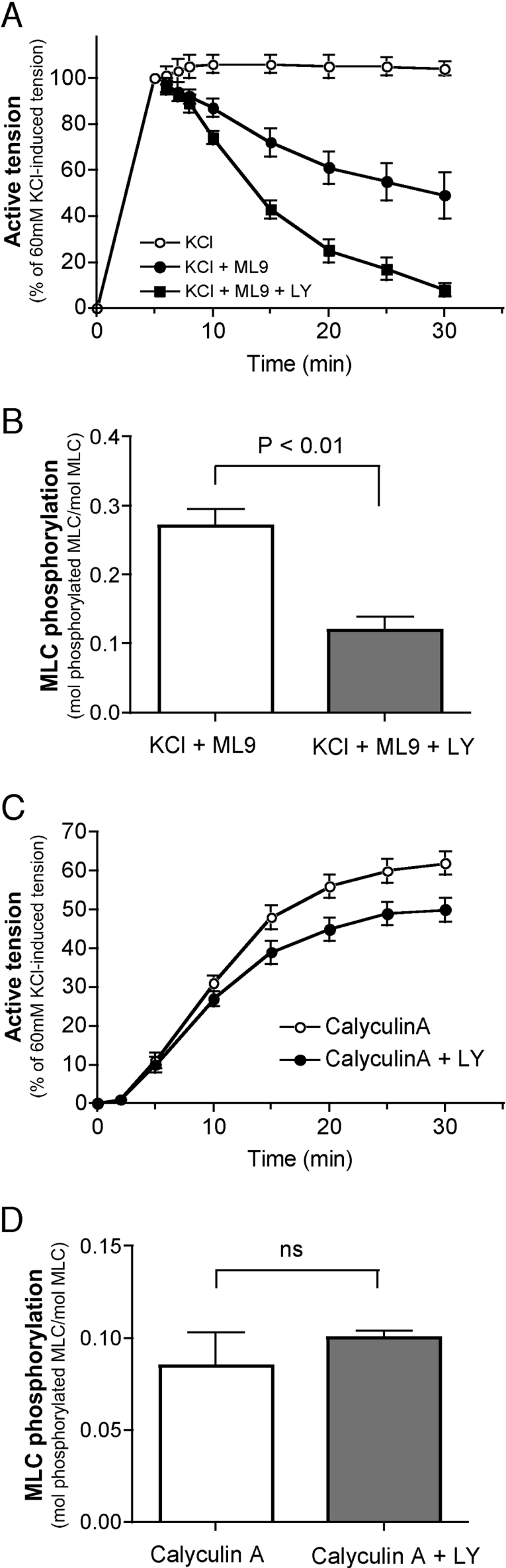
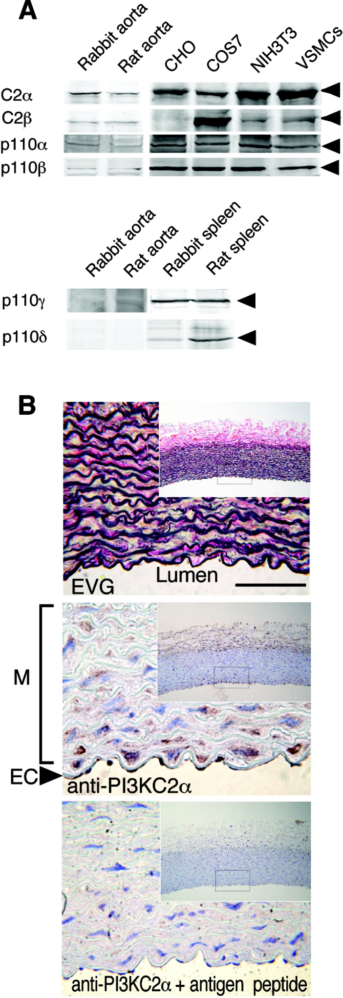
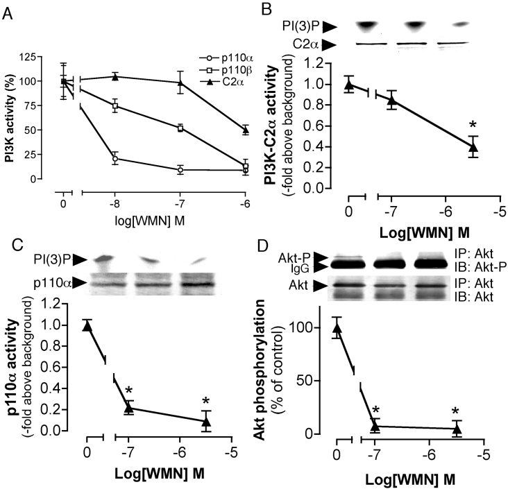
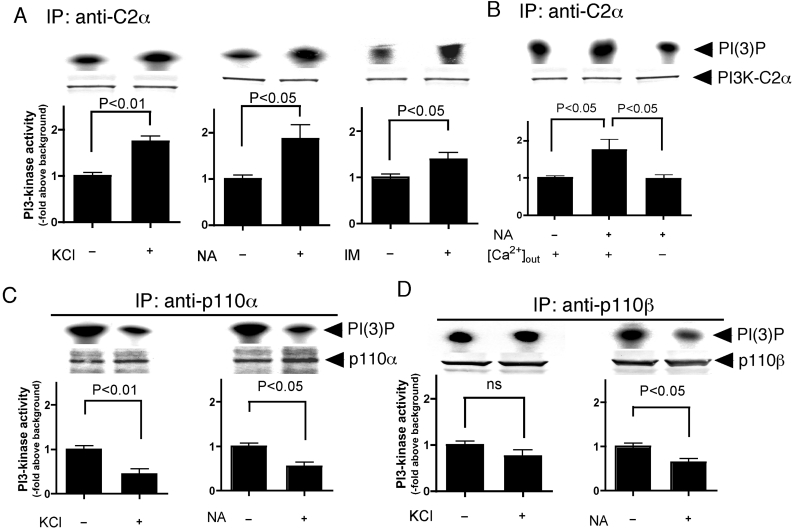
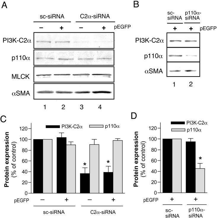
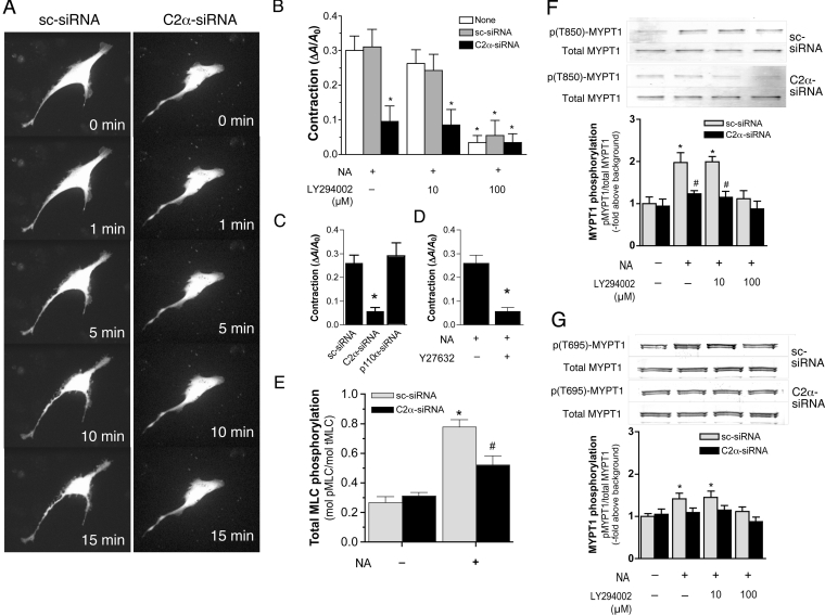
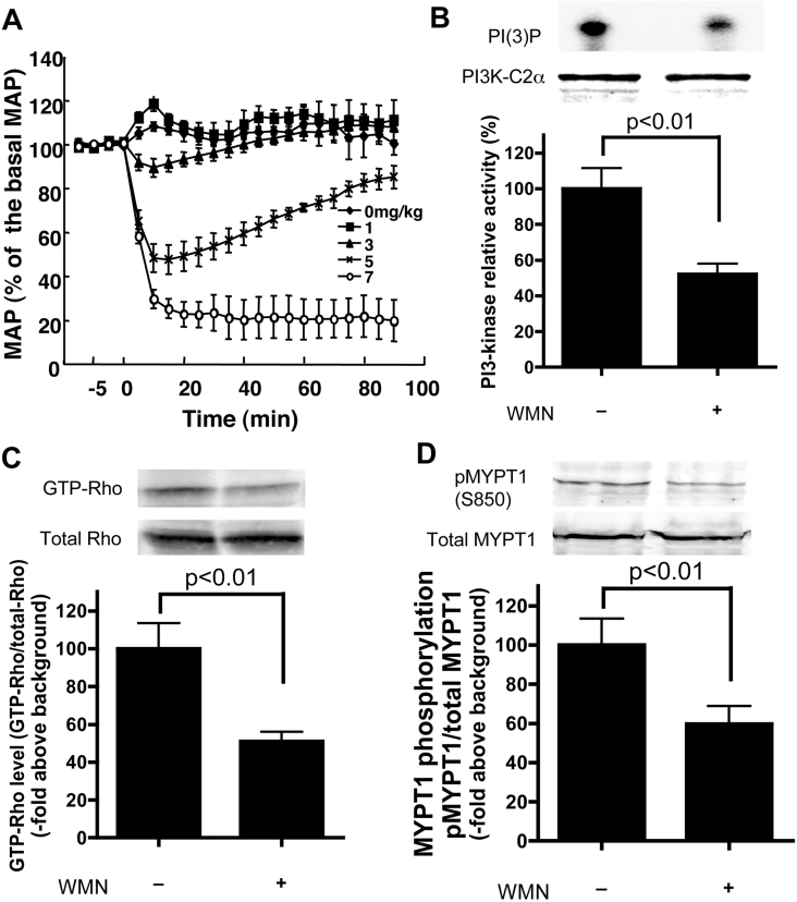
We demonstrated previously that membrane depolarization and excitatory receptor agonists such as noradrenaline induce Ca2+-dependent Rho activation in VSM (vascular smooth muscle), resulting in MP (myosin phosphatase) inhibition through the mechanisms involving Rho kinase-mediated phosphorylation of its regulatory subunit MYPT1. In the present study, we show in de-endothelialized VSM strips that the PI3K (phosphoinositide 3-kinase) inhibitors LY294002 and wortmannin inhibited KCl membrane depolarization- and noradrenaline-induced Rho activation and MYPT1 phosphorylation, with concomitant inhibition of MLC (20-kDa myosin light chain) phosphorylation and contraction. LY294002 also augmented de-phosphorylation of MLC and resultantly relaxation in KCl-contracted VSM, whereas LY294002 was much less effective or ineffective under the conditions in which MP was inhibited by either a phosphatase inhibitor or a phorbol ester in Rho-independent manners. VSM express at least four PI3K isoforms, including the class I enzymes p110alpha and p110beta and the class II enzymes PI3K-C2alpha and -C2beta. The dose-response relationships of PI3K-inhibitor-induced inhibition of Rho, MLC phosphorylation and contraction were similar to that of PI3K-C2alpha inhibition, but not to that of the class I PI3K inhibition. Moreover, KCl and noradrenaline induced stimulation of PI3K-C2alpha in a Ca2+-dependent manner, but not of p110alpha or p110beta. Down-regulation of PI3K-C2alpha expression by siRNA (small interfering RNA) inhibited contraction and phosphorylation of MYPT1 and MLC in VSM cells. Finally, intravenous wortmannin infusion induced sustained hypotension in rats, with inhibition of PI3K-C2alpha activity, GTP-loading of Rho and MYPT1 phosphorylation in the artery. These results indicate the novel role of PI3K-C2alpha in Ca2+-dependent Rho-mediated negative control of MP and thus VSM contraction.


Figures

Figure 1
Figure 1. Inhibition of membrane depolarization-induced contraction and MLC phosphorylation by PI3K inhibitors
Time-dependent changes of KCl (60 mM)-induced contraction (A) and total MLC phosphorylation (B) in the presence and absence of WMN (1 μM) or LY (100 μM). Dose-dependent inhibition of KCl (60 mM)-induced contraction (C) and total MLC phosphorylation (D) at 5 min by PI3K inhibitors. (E) Inhibition of KCl-induced increase in the content of pp-MLC (di-phosphorylated form of MLC) by a high, but not a low, concentration of WMN. VSM was pretreated with the indicated concentrations of WMN and stimulated with 60 mM KCl for 5 min. The content of pp-MLC was analysed by using anti-pp-MLC-specific antibody. A portion of extracts was analysed for the content of total MLC by Western blot analysis using anti-MLC antibody (MY21). In all panels, PI3K inhibitors were added to VSM 30 min before KCl stimulation.
Figure 2
Figure 2. Inhibition of IM- and NA-induced contraction and MLC phosphorylation by PI3K inhibitors
(A) Time-dependent changes of IM (5 μM)-induced contraction of VSM in the presence and absence of WMN and LY as in Figure 1(A). (B) Time-dependent changes of NA (3 μM)-induced contraction in the presence and absence of WMN and LY as in (A). (C) Dose-dependent inhibition of NA- and phorbol-ester-induced contraction by PI3K inhibitors. VSM was stimulated with NA (3 μM) for 5 min or by phorbol 12,13-dibutyrate (PDBu) (1 μM) for 60 min in the presence of various concentrations of WMN or LY. (D) Inhibition of NA (3 μM)- and PDBu (1 μM)-induced total MLC phosphorylation by WMN (1 μM) and LY (100 μM). VSM was treated as in (C). In all panels, PI3K inhibitors were added to VSM 30 min before KCl stimulation.
Figure 3
Figure 3. Inhibition by PI3K inhibitors of KCl- and NA-induced activation of Rho and phosphorylation of MYPT1 and CPI-17
(A) Time-dependent changes of the amounts of GTP-RhoA in KCl-stimulated VSM in the presence of absence of LY. VSM was treated as in Figures 1(A) and 1(B). A portion (1/27) of tissue extracts was subjected to Western blot analysis for evaluating amounts of total RhoA in each sample. (B) Dose-dependent inhibition of KCl-induced RhoA activation by LY. VSM was treated as in Figure 1(C). (C) Inhibition of KCl (60 mM)- and NA (3 μM)-induced Rho activation at 5 min by LY (100 μM) and WMN (1 μM). (D) Inhibition of KCl- and NA-induced Thr850 MYPT1 phosphorylation by LY and WMN. (E) Inhibition of KCl- and NA-induced Thr695 MYPT1 phosphorylation by WMN. In (C) and (D), VSM was treated as in (C), and was analysed for Thr850 and Thr695 phosphorylation of MYPT1 by Western blotting using phosphorylation site-specific anti-phospho-MYPT1 antibodies. (F) Effects of Y-27632 on KCl- and NA-induced Thr850 and Thr695 MYPT1 phosphorylation. VSM was pretreated with 10 μM Y-27632 for 30 min and then stimulated as in (C). (G) Inhibition of KCl- and NA-induced Thr38 CPI-17 phosphorylation by WMN. VSM was treated as in (C). * and #, P<0.05 compared with non-treated control and non-inhibitor-treated stimulation respectively.
Figure 4
Figure 4. Augmentation by a PI3K inhibitor of MLC de-phosphorylation and relaxation
(A) Augmentation by LY of the MLCK inhibitor ML9-induced relaxation of KCl-contracted VSM. At 5 min after KCl (60 mM) stimulation, relaxation was induced by adding ML9 (30 μM) with or without LY (100 μM). (B) Augmentation of ML9-induced MLC de-phosphorylation by LY. VSM was treated as in (A). MLC phosphorylation levels were determined at 25 min after the addition of either ML9 (KCl+ML9) or ML9 plus LY (KCl+ML9+LY). (C) Slight inhibition of the phosphatase inhibitor calyculin A-induced contraction by LY. LY (100 μM) was added 30 min before calyculin A addition. (D) No inhibition of calyculin A-induced MLC phosphorylation by LY. VSM was treated as in (C). ns, no statistical significance.
Figure 5
Figure 5. Expression of PI3K-C2α in the aortae and cultured cells, and its regulation and sensitivity to a PI3K inhibitor
(A) Expression of PI3K isoforms in the aorta and cultured cells. Homogenates of rabbit and rat aortae and spleens (100 μg of protein each) and several different cultured cell types (30 μg of protein each), including rat aortic VSMCs, were analysed by immunoblotting analysis using isoform-specific antibodies. (B) Immunohistochemical detection of PI3K-C2α in VSM and endothelial cells in the rabbit aortic tissue. The aortic tissue sections were stained using the elastica van Gieson (EVG) method (top panel), antigen peptide-non-pre-absorbed (middle panel) and pre-absorbed (bottom panel) anti-PI3K-C2α antibodies. The dotted box areas in the insets were magnified. M, media; EC, endothelium. Scale bar, 20 μm.
Figure 6
Figure 6. The sensitivity of PI3K isoforms and Akt to a PI3K inhibitor
(A) Different in vitro sensitivities of p110α, p110β and PI3K-C2α to WMN. The PI3K isoforms were immunoprecipitated from rabbit aortae, followed by in vitro PI3K assay in the presence of the indicated concentrations of WMN. (B) Lower sensitivity of PI3K-C2α to WMN in vivo in VSM. Intact VSM was treated with the indicated concentrations of WMN for 30 min, and then PI3K-C2α was immunoprecipitated from WMN-treated VSM followed by in vitro PI3K assay. Above the histograms are autoradiographs of the PI(3)P band and protein bands of PI3K-C2α in portions of immunoprecipitates. Sensitivity of (C) p110α and (D) Akt phosphorylation to WMN in vivo in VSM. In (C) and (D), VSM was pretreated with WMN as in (B). In (D), VSM was stimulated with PDGF-B chain (30 ng/ml) for 10 min at 20 min after WMN addition.*, P<0.01 compared with WMN non-treated control.
Figure 7
Figure 7. Regulation of the activities of PI3K isoforms by KCl and NA
(A) Stimulation of PI3K-C2α by KCl and NA. VSM was stimulated by either KCl (60 mM), NA (3 μM) or IM (5 μM) for 5 min. (B) Dependence of NA-induced PI3K-C2α activation on extracellular Ca2+. VSM was incubated in either normal Ca2+-containing or Ca2+-free EGTA (1 mM)-containing buffer for 5 min, and stimulated with NA (3 μM) for 5 min. PI3K-C2α was immunoprecipitated with a specific antibody followed by in vitro PI3K assay. (C) and (D) The effects of KCl and NA respectively on the activities of p110α and p110β. VSM was stimulated with either KCl or NA for 5 min and analysed for PI3K activity as in (A). ns, no statistical significance.
Figure 8
Figure 8. Selective inhibition of PI3K-C2α protein expression by siRNA specific to PI3K-C2α
(A) Effects of C2α-siRNA (PI3K-C2α-specific siRNA) on protein expression. (B) Effects of p110α-siRNA (p110α-specific siRNA) on protein expression. In (A) and (B), the VSMCs were transfected with C2α-siRNA, p110α-siRNA or sc-siRNA with or without the EGFP expression vector pEGFP, and were analysed for PI3K-C2α, p110α, smooth muscle α-actin (αSMA) and MLCK protein expression by Western blotting. (C) and (D) Quantitative data of the expression of PI3K-C2α and p110α proteins. *, P<0.01 compared with sc-siRNA-treated control.
Figure 9
Figure 9. Inhibition of contraction and phosphorylation of MLC and MYPT1 by siRNA-mediated PI3K-C2α silencing, a PI3K inhibitor and a Rho kinase inhibitor
(A) Inhibition of NA-induced contraction by PI3K-C2α silencing. The VSMCs that had been transfected with either C2α-siRNA or sc-siRNA were stimulated with NA (10 μM), and changes in the planar cell surface area were monitored continuously for 15 min. (B) Quantified results of NA-induced contraction of VSMCs transfected with siRNAs and/or treated with LY at the concentrations indicated. The VSMCs were pretreated with LY or left unpretreated for 30 min, and stimulated with NA (10 μM) for up to 15 min. *, P<0.01 compared with sc-siRNA-treated NA-stimulated control without LY. (C) Ineffectiveness of p110α silencing in inhibiting NA-induced contraction. (D) Inhibition of NA-induced contraction by the Rho kinase inhibitor Y-27632. The VSMCs were pretreated with Y27632 (10 μM) or left unpretreated for 30 min, and stimulated with NA (10 μM) for up to 15 min. *, P<0.01 compared with NA-stimulated Y27632-unpretreated control. (E) Inhibition of NA-induced total MLC phosphorylation. The VSMCs that had been transfected with either C2α-siRNA or sc-siRNA were stimulated with NA (10 μM) for 10 min. *, P<0.01 compared with sc-siRNA-treated non-stimulated control. #, P<0.05 compared with sc-siRNA-treated NA-stimulated control. (F) Inhibition of NA-induced Thr850 MYPT1 phosphorylation by siRNA-mediated PI3K-C2α silencing and/or LY. (G) Inhibition on NA-induced Thr695 MYPT1 phosphorylation of siRNA-mediated PI3K-C2α silencing and/or LY. In (F) and (G), the VSMCs that had been transfected with either C2α-siRNA or sc-siRNA were with LY at the concentrations indicated or left unpretreated for 30 min, stimulated with NA (10 μM) for 10 min, and analysed for Thr850 and Thr695 phosphorylation of MYPT1 by Western blotting using phosphorylation site-specific anti-phospho-MYPT1 antibodies. *, P<0.05 compared with sc-siRNA-treated non-stimulation without LY. #, P<0.05 compared with sc-siRNA-treated, NA-stimulated control. Results are means±S.E.M. of values from 15–45 cells.
Figure 10
Figure 10. Depressor response and inhibition of PI3K-C2α, Rho and MYPT1 phosphorylation induced by WMN infusion in rats
(A) Changes in MAP induced by WMN infusion in rats. At zero time, varying doses of WMN were intravenously bolus-infused and the MAP was monitored continuously through a catheter inserted into the femoral artery. (B) Inhibition of aortic PI3K-C2α activity by WMN infusion. Rats were killed 10 min after WMN (5 mg/kg of body mass) infusion, and the aortae were removed. PI3K-C2α was immunoprecipitated and analysed for in vitro PI3K activity. (C) Inhibition of aortic Rho activity by WMN infusion. Rats were treated, and the aortae were removed in as in (B). Amounts of GTP-RhoA in the aortae were determined as in Figures 3(A)–3(C). (D) Inhibition of aortic MYPT1 Thr850 phosphorylation by WMN infusion. Rats were treated, and the aortae were removed in as in (B). Phosphorylation of MYPT1 in the aorta was determined as in Figure 3(D).

Similar articles

Cited by

References

    1. Somlyo A. P., Somlyo A. V. Signal transduction and regulation in smooth muscle. Nature (London) 1994;372:231–236. - PubMed
    1. Hartshorne D. J., Ito M., Ikebe M. Myosin and contractile activity in smooth muscle. Adv. Exp. Med. Biol. 1989;255:269–277. - PubMed
    1. Somlyo A. P., Somlyo A. V. Ca2+ sensitivity of smooth muscle and nonmuscle myosin II: modulated by G proteins, kinases, and myosin phosphatase. Physiol. Rev. 2003;83:1325–1358. - PubMed
    1. Kitazawa T., Gaylinn B. D., Denney G. H., Somlyo A. P. G-protein-mediated Ca2+ sensitization of smooth muscle contraction through myosin light chain phosphorylation. J. Biol. Chem. 1991;266:1708–1715. - PubMed
    1. Noda M., Yasuda-Fukazawa C., Moriishi K., Kato T., Okuda T., Kurokawa K., Takuwa Y. Involvement of Rho in GTPγS-induced enhancement of phosphorylation of 20 kDa myosin light chain in vascular smooth muscle cells: inhibition of phosphatase activity. FEBS. Lett. 1995;367:246–250. - PubMed

Publication types

MeSH terms

Substances