. 2005 Dec;13(12):1829-36.
doi: 10.1016/j.str.2005.08.018.

Incomplete protein packing as a selectivity filter in drug design

Affiliations

Incomplete protein packing as a selectivity filter in drug design

Ariel Fernández. Structure. 2005 Dec.

Abstract

The conservation of structure across paralog proteins promotes alternative protein-ligand associations often leading to side effects in drug-based inhibition. However, sticky packing defects are typically not conserved across paralogs, making them suitable targets to reduce drug toxicity. This observation enables a strategy for the design of highly specific inhibitors involving ligands that wrap nonconserved packing defects. The selectivity of these inhibitors is evidenced in affinity assays on a cancer-related pharmacokinome: a powerful inhibitor is redesigned by using the wrapping technology to enhance its selectivity and affinity for a target kinase. In this way, the packing defects of a soluble protein may be used as selectivity filters for drug design.


Figures

Figure 1
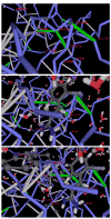
Figure 1. Structure of HIV-1 Protease with an Inhibitor Acting as a Dehydron Wrapper
A dehydron is identified by determining the extent of the intramolecular desolvation, ρ, of the hydrogen bond, quantified as the number of nonpolar groups within its desolvation domain. The desolvation domain consists of two intersecting balls of radii 6.4 Å centered at the α-carbons of the paired residues. Most (~92% of the PDB entries) stable folds have at least two-thirds of backbone hydrogen bonds with ρ = 26.6 ± 7.5. Dehydrons are hydrogen bonds with ρ ≤19 (their r value is below the mean minus one Gaussian dispersion). The figure shows the Indinavir (Crixivan) inhibitor crystallized in complex with HIV-1 protease (PDB: 2BPX). The packing defects of the dimeric protease are shown in their spatial relation to the inhibitor position. The protein chain backbone is represented by blue virtual bonds joining α-carbons, well-wrapped backbone hydrogen bonds are shown as light-gray segments joining the α-carbons of the paired residues, and dehydrons are shown as green segments. The figure shows in detail the protease cavity, the pattern of packing defects, and the inhibitor positioned as a dehydron wrapper.
Figure 2
Figure 2. Inhibitor as a Wrapper of Packing Defects in the Urokinase-Type Plasminogen Activator
(A) Detail of the dehydron pattern of the protein cavity. (B) The inhibitor-protein complexation revealing the position of the inhibitor as a wrapper of the packing defects in the cavity. The only dehydrons in a concave region of the protein surface are: Cys191-Asp194, Asp194-Gly197, and Gln192-Lys143. Upon complexation, the inhibitor wraps all three dehydrons, contributing six nonpolar groups to their desolvation domains. (C) Hydrophobic residues in the cavity region and their mismatch against polar moieties in the inhibitor across the protein-ligand interface. There are three nonpolar residues in the rim of the protein cavity, Ile138, Val213, and Trp215, but none is engaged in hydrophobic interactions with the inhibitor.
Figure 3
Figure 3. The Nonconserved Wrapping across Paralogs Sharing Common Folds with Drug-Targeted Proteins
Dehydron pattern in α-thrombin (PDB: 1A3E), a paralog of the plasminogen activator (PDB: 1C5W) sharing a common domain structure, but different wrapping, together with the location of the inhibitor within the complex. The figure reveals the role of the inhibitor as a wrapper of the packing defects in the cavity. Notice the difference in the dehydron pattern of the cavity, distinguishing the α-thrombin from its paralog shown in Figure 2.
Figure 4
Figure 4. Modifications of Gleevec Geared at Improving Selectivity and Affinity for Brc-Abl
(A) Three possible sites for Gleevec methylation (I–III) aimed at selectively improving the wrapping of packing defects of Brc-Abl (PDB: 1FPU). (B) Structural alignment of Brc-Abl and its six paralogs by using the program Cn3D. The yellow region corresponds to a β hairpin in Brc-Abl covering amino acids 247–257. (C) The modified Gleevec-based molecule methylated at sites I and II and assayed in vitro in this study. (D) Rate of phosphorylation of Brc-Abl (blue), C-kit (green), Lck (red), Chk1 (purple), and Pdk1 (brown) in the presence of Gleevec (triangles) and in the presence of the I-, II-methylated modified Gleevec (squares). The latter compound was designed to better wrap the nonconserved dehydrons in Brc-Abl. Within the means of detection, the kinase phosphorylation rates do not vary appreciably in the range of 0–100 nM inhibitor concentration. Error bars represent the dispersion in measurements over ten repetitions of each kinetic assay.

Similar articles

Cited by

References

    1. Arkin MR, Wells JA. Small-molecule inhibitors of protein-protein interactions: progressing towards the dream. Nat Rev Drug Discov. 2004;3:301–317. - PubMed
    1. Attoub S, Rivat C, Rodrigues S, Van Borexlaer S, Bedin M, Buyneel E, Louvet C, Kornprobst M, Andre T, Mareel M, et al. The c-kit tyrosine kinase inhibitor STI-571 for colorectal cancer therapy. Cancer Res. 2002;62:4879–4883. - PubMed
    1. Cohen MS, Zhang C, Shokat KM, Taunton J. Structural Bioinformatics-based design of selective, irreversible kinase inhibitors. Science. 2005;308:1318–1321. - PMC - PubMed
    1. Deremble C, Lavery R. Macromolecular recognition. Curr Opin Struct Biol. 2005;15:171–175. - PubMed
    1. Dunker KA, Brown C, Obradovic Z. Indentification and function of usefully disordered proteins. Adv Protein Chem. 2002;62:25–49. - PubMed

Publication types

LinkOut - more resources