Comparative Study
. 2005 Dec 7;25(49):11300-12.
doi: 10.1523/JNEUROSCI.2270-05.2005.

Control of dendritic arborization by the phosphoinositide-3'-kinase-Akt-mammalian target of rapamycin pathway

Affiliations
Comparative Study

Control of dendritic arborization by the phosphoinositide-3'-kinase-Akt-mammalian target of rapamycin pathway

Jacek Jaworski et al. J Neurosci. .

Abstract

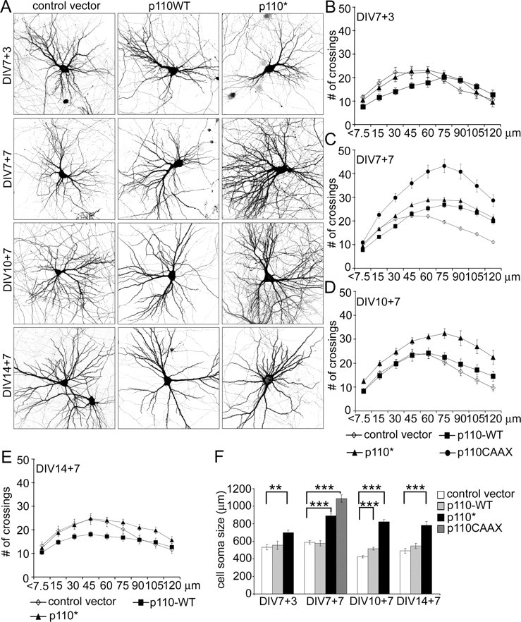
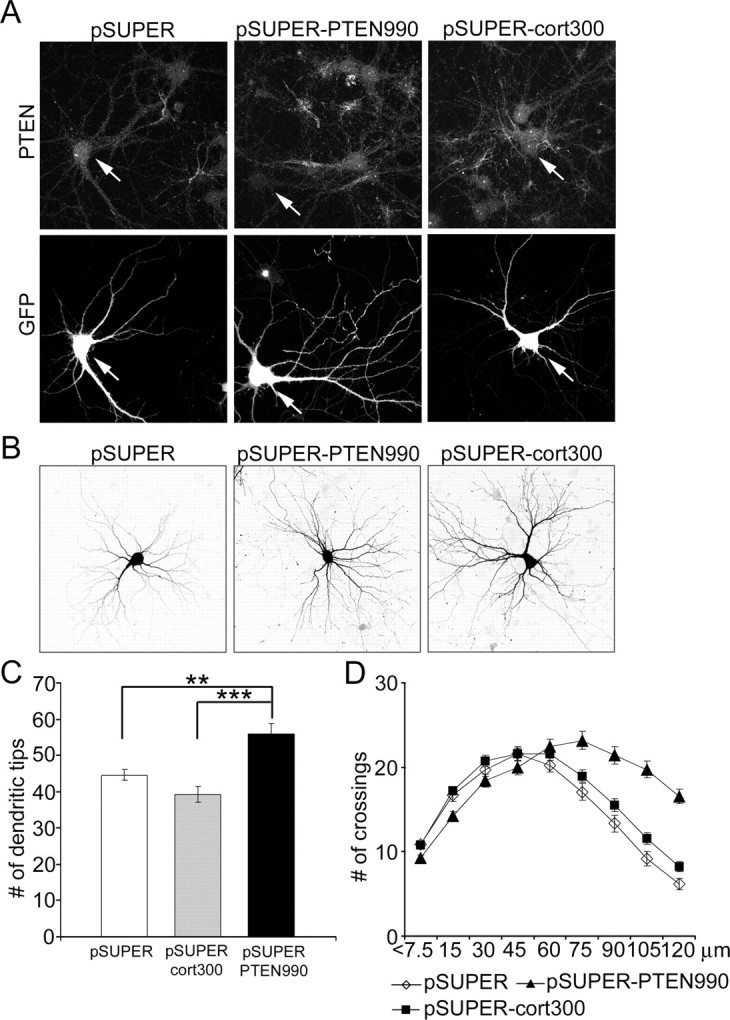
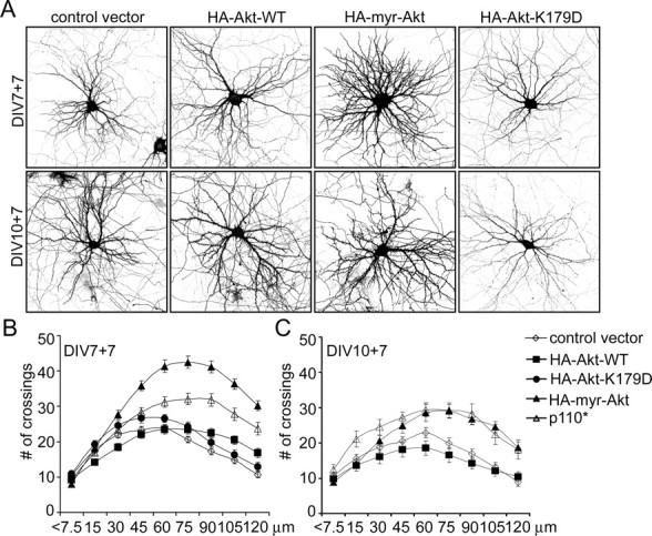
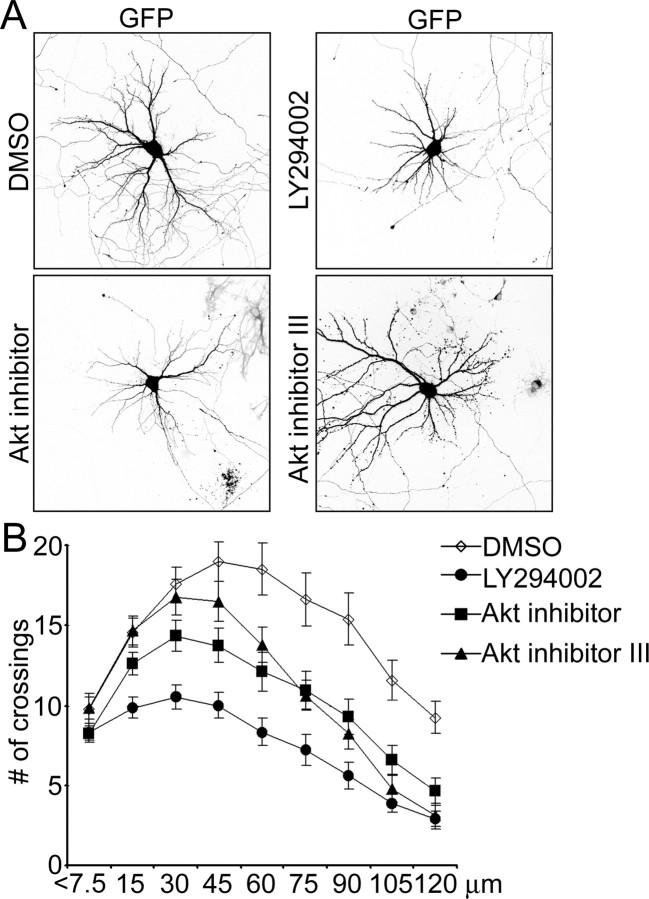
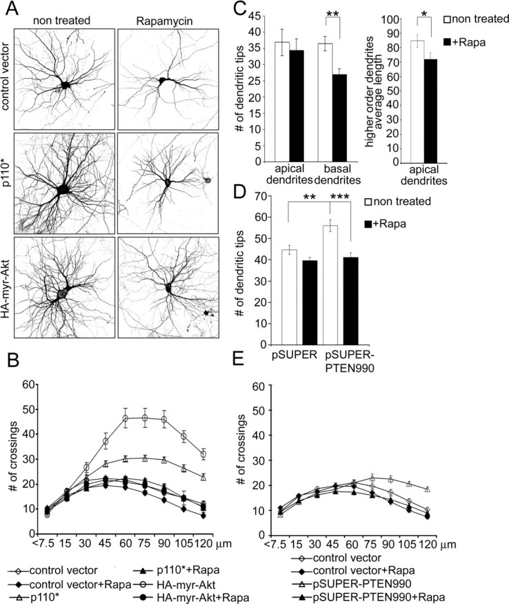
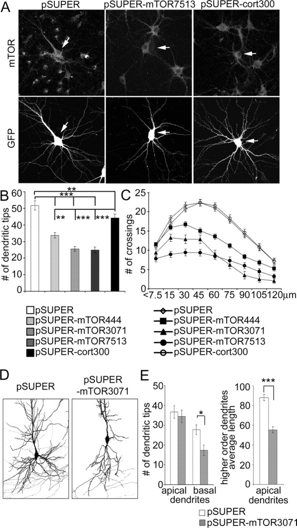
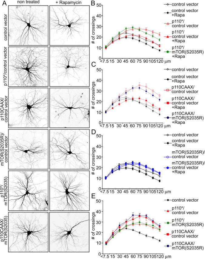
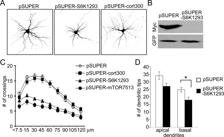
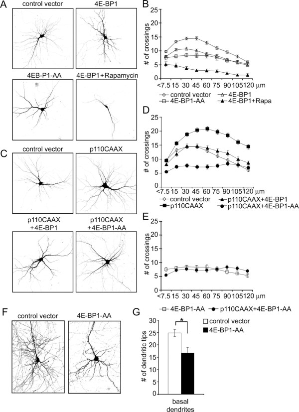
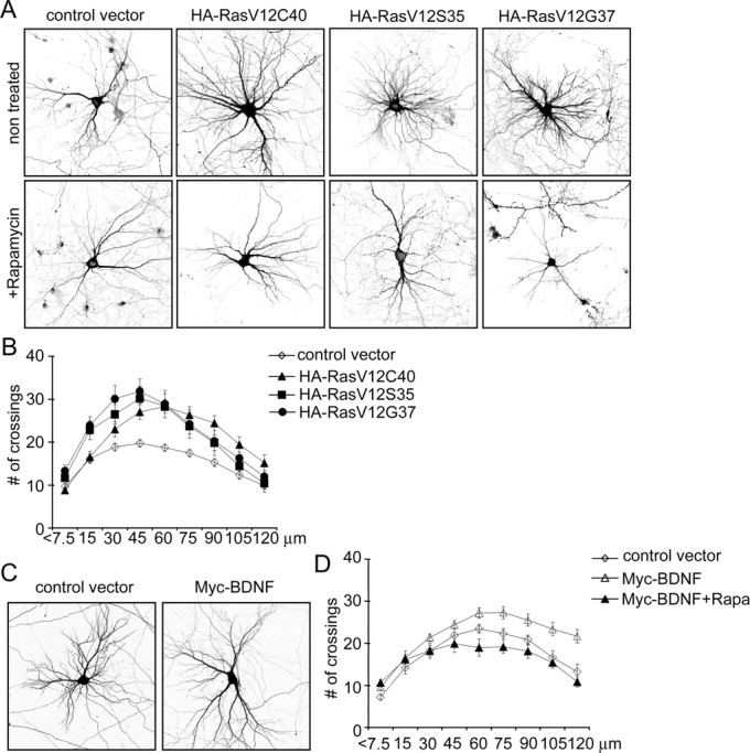
The molecular mechanisms that determine the size and complexity of the neuronal dendritic tree are unclear. Here, we show that the phosphoinositide-3' kinase (PI3K)-Akt-mammalian target of rapamycin (mTOR) signaling pathway promotes the growth and branching of dendrites in cultured hippocampal neurons. Constitutively active mutants of Ras, PI3K, and Akt, or RNA interference (RNAi) knockdown of lipid phosphatase PTEN (phosphatase and tensin homolog deleted on chromosome Ten), induced growth and elaboration of dendrites that was blocked by mTOR inhibitor rapamycin and/or by overexpression of eIF-4E binding protein 1 (4E-BP1), which inhibits translation of 5' capped mRNAs. The effect of PI3K on dendrites was lost in more mature neurons (>14 d in vitro). Dendritic complexity was reduced by inhibition of PI3K and by RNAi knockdown of mTOR or p70 ribosomal S6 kinase (p70S6K, an effector of mTOR). A rapamycin-resistant mutant of mTOR "rescued" the morphogenetic effects of PI3K in the presence of rapamycin. By regulating global and/or local protein translation, and as a convergence point for multiple signaling pathways, mTOR could play a central role in the control of dendrite growth and branching during development and in response to activity.


Figures

Figure 1.
Figure 1.
PI3K promotes dendrite branching in hippocampal neurons. A, Representative micrographs of hippocampal neurons transfected with control vector or wild-type p110α or constitutively active p110*. Neuron morphology was visualized by cotransfected GFP. Age and duration of transfection are indicated (e.g., DIV7 + 3 indicates transfected at 7 DIV and expressed for 3 d). B–E, Sholl analysis of neurons transfected with wild-type or active p110 mutants or control vector at various DIV as indicated. The horizontal axis of the Sholl plot indicates the distance from the cell soma. F, Effect of overexpression of PI3K variants on cell soma size. Age and duration of transfection are indicated. ***p < 0.001; **p < 0.01.
Figure 2.
Figure 2.
Knockdown of PTEN by RNAi increases dendritic branching of hippocampal neurons. A, PTEN immunostaining of hippocampal neurons transfected at 7 DIV for 4 d with pSUPER vector control, pSUPERcort300 (siRNA directed against cortactin), or pSUPER-PTEN990 (siRNA directed against PTEN). GFP was cotransfected to identify transfected cells and to visualize neuronal morphology. Arrows indicate transfected cells. B, Representative images of hippocampal neurons transfected at 7 DIV with pSUPER, pSUPERcort300, or pSUPER-PTEN990 for 5 d. Morphology of transfected cells was visualized by cotransfected GFP. C, Number of dendritic tips in hippocampal neurons transfected at 7 DIV for 5 d with pSUPER, pSUPERcort300, or pSUPER-PTEN990. ***p < 0.001; **p < 0.01. D, Sholl analysis of neurons transfected with pSUPER, pSUPERcort300, or pSUPER-PTEN990.
Figure 3.
Figure 3.
Constitutively active Akt promotes dendritic branching similar to PI3K. A, Representative micrographs of hippocampal neurons transfected with control vector, wild-type Akt (WT), constitutively active Akt (myr-Akt), or kinase-dead Akt (Akt-K179D). Neuronal morphology was visualized by cotransfected GFP. Age and duration of transfection are indicated. B, C, Sholl analysis of neurons transfected with control vector or Akt or PI3K constructs at various DIV as indicated.
Figure 4.
Figure 4.
Inhibition of PI3K and Akt reduces dendritic tree complexity. A, Representative micrographs of hippocampal neurons transfected with Bcl-2 at 7 DIV and treated for 3 d from 9 DIV with either 50 μm LY294002 or 10 μm Akt inhibitor or 25 μm Akt inhibitor III. Neuron morphology was visualized by cotransfected GFP. B, Sholl analysis of hippocampal neurons treated with PI3K or Akt inhibitors or DMSO vehicle as in A.
Figure 5.
Figure 5.
Rapamycin inhibits dendrite branching and the effects of PI3K and Akt. A, Representative micrographs of hippocampal neurons treated with vehicle or 10 nm rapamycin for 6 d. Neurons were transfected with control vector, constitutively active PI3K (p110*), or constitutively active Akt (myr-Akt) 1 d before rapamycin treatment. Neuron morphology was visualized by cotransfected GFP. B, Sholl analysis of neurons transfected with constructs and treated with rapamycin as in A. C, Effect of rapamycin on total number of apical and basal dendritic branches and average length of secondary and tertiary (higher-order) apical branches in organotypic cultures of hippocampus. Dendrites of CA1 neurons were visualized by transfection with GFP. Rapamycin (20 nm) was added 18 h after transfection for 5 d. D, Effect of rapamycin on number of dendrite tips in hippocampal neurons in dissociated culture, transfected at 7 DIV for 5 d with pSUPER or pSUPER-PTEN990. Rapamycin was added 8 h after transfection. ***p < 0.001; **p < 0.01. E, Sholl analysis of neurons transfected with pSUPER or pSUPER-PTEN990, cultured with or without rapamycin.
Figure 6.
Figure 6.
RNAi knockdown of mTOR decreases dendrite branching. A, Representative images of GFP and mTOR staining of hippocampal neurons transfected at 7 DIV for 4 d with pSUPER, pSUPERcort300, or pSUPER-mTOR7513 (RNAi directed against mTOR). GFP was cotransfected to visualize transfected cells (indicated by arrows). B, Number of dendrite tips in hippocampal neurons transfected at 7 DIV for 4 d with pSUPER, pSUPERcort300, or one of pSUPER-mTOR siRNA plasmids (444, 3071, or 7513). C, Sholl analysis of neurons transfected with control or mTOR siRNA plasmids. D, Images of CA1 neurons in organotypic culture transfected for 5 d with pSUPER vector or pSUPER-mTOR3071 siRNA plasmid (only basal dendrites shown in full). GFP was cotransfected for visualization of cell. E, Effect of mTOR3071 siRNA on total number of basal dendrites and average length of secondary and tertiary branches (referred to as higher-order dendrites) of apical dendrites in CA1 neurons of hippocampal organotypic cultures. ***p < 0.001; **p < 0.01; *p < 0.05.
Figure 7.
Figure 7.
Rapamycin inhibition of PI3K effect can be circumvented by rapamycin-insensitive mutant of mTOR. A, Representative micrographs of hippocampal neurons transfected at 7 DIV with the indicated constructs and treated with vehicle or rapamycin (10 nm) for 6 d. Neuronal morphology was visualized by cotransfected GFP. B–E, Sholl analysis of neurons transfected with the indicated constructs in the absence or presence of rapamycin (+Rapa).
Figure 8.
Figure 8.
RNAi knockdown of p70S6K decreases dendrite branching. A, Representative micrographs of hippocampal neurons transfected at 7 DIV for 4 d with pSUPER, pSUPERcort300, or pSUPER-S6K1293 (RNAi directed against p70S6K). GFP was cotransfected for visualization of transfected cells. B, Western blot analysis of effect of pSUPER-S6K1293 on expression of cotransfected Myc-p70S6K protein in COS7 cells. C, Sholl analysis of neurons transfected at 7 DIV for 4 d with pSUPER, pSUPERcort300, or pSUPER-S6K1293. D, Effect of pSUPER-S6K1293 transfection on total number of apical and basal dendrites in CA1 neurons of hippocampal organotypic cultures. *p < 0.05
Figure 9.
Figure 9.
Overexpression of 4E-BP1 inhibits dendritic branching. Representative micrographs of hippocampal neurons transfected at 7 DIV for 1 week with control vector, wild-type 4E-BP1, or 4E-BP1-AA mutant. Rapamycin (10 nm) was added 1 d after transfection, as indicated. Neuron morphology was visualized by cotransfected GFP. B, Sholl analysis of neurons transfected and/or treated as in A. C, Representative micrographs of hippocampal neurons transfected at 7 DIV for 7 d with p110CAAX and 4E-BP1 constructs, as indicated. Neuronal morphology was visualized by cotransfected GFP. D, E, Sholl analysis of neurons transfected with the indicated constructs as described in A and C. F, G, Effect of 4E-BP1-AA mutant over expression on total number of basal dendrites in CA1 neurons of hippocampal organotypic cultures. *p < 0.05.
Figure 10.
Figure 10.
Ras and BDNF regulate dendritic branching in an mTOR-dependent manner. A, Representative micrographs of hippocampal neurons transfected at 7 DIV for 1 week with control vector, PI3K-specific (RasV12C40), MAPK-specific (RasV12S35), or Ral–GDS-specific (RasV12G37) constitutively active mutants of Ras, and treated with vehicle or single dose of 10 nm rapamycin 1 d after transfection. Neuron morphology was visualized by cotransfected GFP. B, Sholl analysis of neurons transfected with different Ras mutants, as indicated. C, Representative micrographs of hippocampal neurons transfected at 7 DIV for 1 week with control vector or myc-BDNF. D, Sholl analysis of neurons transfected with myc-BDNF grown in the absence or presence of 10 nm rapamycin for 6 d.

References

    1. Acebes A, Ferrus A (2000) Cellular and molecular features of axon collaterals and dendrites. Trends Neurosci 23: 557–565. - PubMed
    1. Akama KT, McEwen BS (2003) Estrogen stimulates postsynaptic density-95 rapid protein synthesis via the Akt/protein kinase B pathway. J Neurosci 23: 2333–2339. - PMC - PubMed
    1. Alpar A, Palm K, Schierwagen A, Arendt T, Gartner U (2003) Expression of constitutively active p21H-rasval12 in postmitotic pyramidal neurons results in increased dendritic size and complexity. J Comp Neurol 467: 119–133. - PubMed
    1. Atwal JK, Massie B, Miller FD, Kaplan DR (2000) The TrkB-Shc site signals neuronal survival and local axon growth via MEK and P13-kinase. Neuron 27: 265–277. - PubMed
    1. Atwal JK, Singh KK, Tessier-Lavigne M, Miller FD, Kaplan DR (2003) Semaphorin 3F antagonizes neurotrophin-induced phosphatidylinositol 3-kinase and mitogen-activated protein kinase kinase signaling: a mechanism for growth cone collapse. J Neurosci 23: 7602–7609. - PMC - PubMed

Publication types

MeSH terms