Comparative Study
. 2005 Dec 7;25(49):11349-56.
doi: 10.1523/JNEUROSCI.3328-05.2005.

Activation of gonadotropin-releasing hormone neurons by kisspeptin as a neuroendocrine switch for the onset of puberty

Affiliations
Comparative Study

Activation of gonadotropin-releasing hormone neurons by kisspeptin as a neuroendocrine switch for the onset of puberty

Seong-Kyu Han et al. J Neurosci. .

Abstract

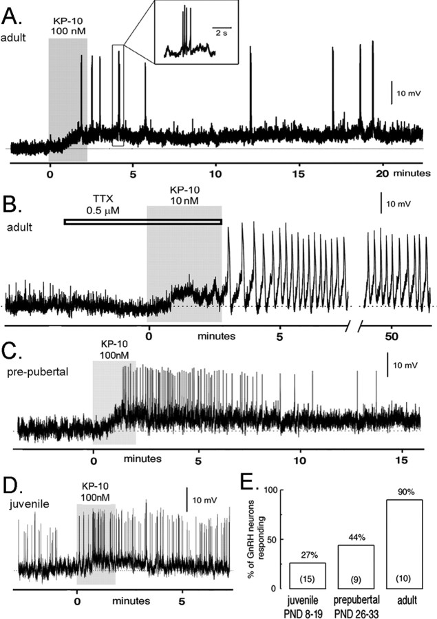
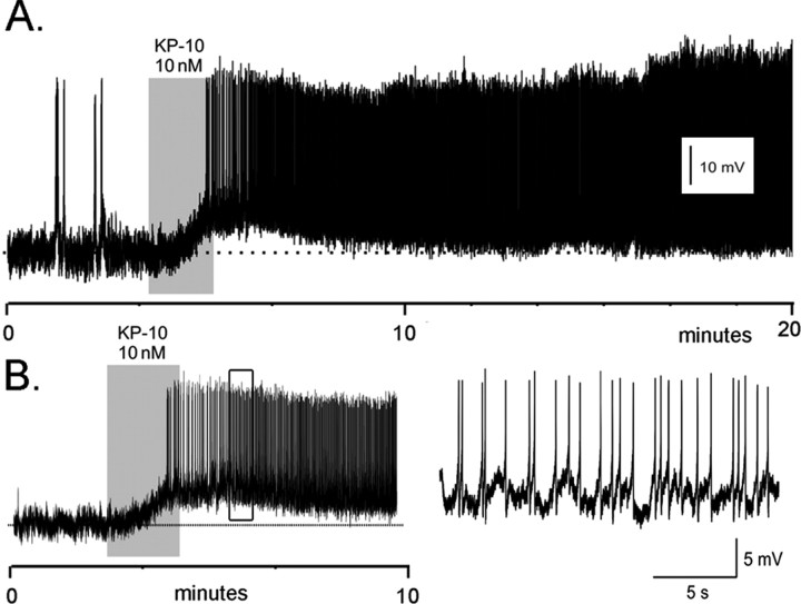
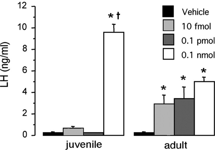
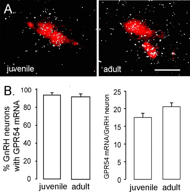
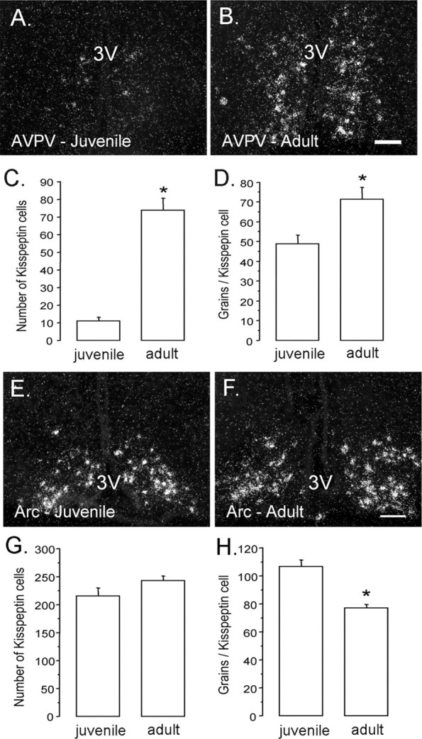
We examined the role of kisspeptin and its receptor, the G-protein-coupled receptor GPR54, in governing the onset of puberty in the mouse. In the adult male and female mouse, kisspeptin (10-100 nM) evoked a remarkably potent, long-lasting depolarization of >90% of gonadotropin-releasing hormone (GnRH)-green fluorescent protein neurons in situ. In contrast, in juvenile [postnatal day 8 (P8) to P19] and prepubertal (P26-P33) male mice, kisspeptin activated only 27 and 44% of GnRH neurons, respectively. This developmental recruitment of GnRH neurons into a kisspeptin-responsive pool was paralleled by an increase in the ability of centrally administered kisspeptin to evoke luteinizing hormone secretion in vivo. To learn more about the mechanisms through which kisspeptin-GPR54 signaling at the GnRH neuron may change over postnatal development, we performed quantitative in situ hybridization for kisspeptin and GPR54 transcripts. Approximately 90% of GnRH neurons were found to express GPR54 mRNA in both juvenile and adult mice, without a detectable difference in the mRNA content between the age groups. In contrast, the expression of KiSS-1 mRNA increased dramatically across the transition from juvenile to adult life in the anteroventral periventricular nucleus (AVPV; p < 0.001). These results demonstrate that kisspeptin exerts a potent depolarizing effect on the excitability of almost all adult GnRH neurons and that the responsiveness of GnRH neurons to kisspeptin increases over postnatal development. Together, these observations suggest that activation of GnRH neurons by kisspeptin at puberty reflects a dual process involving an increase in kisspeptin input from the AVPV and a post-transcriptional change in GPR54 signaling within the GnRH neuron.


Figures

Figure 1.
Figure 1.
Kisspeptin exerts a potent, direct depolarizing action on GnRH neurons that is regulated developmentally. Perforated-patch, voltage recordings from GnRH–GFP neurons in the acute brain slice preparation of male mice. A, An adult male GnRH neuron (RMP, –63 mV) is depolarized by 100 nm kisspeptin-10 (KP-10) for >20 min and exhibits short bursts of action potentials (inset). B, Adult male GnRH neuron (RMP, –60 mV) exhibiting a depolarizing response and pronounced membrane oscillations to 10 nm kisspeptin-10 (KP-10) in the presence of TTX. This cell continued to display membrane oscillations for >50 min before the recording was lost. C, A prepubertal (P30; RMP, –63 mV) GnRH neuron exhibiting a depolarizing response associated with increased firing in response to 100 nm kisspeptin-10 (KP-10). The depolarizing response in this cell lasted for 20 min. D, Spontaneously active juvenile (P9; RMP, –60 mV) GnRH neuron responding to kisspeptin-10 (KP-10) with a transient depolarization. E, Developmental recruitment of kisspeptin-sensitive GnRH neurons across puberty. The histogram shows the percentage of male juvenile (P8–P19), prepubertal (P26–P33), and adult (older than P60) GnRH neurons activated by KP-10 (n in parentheses). PND, Postnatal day.
Figure 2.
Figure 2.
Kisspeptin exerts a potent activational effect on GnRH neurons in adult female proestrous mice. Perforated-patch, voltage recordings from proestrous female GnRH–GFP neurons in the acute brain slice demonstrate a remarkably intense and prolonged activation by 10 nm kisspeptin (KP-10). A, Adult female GnRH neuron (RMP, –75 mV) that was activated by KP-10 for >80 min, at which time the recording was lost. B, Adult female GnRH neuron (RMP, –68 mV) activated by KP-10. The firing pattern occurring within the open box is shown at an expanded time scale on the right.
Figure 3.
Figure 3.
Developmental differences in the ability of kisspeptin to elicit LH release in vivo. Comparison of the LH response to kisspeptin-52 between P18 and adult male mice. Various doses of kisspeptin were administered into the lateral cerebral ventricle of the brain (intracerebroventricularly) of P18 and adult male mice (n = 6 per group). Serum was collected 30 min later. *p < 0.01 versus vehicle control; p < 0.001 versus adult receiving 0.1 nmol. Error bars represent SEM.
Figure 4.
Figure 4.
Expression of GPR54 mRNA in GnRH neurons across development. A, Representative photomicrographs of cells coexpressing GPR54 mRNA (reflected by silver grains, appearing as clusters of white dots) and GnRH mRNA (labeled with Vector Red) in juvenile (left) and intact adult (right) male mice. Scale bar, 20 μm. B, Quantitative analysis of GPR54 mRNA in GnRH neurons demonstrated that neither the percentage of double-labeled cells (left) nor the relative expression of GPR54 mRNA in GnRH neurons (reflected by the number of silver grains per GnRH neuron; right) differed significantly between P18 and adult mice (n = 6/7 per group). Errorbars represent SEM.
Figure 5.
Figure 5.
Developmental changes in KiSS-1 mRNA expression within the anteroventral periventricular and arcuate nuclei. A–D, KiSS-1 mRNA in the AVPV. Photomicrographs show AVPV cells possessing KiSS-1 mRNA (reflected by silver grains, appearing as clusters of white dots) in juvenile (left) and adult (right) male mice. Quantitative analysis of kisspeptin mRNA revealed that both cell number (C; p < 0.0001) and cell content (D; p < 0.05) of kisspeptin mRNA increased significantly from juvenile to adult mice (n = 6/7 per group). E, F, KiSS-1 mRNA in the arcuate nucleus (Arc). Photomicrographs show KiSS-1 mRNA in the arcuate nucleus (Arc) of juvenile (E) and adult (F) male mice (n = 6/7 per group). Quantitative analysis of KiSS-1 mRNA in the Arc revealed that the number of positive cells did not differ between juvenile and adult mice (G); however, the per cell content of KiSS-1 mRNA (reflected by the number of silver grains per cell) was reduced in adult mice (H; p < 0.001). The asterisks denote statistical significance. Scale bars, 100 μm. 3V, Third ventricle. Error bars represent SEM.

References

    1. Chappel PE, Lydon JP, Conneely OM, O'Malley BT, Levine JE (1997) Endocrine defects in mice carrying a null mutation for the progesterton receptor gene. Endocrinology 138: 4147–4152. - PubMed
    1. de Roux N, Genin E, Carel JC, Matsuda F, Chaussain JL, Milgrom E (2003) Hypogonadotropic hypogonadism due to loss of function of the KiSS1-derived peptide receptor GPR54. Proc Natl Acad Sci USA 100: 10972–10976. - PMC - PubMed
    1. Funes S, Hedrick JA, Vassileva G, Markowitz L, Abbondanzo S, Golovko A, Yang S, Monsma FJ, Gustafson EL (2003) The KiSS-1 receptor GPR54 is essential for the development of the murine reproductive system. Biochem Biophys Res Commun 312: 1357–1363. - PubMed
    1. Gonzales C, Voirol MJ, Giacomini M, Gaillard RC, Pedrazzini T, Pralong FP (2004) The neuropeptide Y Y1 receptor mediates NPY-induced inhibition of the gonadotrope axis under poor metabolic conditions. FASEB J 18: 137–139. - PubMed
    1. Gottsch ML, Cunningham MJ, Smith JT, Popa SM, Acohido BV, Crowley WF, Seminara S, Clifton DK, Steiner RA (2004) A role for kisspeptins in the regulation of gonadotropin secretion in the mouse. Endocrinology 145: 4073–4077. - PubMed

Publication types

MeSH terms