Comparative Study
. 2005 Dec 7;25(49):11374-84.
doi: 10.1523/JNEUROSCI.3871-05.2005.

Modulation of NMDA receptors by pituitary adenylate cyclase activating peptide in CA1 neurons requires G alpha q, protein kinase C, and activation of Src

Affiliations
Comparative Study

Modulation of NMDA receptors by pituitary adenylate cyclase activating peptide in CA1 neurons requires G alpha q, protein kinase C, and activation of Src

D S Macdonald et al. J Neurosci. .

Erratum in

  • J Neurosci. 2005 Dec 14;25(50):table of contents

Abstract

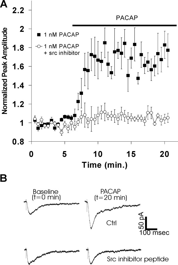
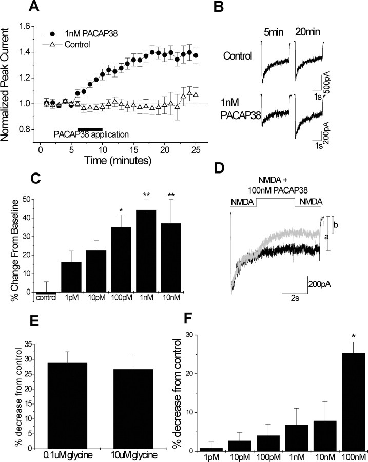
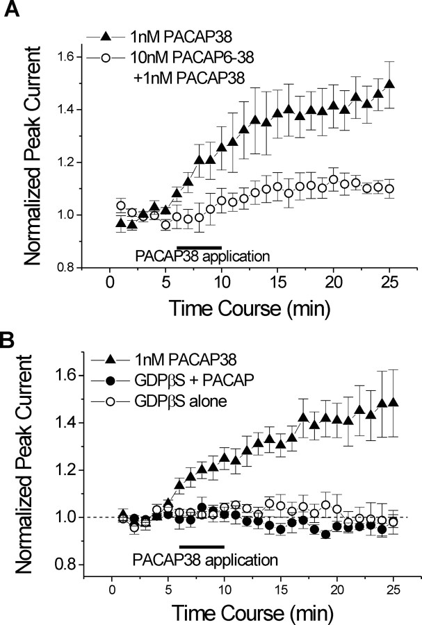
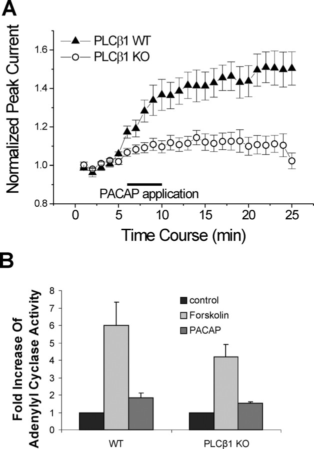
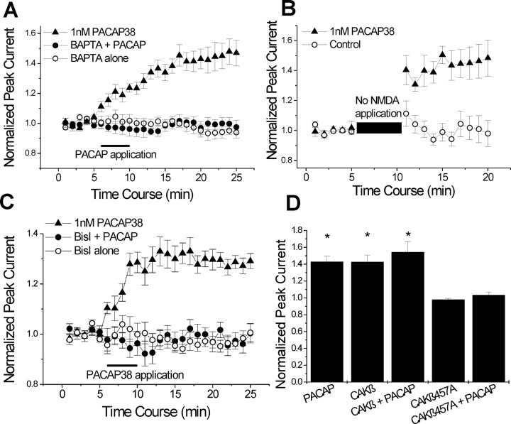
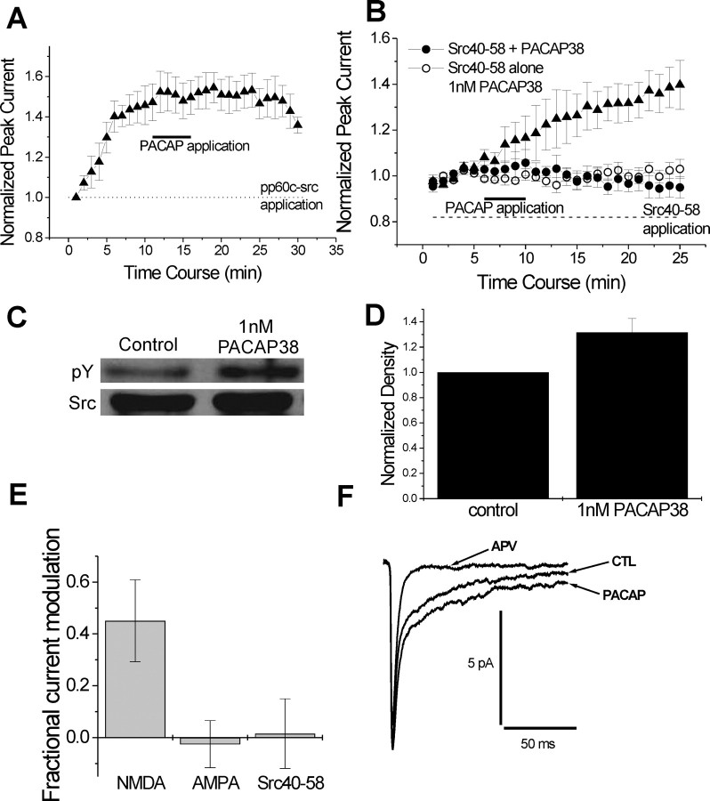
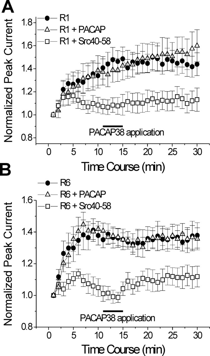
At CA1 synapses, activation of NMDA receptors (NMDARs) is required for the induction of both long-term potentiation and depression. The basal level of activity of these receptors is controlled by converging cell signals from G-protein-coupled receptors and receptor tyrosine kinases. Pituitary adenylate cyclase activating peptide (PACAP) is implicated in the regulation of synaptic plasticity because it enhances NMDAR responses by stimulating Galphas-coupled receptors and protein kinase A (Yaka et al., 2003). However, the major hippocampal PACAP1 receptor (PAC1R) also signals via Galphaq subunits and protein kinase C (PKC). In CA1 neurons, we showed that PACAP38 (1 nM) enhanced synaptic NMDA, and evoked NMDAR, currents in isolated CA1 neurons via activation of the PAC1R, Galphaq, and PKC. The signaling was blocked by intracellular applications of the Src inhibitory peptide Src(40-58). Immunoblots confirmed that PACAP38 biochemically activates Src. A Galphaq pathway is responsible for this Src-dependent PACAP enhancement because it was attenuated in mice lacking expression of phospholipase C beta1, it was blocked by preventing elevations in intracellular Ca2+, and it was eliminated by inhibiting either PKC or cell adhesion kinase beta [CAKbeta or Pyk2 (proline rich tyrosine kinase 2)]. Peptides that mimic the binding sites for either Fyn or Src on receptor for activated C kinase-1 (RACK1) also enhanced NMDAR in CA1 neurons, but their effects were blocked by Src(40-58), implying that Src is the ultimate regulator of NMDARs. RACK1 serves as a hub for PKC, Fyn, and Src and facilitates the regulation of basal NMDAR activity in CA1 hippocampal neurons.


Figures

Figure 1.
Figure 1.
PACAP38 enhances NMDA currents in hippocampal slice. A, Application of 1 nm PACAP38 to hippocampal slices caused increased amplitude in NMDA currents. Pyramidal cells were recorded in a whole-cell configuration. NMDA peak currents reached a maximal increase ∼8 min after application. Normalized peak amplitude for PACAP-treated cells was 167 ± 10% compared with baseline (n = 6). When Src(40-58) (25 μg/ml) was included in the patch pipette, PACAP38 failed to elicit a response [normalized peak current, 102 ± 2% (n = 6); p < 0.005, unpaired t test; data obtained at 20 min of recording]. The black bar indicates time and duration of 1 nm PACAP38 application. B, Sample traces from individual cells with and without Src(40-58) in the patch pipette at baseline (t = 0 min) and after PACAP38 application (t = 20 min).
Figure 2.
Figure 2.
PACAP38 enhances peak currents in isolated CA1 pyramidal neurons. A, Application of PACAP38 (1 nm) to acutely isolated CA1 pyramidal neurons resulted in an increase of NMDA-evoked peak currents that outlasted the period of application, did not reverse during washout, and persisted throughout the recording period. NMDA-evoked peak currents in control cells were unchanged throughout the time course of the experiment. Cells treated with 1 nm PACAP38 had significantly larger NMDA-evoked peak currents (control, 98 ± 7%, n = 9; 1 nm PACAP38, 140 ± 5%, n = 9; p < 0.001, unpaired t test; data obtained at 20 min of recording). The black bar indicates time and duration of 1 nm PACAP38 application. B, Sample traces of NMDA-evoked currents for control and PACAP38-treated cells. Traces represent points immediately before PACAP38 application (t = 5 min) and 10 min after PACAP38 application (t = 20 min). C, PACAP38 modulation of NMDA peak currents was dose dependent. Change from baseline: control, -1 ± 7%, n = 9; 1 pm,16 ± 6%, n = 4; 10 pm,23 ± 5%, n = 5; 100 pm,35 ± 7%, n = 5; 1 nm,44 ± 5%, n = 9; 10 nm,37 ± 13%, n = 6; data obtained at 20 min of recording. *p < 0.05, **p < 0.001 versus control, ANOVA followed by post hoc Tukey's test. D, PACAP38 at 100 nm directly inhibits NMDA currents. Sample traces from the same cell are shown without 100 nm PACAP38 application (black trace) and with 100 nm PACAP38 application (gray trace) during middle 3 s of NMDA-evoked current. Inhibition by PACAP38 was measured as the percentage that the steady-state decreased. E, Inhibition by 100 nm PACAP was not altered by the concentration of glycine. PACAP38 at 100 nm caused equal levels of inhibition in 0.1 and 10 μm glycine (low glycine, 29 ± 4%, n = 6; high glycine, 27 ± 4%, n = 6; p > 0.3, unpaired t test). F, NMDA currents were only inhibited by 100 nm PACAP38. With doses from 1 pm to 100 nm, only 100 nm PACAP38 showed a significant inhibition of NMDA currents (ANOVA followed by post hoc Tukey's test, p < 0.05).
Figure 3.
Figure 3.
Effect of PACAP38 on NMDA-evoked currents requires the G-protein-coupled PAC1 receptor. A, Addition of 10 nm PACAP(6-38), a potent antagonist of PACAP38 specific for the PAC1 receptor, in all extracellular solutions for the duration of the experiment significantly attenuated the increase in NMDA-evoked currents by 1 nm PACAP38 (1 nm PACAP38, 141 ± 8%, n = 6; 10 nm PACAP(6-38) plus 1 nm PACAP38, 113 ± 5%, n = 7; p < 0.05, unpaired t test; data obtained at 20 min of recording). The black bar indicates time and duration of 1 nm PACAP38 application. B, Increased NMDA-evoked peak currents are blocked by GDP-β-S. Intracellular application of 20 μm GDP-β-S blocked the increase in NMDA-evoked peak currents by 1 nm PACAP38 (1 nm PACAP38, 141 ± 7%, n = 7; 1 nm PACAP38 plus 20 μm GDP-β-S, 96 ± 2%, n = 6; 20 μm, p < 0.001, un paired t test; data obtained at 20 min of recording). Comparatively, GDP-β-S alone had no effect on NMDA-evoked currents (103 ± 6% of baseline; n = 5). The black bar indicates time and duration of 1 nm PACAP38 application.
Figure 4.
Figure 4.
PLCβ1 is required for the PACAP38 response. A, PACAP38 (1 nm) failed to elicit a response in PLCβ1 knock-out mice but produced a robust increase in NMDA-evoked peak currents in wild-type mice (wild-type mice, 144 ± 8%, n = 10; knock-out mice, 110 ± 5%, n = 14; p < 0.001, unpaired t test; data obtained at 20 min of recording). The black bar indicates time and duration of 1 nm PACAP38 application. B, Adenylyl cyclase activity was unchanged in PLCβ1 knock-out mice. Hippocampal tissue was extracted from wild-type or PLCβ1 knock-out mice, and tissue from three to five mice were pooled. Forskolin (30 μm) induced a similar fold increase in both wild-type (6.0 ± 1.3-fold increase; n = 4) and knock-out (4.2 ± 0.7-fold increase, n = 4; p > 0.05, unpaired t test) animals. Compared with vehicle-treated tissue, PACAP38-treated hippocampal tissue from wild-type mice had a 1.8 ± 0.3-fold (n = 4) increase in AC activity. In hippocampal tissue from PLCβ knock-out mice, PACAP induced a similar fold increase in AC activity (1.5 ± 0.1-fold increase, n = 4; p > 0.05, unpaired t test).
Figure 5.
Figure 5.
Intracellular calcium and calcium-activated kinases are required for the PACAP38 response. A, Intracellular administration of BAPTA (20 mm) blocked the PACAP38 effect and was similar to BAPTA alone (BAPTA plus PACAP38, 97 ± 4%, n = 6; BAPTA alone, 95 ± 4%, n = 6; p > 0.2, unpaired t test; data obtained at 20 min of recording; 1 nm PACAP38, 143 ± 7%, n = 6). The black bar indicates time and duration of 1 nm PACAP38 application. B, Blocking the entry of external calcium did not prevent PACAP-mediated enhancement of NMDA peak currents. To block external calcium entry, no NMDA was applied during the application of 1 nm PACAP38 (control, 98 ± 9%, n = 6; 1 nm PACAP38, 148 ± 12%, n = 5; p < 0.01, unpaired t test; data obtained at 20 min of recording). C, Protein kinase C is required for the PACAP38 response. Application of the PKC inhibitor bisindolylmaleimide I (500 nm) applied through internal and external solution blocked the effect of PACAP38 on NMDA-evoked currents and had no effect by itself (1 nm PACAP38, 130 ± 4%, n = 8; PACAP38 plus bisindolylmaleimide I, 103 ± 5%, n = 12; p<0.005, unpaired t test; bisindolylmaleimide I alone, 97 ± 2%, n = 5). The black bar indicates time and duration of 1 nm PACAP38 application. D, Pyk2/CAKβ is required for the PACAP38 response. Intracellular application of wild-type CAKβ (0.5 μg/ml) caused an increase in NMDA-evoked peak currents, reaching a plateau after 7 min. Five minute application of PACAP38 (1 nm) on top of the plateau caused no significant increase (CAKβ before PACAP38, 142 ± 8%, n = 6; 5 min after PACAP38 application, 154 ± 12%, n = 6; p > 0.3, paired t test; data obtained at 10 and 25 min of recording). The PACAP38 response could be blocked by a kinase inactive mutant version of the enzyme (lysine 457 to alanine). Intracellular application of CAKβ 457A (0.5 μg/ml) blocked the effect of PACAP38 (1 nm) and was similar to CAKβ 457A applied by itself (CAKβ 457A plus 1 nm PACAP38, 103 ± 3%, n = 6; CAKβ alone, 98 ± 2%, n = 5; data obtained at 20 min of recording; *p < 0.01 vs CAKβ 457A and CAKβ 457A plus 1 nm PACAP38, ANOVA followed by post hoc Tukey's test).
Figure 6.
Figure 6.
Nonreceptor tyrosine kinase Src is required for PACAP38 modulation of NMDA currents. A, Inclusion of 30 U/ml p60c-src in the intracellular solution caused a rapid and sustained increase in NMDA-evoked currents that occluded the effect of 1 nm PACAP38 (before PACAP38 application, 146 ± 10%, n = 6; 10 min after PACAP38 application, 153 ± 8%, n = 6; p > 0.5, paired t test; data obtained at 10 and 25 min of time recording). The black bar indicates time and duration of 1 nm PACAP38 application. B, Inclusion of the selective Src kinase peptide inhibitor Src(40-58) at 25 μg/ml in the patch pipette completely blocked the PACAP38 response (1 nm PACAP38, 132 ± 8%, n = 6; Src(40-58) plus 1 nm PACAP38, 100 ± 3%, n = 8; p < 0.005, unpaired t test; data obtained at 20 min of recording). Inclusion of Src(40-58) by itself had no effect [Src(40-58) alone, 97 ± 4%, n = 6; data obtained at 20 min of recording]. C, D, Activation of Src by 1 nm PACAP38 was determined by phosphorylation levels of Src. Microdissected CA1 hippocampal tissue was treated with vehicle or 1 nm PACAP38 for 10 min. Src was immunoprecipitated and probed with a phosphotyrosine antibody. Phosphorylation levels were normalized to the total amount of Src loaded and measured as a relative change compared with control. PACAP38 treatment increased tyrosine phosphorylation levels 32 ± 11% (n = 3) above vehicle-treated tissue. E, PACAP38 increased the NMDA component of mEPSPs, and the increase was Src dependent. Application of PACAP38 (1 nm) to cultured hippocampal neurons for 5 min increased the NMDA component of miniature EPSPs by 45 ± 16% (n = 6). Comparatively, the AMPA component of the mEPSPs was unchanged, with a fractional current modulation of -3 ± 9% (n = 6). Inclusion of the selective Src kinase peptide inhibitor Src(40-58) at 25 μg/ml in the patch pipette completely blocked the PACAP38 response (fractional change, 1.5 ± 13%; n = 5). F, Sample traces are shown for control, APV treated (i.e., AMPA component), and after PACAP38 treatment.
Figure 7.
Figure 7.
Binding of Src to the scaffolding protein RACK1 is required for PACAP modulation. A, Inclusion of 100 μm R1 in the patch pipette caused an increase in normalized NMDA peak currents. In a separate group of cells, the external application of 1 nm PACAP38 caused no additional enhancement of NMDA peak currents. When the selective Src kinase peptide inhibitor Src(40-58) at 25 μg/ml was included in the patch along with 100 μm R1, the potentiation of peak currents was severely attenuated: R1 alone, 147 ± 9%, n = 7; R1 plus PACAP38, 150± 12%, n = 7; R1 plus Src(40-58), 112 ± 9%, n = 9; data obtained at 25 min of recording. *p< 0.05 versus R1 plus Src(40-58), ANOVA followed by post hoc Tukey's test. The black bar indicates application of PACAP38. B, Inclusion of 100 μm R6 resulted in a similar pattern as that for experiments with the R1 peptide: R6 alone, 137 ± 8%, n = 7; R6 plus PACAP38, 138 ± 6%, n = 6; R6 plus Src(40-58), 112 ± 6%, n = 6; data obtained at 25 min of recording. *p < 0.05 versus R6 plus Src(40-58), ANOVA followed by post hoc Tukey's test. The black bar indicates application of PACAP38.

References

    1. Alier KA, Morris BJ (2005) Divergent regulation of Pyk2/CAKbeta phosphorylation by Ca2+ and cAMP in the hippocampus. Biochim Biophys Acta - PubMed
    1. Arimura A, Somogyvari-Vigh A, Weill C, Fiore RC, Tatsuno I, Bay V, Brenneman DE (1994) PACAP functions as a neurotrophic factor. Ann NY Acad Sci 739: 228-243. - PubMed
    1. Benquet P, Gee CE, Gerber U (2002) Two distinct signaling pathways upregulate NMDA receptor responses via two distinct metabotropic glutamate receptor subtypes. J Neurosci 22: 9679-9686. - PMC - PubMed
    1. Chang BY, Conroy KB, Machleder EM, Cartwright CA (1998) RACK1, a receptor for activated C kinase and a homolog of the beta subunit of G proteins, inhibits activity of src tyrosine kinases and growth of NIH 3T3 cells. Mol Cell Biol 18: 3245-3256. - PMC - PubMed
    1. Chang BY, Chiang M, Cartwright CA (2001) The interaction of Src and RACK1 is enhanced by activation of protein kinase C and tyrosine phosphorylation of RACK1. J Biol Chem 276: 20346-20356. - PubMed

Publication types

MeSH terms

Substances

LinkOut - more resources