Comparative Study
. 2005 Dec 7;25(49):11444-54.
doi: 10.1523/JNEUROSCI.1711-05.2005.

Parsing molecular and behavioral effects of cocaine in mitogen- and stress-activated protein kinase-1-deficient mice

Affiliations
Comparative Study

Parsing molecular and behavioral effects of cocaine in mitogen- and stress-activated protein kinase-1-deficient mice

Karen Brami-Cherrier et al. J Neurosci. .

Erratum in

  • J Neurosci. 2006 Jan 18;26(3):table of contents. Simon, Arthur J [corrected to Arthur, Simon J]

Abstract

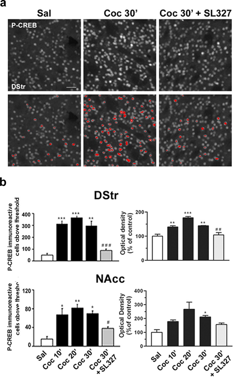
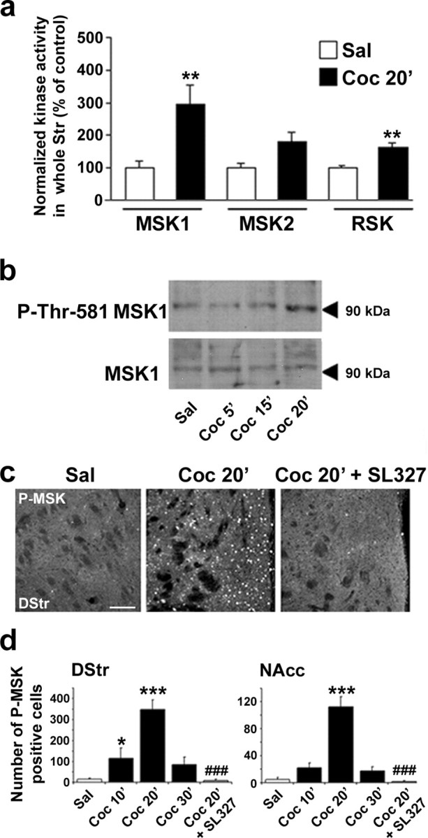
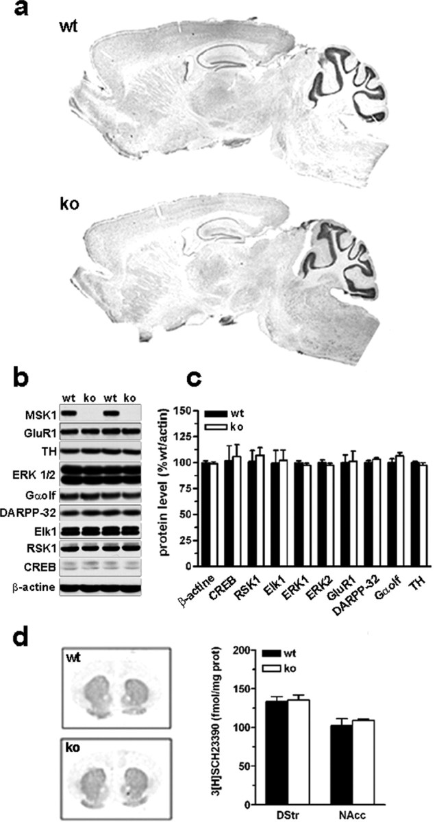
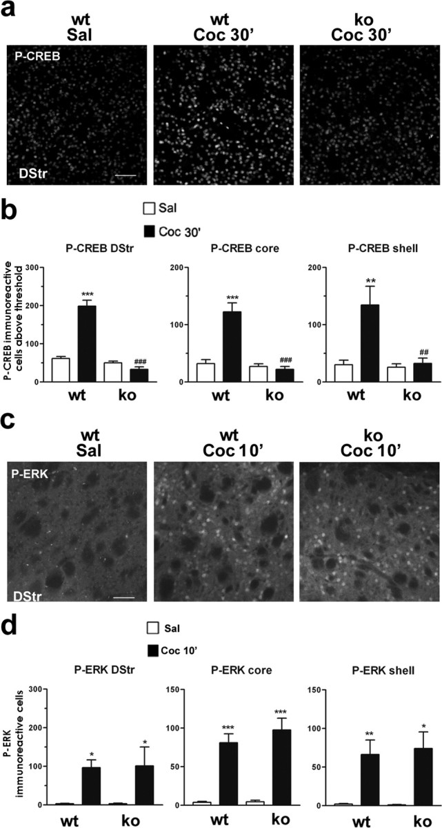
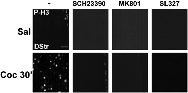
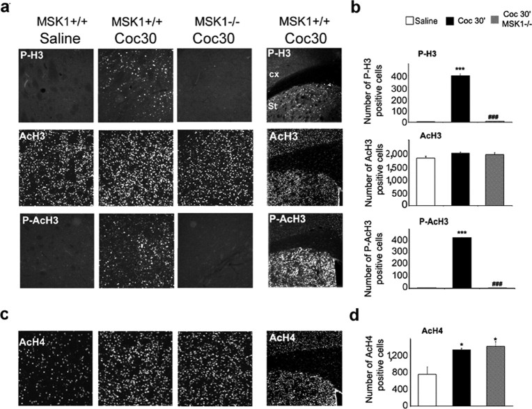
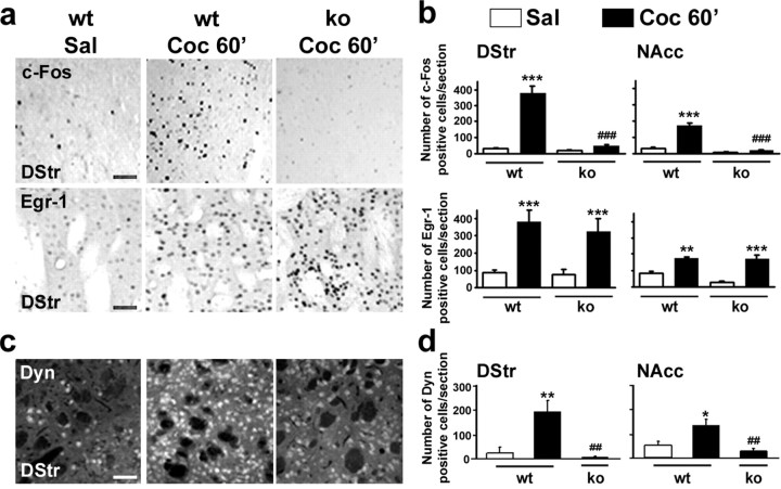
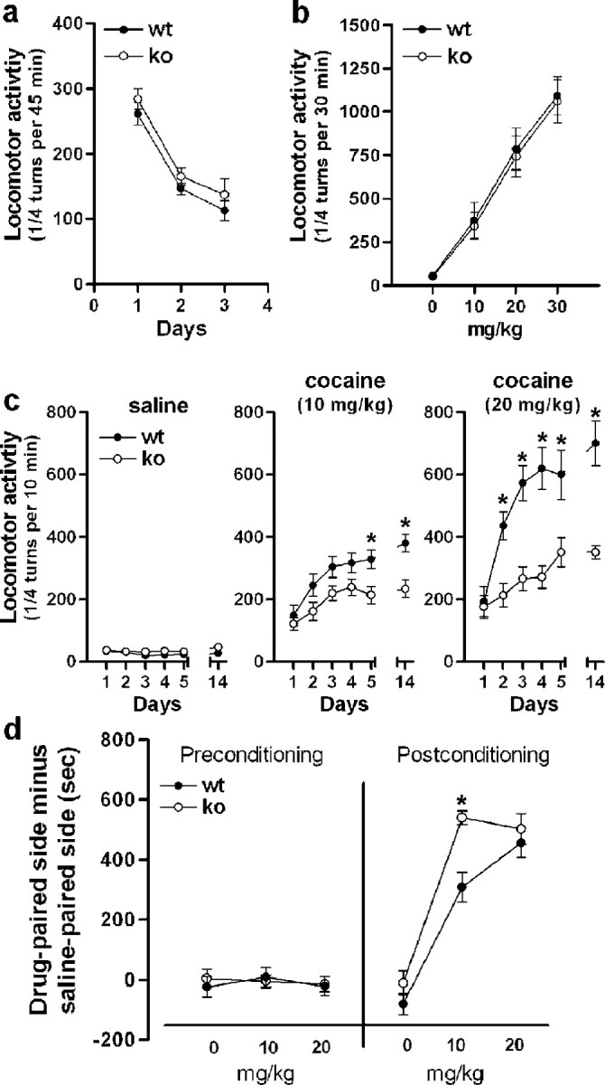
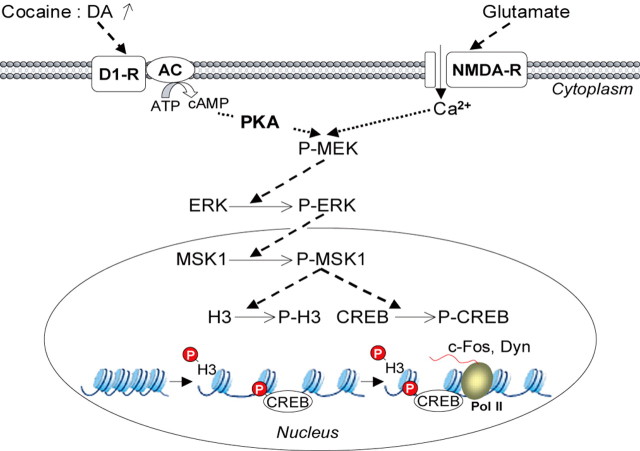
Although the induction of persistent behavioral alterations by drugs of abuse requires the regulation of gene transcription, the precise intracellular signaling pathways that are involved remain mainly unknown. Extracellular signal-regulated kinase (ERK) is critical for the expression of immediate-early genes in the striatum in response to cocaine and Delta9-tetrahydrocannabinol and for the rewarding properties of these drugs. Here we show that in mice a single injection of cocaine (10 mg/kg) activates mitogen- and stress-activated protein kinase 1 (MSK1) in dorsal striatum and nucleus accumbens. Cocaine-induced phosphorylation of MSK1 threonine 581 and cAMP response element-binding protein (CREB) serine 133 (Ser133) were blocked by SL327, a drug that prevents ERK activation. Cocaine increased the acetylation of histone H4 lysine 5 and phosphorylation of histone H3 Ser10, demonstrating the existence of drug-induced chromatin remodeling in vivo. In MSK1 knock-out (KO) mice CREB and H3 phosphorylation in response to cocaine (10 mg/kg) were blocked, and induction of c-Fos and dynorphin was prevented, whereas the induction of Egr-1 (early growth response-1)/zif268/Krox24 was unaltered. MSK1-KO mice had no obvious neurological defect but displayed a contrasted behavioral phenotype in response to cocaine. Acute effects of cocaine and dopamine D1 or D2 agonists were unaltered. Sensitivity to low doses, but not high doses, of cocaine was increased in the conditioned place preference paradigm, whereas locomotor sensitization to repeated injections of cocaine was decreased markedly. Our results show that MSK1 is a major striatal kinase, downstream from ERK, responsible for the phosphorylation of CREB and H3 and is required specifically for the induction of c-Fos and dynorphin as well as for locomotor sensitization.


Figures

Figure 1.
Figure 1.
Cocaine-induced CREB phosphorylation is ERK dependent in the striatum. Mice were injected intraperitoneally with saline or cocaine (10 mg/kg) in the absence or presence of SL327 (50 mg/kg) before the cocaine. a, Top panels present immunocytochemical detection of phospho-Ser133-CREB in striatal neurons from mice injected with saline, cocaine, and SL327 before the cocaine. The bottom panels are a computerized representation of P-CREB-positive cells after the application of a threshold determined from saline-treated striatal sections. Cells with fluorescence above this threshold were counted. Scale bar, 40 μm. b, P-CREB immunoreactivity above a threshold (left panels) or global P-CREB immunofluorescence from the striatal sections (right panels) were analyzed in the dorsal striatum (DStr) and in the nucleus accumbens (NAcc) at 10, 20, or 30 min after cocaine injection (Coc 10′, Coc 20′, Coc 30′) in the absence or in the presence of SL327 (50 mg/kg; Coc 30′ plus SL327). Statistical comparisons in the left panels (means ± SEM; 4 mice per group) include one-way ANOVA (in DStr, F(4, 14) = 30.04 and p < 0.001; in NAcc, F(4, 14) = 11.90 and p < 0.001), followed by post hoc comparison (Scheffé's test; *p < 0.05, **p < 0.01, and ***p < 0.001 Sal vs Coc; #p < 0.05 and ###p < 0.001 Coc 30′ vs Coc 30′ plus SL327). Statistical comparisons in the right panels (means ± SEM; 3-4 mice per group) include one-way ANOVA (in DStr, F(4, 14) = 22.36 and p < 0.001; in NAcc, F(4, 14) = 5.82 and p < 0.05), followed by post hoc comparison (Scheffé's test; *p < 0.05, **p < 0.01, and ***p < 0.001 Sal vs Coc; and ##p < 0.001 Coc 30′ vs Coc 30′ plus SL327).
Figure 2.
Figure 2.
Cocaine-induced MSK1 activation is ERK-dependent in the striatum. a, Kinase activity of MSK1, MSK2, and RSK was measured in striatal extracts from mice treated with saline (Sal) or 10 mg/kg cocaine (Coc 20′) 20 min before death. Data (means ± SEM; 6 mice per group) were analyzed by Student's t test (for MSK1, t10 = 3.33 and **p < 0.01; for MSK2, t10 = 2.145, not significant; for RSK, t10 = 4.619 and **p < 0.01). b, Immunoblot analysis of striatal extracts obtained 5, 15, or 20 min after cocaine injection (Coc 5′, Coc 15′, Coc 20′), using antibodies reacting with phospho-MSK1 (P-Thr581-MSK1) or MSK1 independently of its phosphorylation. A 90 kDa immunoreactive band with the expected molecular mass of MSK1 was observed with both antibodies. Similar results were obtained in three independent experiments. c, Phospho-MSK1 (P-MSK) immunohistochemical detection from dorsal striatum (DStr) of saline-treated (Sal) or cocaine-treated mice in the absence (Coc 20′) or presence of SL327 (50 mg/kg; Coc 20′ plus SL327). Scale bar, 80 μm. d, P-MSK1-immunoreactive cells were counted as described for P-CREB in the legend for Figure 1. Data (means ± SEM; 3-4 mice per group) were analyzed with one-way ANOVA (in DStr, F(4,14) = 18.48 and p < 0.001; in NAcc, F(4,14) = 36.65 and p < 0.001), followed by post hoc comparison (Scheffé's test; *p < 0.05 and ***p < 0.001 Sal vs Coc; ###p < 0.001 Coc 30′ vs Coc 30′ plus SL327).
Figure 3.
Figure 3.
Brain morphology and protein expression in MSK1 knock-out mice. a, Brain morphology was analyzed by using cresyl violet staining in wild-type (wt) and MSK1-KO (ko) mice. b, Expression levels of some important proteins for cell signaling studied by immunoblotting in striatal extracts. MSK1 is expressed strongly in the striatum of wild-type mice. None of the analyzed proteins was modified in MSK1-KO mice. c, Quantification of protein expression levels in wt and ko mice (means ± SEM; n = 7 mice per group). d, D1 receptor binding was studied and quantified by using [3H]SCH23390 as a radioligand (means ± SEM; n = 3 mice per group).
Figure 4.
Figure 4.
Cocaine-induced CREB phosphorylation is impaired in MSK1 knock-out mice. a, Illustration of P-CREB immunoreactivity 30 min after saline (Sal) or cocaine (10 mg/kg; Coc 30′) injection in the dorsal striatum of wild-type (wt) or MSK1-KO (ko) mice. Scale bar, 80 μm. b, P-CREB-immunoreactive cells below the threshold were counted in the dorsal striatum (DStr) and in the nucleus accumbens core and shell 30 min after saline or cocaine injection in wt and ko mice. Data (means ± SEM; 5 mice per group) were analyzed with one-way ANOVA (in DStr, F(5, 21) = 63.82 and p < 0.00 1; in core, F(5, 21) = 24.54 and p < 0.0001; in shell, F(5, 21) = 7.743 and p < 0.005), followed by post hoc comparison (Scheffé's test; **p < 0.01 and ***p < 0.001 Sal vs Coc in wt mice; ##p < 0.01 and ###p < 0.001 wt vs ko mice). c, Cocaine-induced phospho-ERK (P-ERK) is unaltered in ko mice. P-ERK immunoreactivity was detected 10 min after saline (Sal) or cocaine (10 mg/kg; Coc 10′) injection in wt or ko mice. Scale bar, 80 μm. d, P-ERK-immunoreactive cells were counted from wt and ko mice 10 min after cocaine administration in the dorsal (DStr) and ventral parts (core and shell) of the striatum. Data (means ± SEM; 3-4 mice per group) were analyzed with one-way ANOVA (in DStr, F(3,12) = 6.21 and p < 0.05; in core, F(3,12) = 37.38 and p < 0.0001; in shell, F(3,12) = 11.02 and p < 0.005), followed by post hoc comparison (Scheffé's test; *p < 0.05, **p < 0.01, and ***p < 0.001 Sal vs Coc in wt and ko mice).
Figure 5.
Figure 5.
Cocaine-induced histone H3 phosphorylation depends on D1 and NMDA receptor stimulation and ERK activation. Histone modification was analyzed 30 min after an injection of saline (Sal) or cocaine (10 mg/kg; Coc 30′) by immunofluorescence, using a phospho-Ser10 H3 (P-H3) antibody. These experiments were performed in the absence of (-) or after pretreatment with a selective D1 receptor antagonist (SCH23390; 0.1 mg/kg 15 min before cocaine), NMDA receptor antagonist (MK801; 0.15 mg/kg 30 min before cocaine), or an MEK inhibitor (SL327; 50 mg/kg 60 min before cocaine), as indicated. Scale bar, 20 μm.
Figure 6.
Figure 6.
Cocaine-induced histone modifications in MSK1 knock-out mice. Histone modifications were analyzed 30 min after cocaine injection (10 mg/kg; Coc 30′) in wild-type (wt) and MSK1-KO (ko) mice. a, Immunofluorescence labeling of phospho-Ser10 H3 (P-H3), acetylated Lys14 H3 (AcH3), and phospho-Ser10/acetylated Lys14 H3 (P-AcH3). Note that histone H3 phosphorylation and phosphoacetylation were restricted to the striatum (Str; right panels). b, Data (means ± SEM; 3 mice per group) were analyzed in the dorsal striatum (DStr) with one-way ANOVA (for P-H3, F(2,6) = 571.84 and p < 0.001; for AcH3, F(2,6) = 1.79, not significant; for P-AcH3, F(2, 6) = 11194.95 and p < 0.001), followed by post hoc comparison (Scheffé's test; ***p < 0.001 Sal vs Coc; ###p < 0.001 wt vs ko). c, Immunohistochemical detection of acetylated Lys5 H4 (AcH4) in the DStr. d, Data (means ± SEM; 3 mice per group) were analyzed in the DStr with one-way ANOVA (for AcH4, F(2, 6) = 8.91 and p < 0.05), followed by post hoc comparison (Scheffé's test; *p < 0.05 Sal vs Coc).
Figure 7.
Figure 7.
Cocaine-induced expression of c-Fos and dynorphin, but not Egr-1, is impaired in MSK1 knock-out mice. a, Immunocytochemical detection of c-Fos and Egr-1 expression in the striatum of wild-type (wt) and MSK1-KO (ko) mice 60 min after the administration of saline (Sal) or cocaine (10 mg/kg; Coc 60′). Scale bars, 40 μm. b, Quantification of c-Fos- and Egr-1-immunoreactive cells in the dorsal striatum (DStr) and in the nucleus accumbens (NAcc). Data (means ± SEM; 3-6 mice per group) were analyzed with one-way ANOVA: for c-Fos (in DStr, F(3, 12) = 8.16 and p < 0.001; in NAcc, F(3, 12) = 31.20 and p < 0.001); for Egr-1 (in DStr, F(3, 12) = 40.53 and p < 0.001; in NAcc, F(3, 12) = 16.87 and p < 0.01). c, Immunocytochemical detection of dynorphin expression was performed as in a. Scale bar, 40 μm. d, Quantification of dynorphin-immunoreactive cells. Data (means ± SEM; 3-6 mice per group) were analyzed with one-way ANOVA (in DStr, F(2, 12) = 8.62 and p < 0.001; in NAcc, F(2, 12) = 9.87 and p < 0.001). Post hoc comparisons were made with Scheffé's test: *p < 0.05, **p < 0.01, and ***p < 0.001 Sal versus Coc; ##p < 0.01 and ###p < 0.001 wt versus ko.
Figure 8.
Figure 8.
Locomotor and rewarding effects of cocaine in MSK1 knock-out mice. a, Spontaneous locomotor activity of wild-type (wt) and MSK1-KO (ko) mice for 3 d consecutively. b, Locomotor activity induced by a first intraperitoneal injection of cocaine at the indicated doses. c, Locomotor sensitization to repeated cocaine injections (10 and 20 mg/kg; n = 8-10 per group). After habituation, the wild-type and MSK1-KO mice were injected daily for 5 d consecutively with cocaine or saline, as indicated. They also were injected with saline or cocaine on day 14 after a 10 d withdrawal. Data were analyzed with a mixed factor ANOVA (repeated measure over time): for saline (effect of time, F(5, 54) = 2.561, not significant; effect of genotype, F(1, 54) = 1.012, not significant); for 10 mg/kg cocaine (effect of time, F(5, 54) = 12.87 and p < 0.01; effect of genotype, F(1, 54) = 9.748 and p < 0.01); for 20 mg/kg cocaine (effect of time, F(5,36) = 39.68 and p < 0.01; effect of genotype, F(1,36) = 27.31 and p < 0.01). In the posthoc comparison (Bonferroni test), *p < 0.01 wt versus ko. d, Conditioned place preference induced by cocaine (n = 7-8 mice per group). The left side of the graph (preconditioning) shows the lack of initial preference for either side by any experimental group (effect of treatment, F(3, 53) = 0.184, not significant; effect of genotype, F(1, 53) = 0.065, not significant) (note that the abscissa labels indicate the doses of cocaine used for each group in the later phase of the test). The right side of the graph shows that MSK1-KO mice displayed an enhanced preference for the side paired with 10 mg/kg cocaine as compared with wild-type mice. At 20 mg/kg of cocaine both wt and ko displayed an identical preference for the cocaine-paired side (effect of treatment, F(3, 53) = 83.75 and p < 0.01; effect of genotype, F(1, 53) = 10.05 and p < 0.01). In the post hoc comparison (Bonferroni test) *p < 0.01 wt versus ko.
Figure 9.
Figure 9.
Schematic representation of the role of MSK1 in neuronal adaptation in response to cocaine. Both dopamine D1 and glutamate NMDA receptors are involved in cocaine-induced ERK activation and immediate-early gene regulation (Valjent et al., 2000, 2005). D1 receptor stimulation increases intracellular levels of cAMP, which activates PKA. NMDA receptor stimulation causes increases in intracellular calcium (Ca2+) levels. Both events converge on the MEK/ERK signaling cascade (P-MEK and P-ERK). Phosphorylated ERKs (P-ERKs) translocate to the nucleus, where they phosphorylate MSK1 (P-MSK1), which controls gene expression via its dual (P-MEK and P-ERK) role on histone H3 and CREB phosphorylation. (1) Histone H3 phosphorylation (P-H3) by MSK1 leads to chromatin remodeling, facilitating DNA accessibility. (2) Phosphorylation of CREB (P-CREB) by MSK1 induces transcription of the immediate-early genes c-fos or preprodynorphin (Dyn) by RNA polymerase II (Pol II).

References

    1. Alarcon JM, Malleret G, Touzani K, Vronskaya S, Ishii S, Kandel ER, Barco A (2004) Chromatin acetylation, memory, and LTP are impaired in CBP+/- mice: a model for the cognitive deficit in Rubinstein-Taybi syndrome and its amelioration. Neuron 42: 947-959. - PubMed
    1. Arthur JS, Cohen P (2000) MSK1 is required for CREB phosphorylation in response to mitogens in mouse embryonic stem cells. FEBS Lett 482: 44-48. - PubMed
    1. Atkins CM, Selcher JC, Petraitis JJ, Trzaskos JM, Sweatt JD (1998) The MAPK cascade is required for mammalian associative learning. Nat Neurosci 1: 602-609. - PubMed
    1. Berke JD, Hyman SE (2000) Addiction, dopamine, and the molecular mechanisms of memory. Neuron 25: 515-532. - PubMed
    1. Berke JD, Paletzki RF, Aronson GJ, Hyman SE, Gerfen CR (1998) A complex program of striatal gene expression induced by dopaminergic stimulation. J Neurosci 18: 5301-5310. - PMC - PubMed

Publication types

MeSH terms

Substances