Comparative Study
. 2005 Dec 7;25(49):11468-78.
doi: 10.1523/JNEUROSCI.3484-05.2005.

Molecular and cellular diversity of neuronal G-protein-gated potassium channels

Affiliations
Comparative Study

Molecular and cellular diversity of neuronal G-protein-gated potassium channels

Lev Koyrakh et al. J Neurosci. .

Abstract

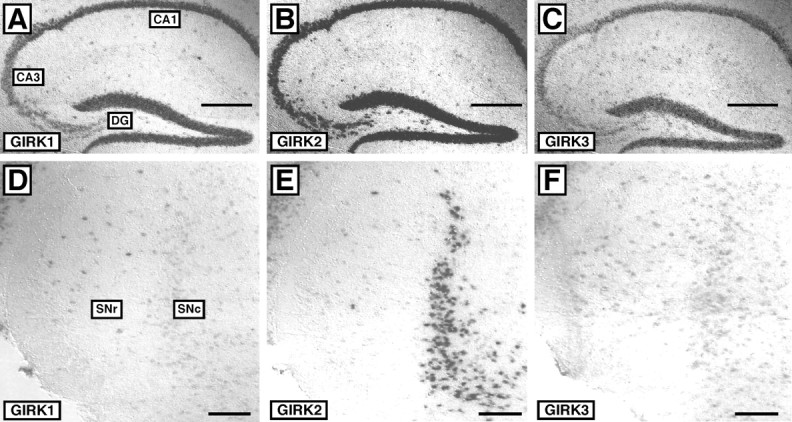
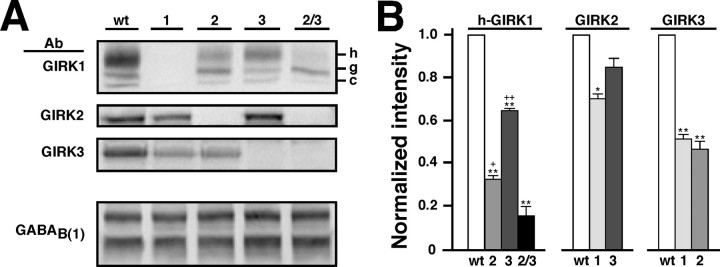
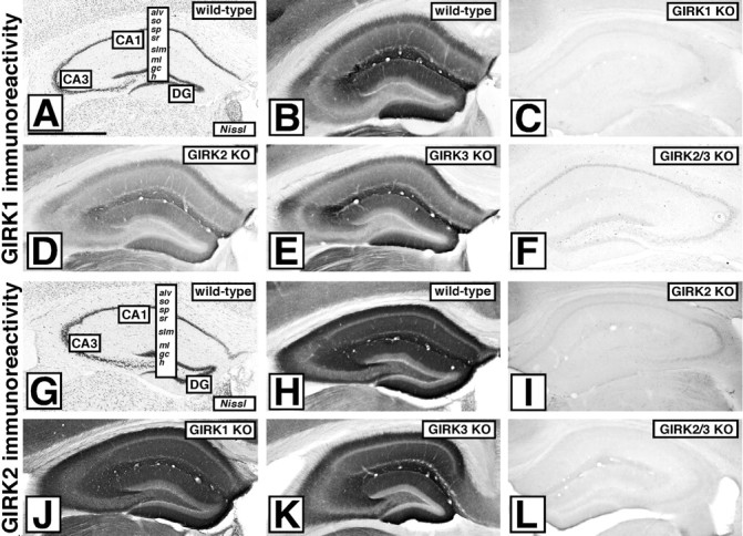
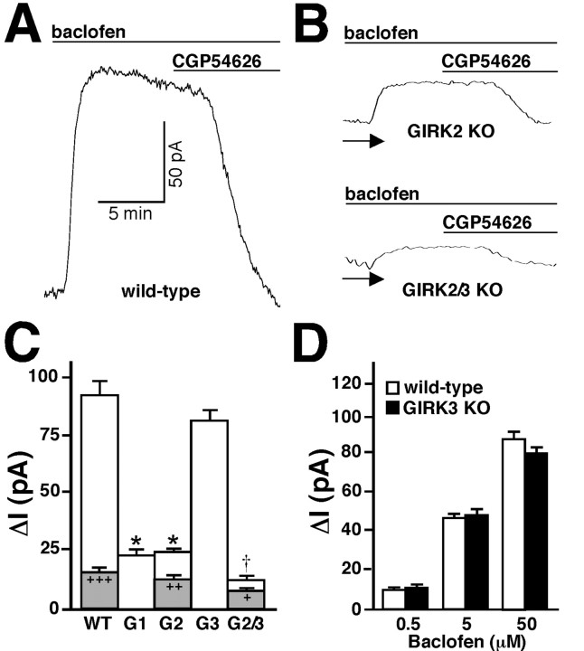
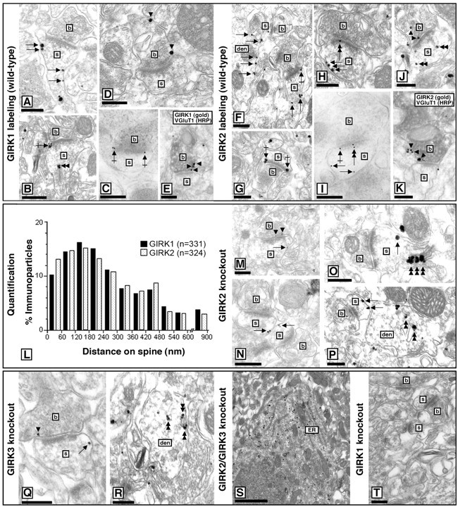
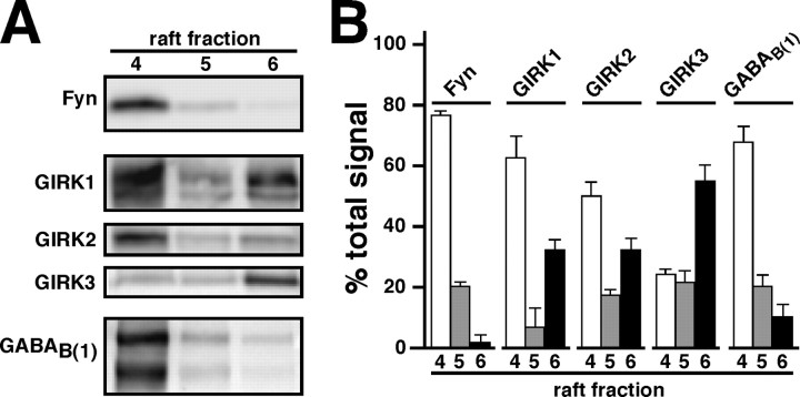
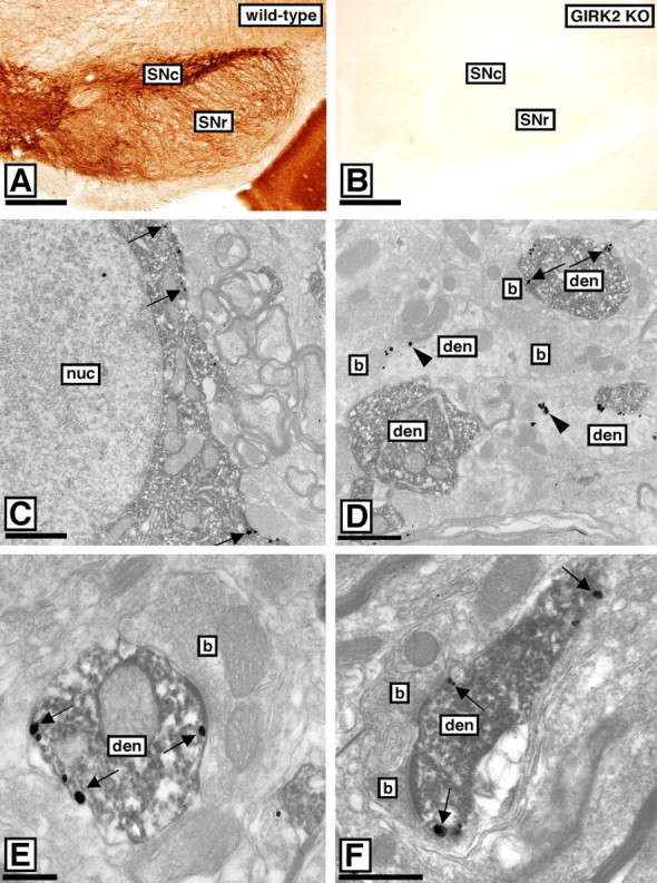
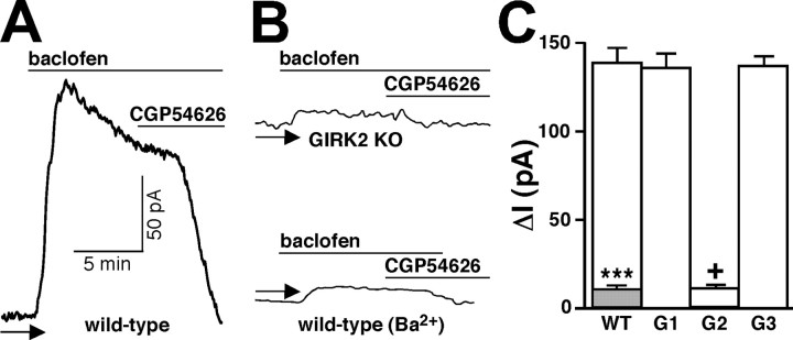
Neuronal G-protein-gated potassium (GIRK) channels mediate the inhibitory effects of many neurotransmitters. Although the overlapping distribution of GIRK subunits suggests that channel composition varies in the CNS, little direct evidence supports the existence of structural or functional diversity in the neuronal GIRK channel repertoire. Here we show that the GIRK channels linked to GABAB receptors differed in two neuron populations. In the substantia nigra, GIRK2 was the principal subunit, and it was found primarily in dendrites of neurons in the substantia nigra pars compacta (SNc). Baclofen evoked prominent barium-sensitive outward current in dopamine neurons of the SNc from wild-type mice, but this current was completely absent in neurons from GIRK2 knock-out mice. In the hippocampus, all three neuronal GIRK subunits were detected. The loss of GIRK1 or GIRK2 was correlated with equivalent, dramatic reductions in baclofen-evoked current in CA1 neurons. Virtually all of the barium-sensitive component of the baclofen-evoked current was eliminated with the ablation of both GIRK2 and GIRK3, indicating that channels containing GIRK3 contribute to the postsynaptic inhibitory effect of GABAB receptor activation. The impact of GIRK subunit ablation on baclofen-evoked current was consistent with observations that GIRK1, GIRK2, and GABAB receptors were enriched in lipid rafts isolated from mouse brain, whereas GIRK3 was found primarily in higher-density membrane fractions. Altogether, our data show that different GIRK channel subtypes can couple to GABAB receptors in vivo. Furthermore, subunit composition appears to specify interactions between GIRK channels and organizational elements involved in channel distribution and efficient receptor coupling.


Figures

Figure 1.
Figure 1.
GIRK subunit mRNA distribution in the hippocampus and substantia nigra. GIRK mRNA distributions were evaluated by in situ hybridization in sections from WT and GIRKKO mice. In sections from WT mice, mRNAs for GIRK1 (A), GIRK2 (B), and GIRK3 (C) were clearly evident in CA1 and CA3 pyramidal neurons, as well as granule cells of the dentate gyrus (DG). There was no specific staining for GIRK mRNAs in sections from the appropriate GIRK KO mouse (data not shown). Images are representative of data obtained from three different WT and GIRK KO panels. The WT mRNA distributions of GIRK1 (D), GIRK2 (E), and GIRK3 (F) were also examined in the SN. Scale bars: A-C, 500 μm; D-F, 100 μm.
Figure 2.
Figure 2.
GIRK subunit proteins are found in the mouse hippocampus. A, Representative immunoblots of hippocampal membrane protein samples from WT, GIRK1 KO (1), GIRK2 (2), GIRK3 (3), and GIRK2/GIRK3 (2/3) KO mice. Blots were probed with antibodies (Ab) for GIRK1, GIRK2, GIRK3, and GABAB(1a/b). GABAB immunoreactivity was observed as two prominent bands at ∼140 and 100 kDA (Malitschek et al., 1998; Fritschy et al., 1999). As described previously (Kennedy et al., 1996, 1999; Marker et al., 2002; Torrecilla et al., 2002), GIRK1 immunoreactivity was visualized as three bands, the lower molecular weight versions thought to represent core (c) and core-glycosylated (g) species. Reductions in the level of the heavily glycosylated (h) GIRK1 species were correlated with the absence of GIRK2 and GIRK3, respectively. The levels of GIRK2 and GIRK3 were also lower in samples from KO mice. B, Densitometric analysis of the impact of GIRK subunit ablation on residual GIRK protein levels in the hippocampus. Hippocampal protein samples were obtained from three separate and complete panels of WT and GIRK KO mice, and the levels of GIRK1 (heavily glycosylated form, h-GIRK1), GIRK2, GIRK3, and GABAB(1) (both isoforms) were determined. There was no effect of GIRK subunit ablation GABAB(1) receptor levels (F(4,10) = 1.764; p = 0.213) (data not shown). *p < 0.05; **p < 0.01 versus WT; +p < 0.05; ++p < 0.01 versus GIRK2/GIRK3 KO.
Figure 3.
Figure 3.
Distribution of GIRK1 and GIRK2 subunits in the hippocampus. Immunohistochemical detection of GIRK1 (A-F) and GIRK2 (G-L) in the hippocampal formations of WT, GIRK1, GIRK2, GIRK3, and GIRK2/GIRK3 (GIRK2/3) KO mice is shown. Staining patterns are representative of data obtained from three different and complete panels of adult WT and GIRK KO mice. A, G, Nissl staining of GIRK1 and GIRK2 in WT hippocampi, respectively. alv, Alveus; so, stratum oriens; sp, stratum pyramidale; sr, stratum radiatum; slm, stratum lacunosum-moleculare; ml, molecular layer; gc, granule cell; h, hilus; DG, dentate gyrus. B, C, GIRK1 immunolabeling in a WT (B) and GIRK1 KO (C) hippocampus. D-F, Residual GIRK1 immunoreactivity in sections from GIRK KO mice. H, GIRK2 immunolabeling in a WT hippocampus. I, L, GIRK2 labeling in GIRK2 KO (I) and GIRK2/GIRK3 KO (L) mice. J, K, No significant changes in immunoreactivity for GIRK2 were detected in the hippocampal formations of GIRK1 KO (J) and GIRK3 KO (K) mice. Scale bar, 1 mm.
Figure 4.
Figure 4.
Baclofen-induced current in CA1 neurons from WT and GIRK KO mice. Currents evoked by baclofen were measured in hippocampal slices from WT and GIRK KO mice. Holding potential was -62 mV. A, Typical current evoked by baclofen (50 μm) in a CA1 neuron from a WT mouse. The outward current was reversed with the GABAB receptor antagonist CGP54626 (1 μm). The holding current before baclofen application in this experiment was 90 pA. B, Typical baclofen-evoked currents in CA1 neurons from GIRK2 and GIRK2/GIRK3 (GIRK2/3) KO mice. The arrow identifies the zero current levels. C, Average peak current evoked by 50 μm baclofen in CA1 neurons from WT (n = 18), GIRK1 (G1; n = 9), GIRK2 (G2; n = 11), GIRK3 (G3; n = 7), and GIRK2/GIRK3 (G2/3; n = 7) KO mice. For some WT (n = 7), GIRK2 KO (n = 8), and GIRK2/GIRK3 double-knock-out (n = 4) recordings, baclofen-evoked currents were measured in the presence of 1 mm Ba2+ (peak currents are shown in gray). *p < 0.05 versus WT; p < 0.01 versus GIRK1 and GIRK2 KO; +p < 0.05; ++p < 0.01; +++p < 0.001 versus control (within genotype). D, Average peak currents evoked by 0.5, 5, and 50 μm baclofen in CA1 neurons from WT (n = 8) and GIRK3 KO (n = 7) mice.
Figure 5.
Figure 5.
Subcellular distributions of GIRK subunits in the hippocampus. Electron micrographs show immunolabeling for GIRK1 and GIRK2 in the stratum radiatum of the CA1 area of WT and GIRK KO mice. den, Dendritic shaft; b, bouton; s, dendritic spine. A, B, GIRK1 immunoparticles were observed primarily along the extrasynaptic plasma membrane (arrows) of dendritic spines of CA1 neurons from WT mice, although perisynaptic labeling (crossed arrow) was also frequently observed. GIRK1 labeling was also found associated with ER cisterna of dendritic shafts and with the spine apparatus (double arrowheads) of spines. C, GIRK1 labeling was never detected within the postsynaptic specialization, as demonstrated with postembedding techniques. D, E, GIRK1 immunoparticles at presynaptic sites were localized to the extrasynaptic plasma membrane (arrowheads) and occasionally to the presynaptic membrane specialization of axon terminals establishing putative excitatory synapses on spines, as confirmed in double-labeling experiments with VGluT1. F-H, GIRK2 immunoparticles were found along the extrasynaptic plasma membrane (arrows) of dendritic shafts of CA1 cells from WT mice, mainly associated with dendritic spines. Both perisynaptic (G, crossed arrows) and synaptic (H, double arrow) labeling at asymmetrical synapses was detected, the latter confirmed using postembedding techniques (I). J, K, GIRK2 immunoparticles were detected at presynaptic sites (arrowheads), mainly in the extra synaptic plasma membrane, and occasionally in the presynaptic membrane specialization of axon terminals establishing putative excitatory synapses on spines, as confirmed using double-labeling experiments with VGluT1. Note also the GIRK2 immunoreactivity associated with ER cisterna of dendritic shafts and the spine apparatus of dendritic spines (J, double arrowheads). L, Distribution of GIRK1 and GIRK2 on dendritic spines of CA1 neurons. Data are displayed as percentage frequency of particles in 60-nm-wide bins, starting at the edge of the postsynaptic specialization. M-T, Electron micrographs showing GIRK1 immunoreactivity in the stratum radiatum of the CA1 area in sections taken from GIRK2 KO (M-P), GIRK3 KO (Q, R), GIRK2/GIRK3 KO (S), and GIRK1 KO (T) mice. Note that GIRK1 immunoreactivity was still observed along the extrasynaptic plasma membrane (arrows) of dendritic spines and shafts of CA1 neurons from GIRK2 KO and GIRK3 KO mice. Immunoparticles for GIRK1 were also observed at perisynaptic (data not shown) positions and at presynaptic sites along the extrasynaptic plasma membrane (arrowheads) of axon terminals establishing putative excitatory synapses on dendritic spines. A higher proportion of immunoparticles for GIRK1 was detected in the ER cisterna of dendritic shafts and the spine apparatus (double arrowhead). Immunoreactivity for GIRK1 was primarily restricted to the ER of CA1 cells in the hippocampus of GIRK2/GIRK3 KO mice and was absent in sections from a GIRK1 KO mouse. Scale bars, 0.2 μm.
Figure 6.
Figure 6.
Compartmentalization of GABAB/GIRK signaling. A, Representative immunoblots showing the distribution of GIRK subunits and GABAB(1) in lipid rafts isolated from WT mice. The lipid raft marker and tyrosine kinase Fyn were consistently observed in fraction 4 after sucrose gradient centrifugation of Triton X-100-insoluble membranes (Delling et al., 2002). B, Densitometric analysis of target distribution in fractions from lipid raft preparations. Three isolations of lipid rafts were performed, and qualitatively similar observations were obtained each time. The histogram shows intensity observed in each lane represented as a percentage of total signal in lanes 4-6 (percentage of total signal).
Figure 7.
Figure 7.
Distribution of GIRK2 in the SNc. A, In WT sections containing the SN, GIRK2 immunoreactivity was most prominent in SNc neuron dendrites found in the SNr. B, No GIRK2 staining was observed in sections from GIRK2 KO mice. C, Colocalization of the GIRK2 subunit and TH in the SNc of WT mice, as revealed using preembedding methods. The peroxidase reaction product (TH immunoreactivity) filled somata and dendritic shafts, whereas immunoparticles (GIRK2 immunoreactivity) were located along the extrasynaptic plasma membrane (arrows). D, Immunoparticles for GIRK2 (arrowheads) were also detected along the extrasynaptic plasma membrane of dendritic shafts immunonegative for TH, likely belonging to GABAergic neurons. E, F, Electron micrographs showing colocalization of the GIRK2 and GABAB(1) in the SNc of WT mice. A peroxidase reaction product (GABAB(1) immunoreactivity) filled dendritic shafts establishing asymmetrical synapses with axon terminals, whereas immunoparticles (GIRK2 immunoreactivity) were located along the extrasynaptic plasma membrane (arrows). b, Bouton; den, dendritic shaft; nuc, nucleus. Scale bars: A, B, 200 μm; C-F, 0.2 μm.
Figure 8.
Figure 8.
Baclofen-induced current in DA neurons of the SNc from WT and GIRK KO mice. A, B, Outward currents induced by baclofen (50 μm) in SNc DA neurons from WT and GIRK2 KO mice. Currents were reversed with CGP54626 (CGP; 1 μm) and dramatically reduced in the presence of 1 mm Ba2+. The arrow identifies the zero current levels. C, Average peak currents evoked by baclofen in SNc DA neurons from WT (n = 12), GIRK1 (G1; n = 6), GIRK2 (G2; n = 4), and GIRK3 (G3; n = 7) KO mice. The gray inset shows the mean peak response of WT neurons to baclofen in the presence of 1 mm Ba2. +p < 0.05 versus WT; ***p < 0.001 versus control (no barium).

References

    1. Alvarez FJ, Villalba RM, Zerda R, Schneider SP (2004) Vesicular glutamate transporters in the spinal cord, with special reference to sensory primary afferent synapses. J Comp Neurol 472: 257-280. - PubMed
    1. Barr J, Van Bockstaele EJ (2005) Vesicular glutamate transporter-1 colocalizes with endogenous opioid peptides in axon terminals of the rat locus coeruleus. Anat Rec A Discov Mol Cell Evol Biol 284: 466-474. - PubMed
    1. Bartsch U, Bartsch S, Dorries U, Schachner M (1992) Immunohistological localization of tenascin in the developing and lesioned adult mouse optic nerve. Eur J Neurosci 4: 338-352. - PubMed
    1. Bettahi I, Marker CL, Roman MI, Wickman K (2002) Contribution of the Kir3.1 subunit to the muscarinic-gated atrial potassium channel IKACh. J Biol Chem 277: 48282-48288. - PubMed
    1. Blednov YA, Stoffel M, Chang SR, Harris RA (2001) GIRK2 deficient mice. Evidence for hyperactivity and reduced anxiety. Physiol Behav 74: 109-117. - PubMed

Publication types

Substances