. 2006 Feb;17(2):779-88.
doi: 10.1091/mbc.e05-08-0704. Epub 2005 Dec 7.

Contractile ring-independent localization of DdINCENP, a protein important for spindle stability and cytokinesis

Affiliations

Contractile ring-independent localization of DdINCENP, a protein important for spindle stability and cytokinesis

Qian Chen et al. Mol Biol Cell. 2006 Feb.

Abstract

Dictyostelium DdINCENP is a chromosomal passenger protein associated with centromeres, the spindle midzone, and poles during mitosis and the cleavage furrow during cytokinesis. Disruption of the single DdINCENP gene revealed important roles for this protein in mitosis and cytokinesis. DdINCENP null cells lack a robust spindle midzone and are hypersensitive to microtubule-depolymerizing drugs, suggesting that their spindles may not be stable. Furthermore DdCP224, a protein homologous to the microtubule-stabilizing protein TOGp/XMAP215, was absent from the spindle midzone of DdINCENP null cells. Overexpression of DdCP224 rescued the weak spindle midzone defect of DdINCENP null cells. Although not required for the localization of the myosin II contractile ring and subsequent formation of a cleavage furrow, DdINCENP is important for the abscission of daughter cells at the end of cytokinesis. Finally, we show that the localization of DdINCENP at the cleavage furrow is modulated by myosin II but it occurs by a mechanism different from that controlling the formation of the contractile ring.


Figures

Figure 1.
Figure 1.
DdINCENP displays dynamic localization during mitosis. (A-G) Fluorescence images of AX2 cells expressing GFP-DdINCENP (green). DNA is shown in blue and tubulin is shown in red. (A and B) During prometaphase, GFP-DdINCENP concentrated at a few dots adjacent to the condensed chromosomes. These dots are consistent with a centromeric localization of DdINCENP. (C and D) During metaphase, GFP-DdINCENP concentrated on a single dot, which was surrounded by the chromosomes aligned on the metaphase plate. The dot may represent the conglomerate of the kinetochores at metaphase. The cell in C was imaged from one pole of the mitotic spindle. The cell in D was imaged from the side of the spindle. Notice that DdINCENP is not found at the spindle poles at this stage. (E-G) From anaphase to telophase, GFP-DdINCENP localized at the spindle poles and the spindle midzone. Also see Video 1. Bar, 5 μm.
Figure 2.
Figure 2.
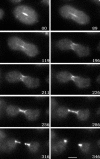
DdINCENP localizes at the cleavage furrow during cytokinesis. An AX2 cell expressing GFP-DdINCENP was followed by time-lapse video microcopy from early telophase. GFP-DdINCENP was especially concentrated on the cortex region of the furrow and the intracellular bridge during cytokinesis. The numbers in images indicate time in seconds. Bar, 5 μm.
Figure 3.
Figure 3.
DdINCENP null cells have mitosis and cytokinesis defects. (A) Immunoblot analysis of whole lysates of wild-type (WT) and DdINCENP null cells (Null) probed with anti-DdINCENP antibodies. The arrow points to the position of the DdINCENP protein (152 kDa) and the asterisk (*) indicates the positions of two unspecific bands recognized by the polyclonal antibodies. (B) Growth curve of the type cells (square solid line) and DdINCENP null cells (triangle dash line) in suspension cultures. DdINCENP null cells grew much more slowly than wild-type cells. (C-C′) A typical population of DdINCENP null cells. The cells were visualized by Differential interference contrast (DIC) microscopy (C) and stained with DAPI to visualize their nuclei (C′). Although some cells contained a single normal nucleus, many cells had multiple nuclei or enlarged nuclei. Bar, 15 μm.
Figure 4.
Figure 4.
DdINCENP null cells have chromosome segregation defect. Time-lapse video microscopy of wild-type (top) and DdINCENP null cells (bottom) during mitosis, both of which expressed GFP-histone 2B. The micrographs were taken by fluorescence microscopy to show the movement of chromosomes (as indicated by GFP-histone 2B) from metaphase to anaphase in these two cells. The times are indicated in seconds. As shown, the wild-type cell segregated its chromosomes normally in a short time (86 s; see Video 5). However, the DdINCENP null cell displayed lagging chromosomes (arrows) during the segregation and stalled at anaphase (see Video 4). Other cells were arrested at metaphase for the entire observation period (see Video 3). Bar, 5 μm.
Figure 5.
Figure 5.
DdINCENP null cells have a defective spindle midzone. (A-E) Merged immunofluorescence images of wild-type (A and B) and DdINCENP null cells (C-E) during anaphase, showing DNA in blue and tubulin in red. The arrows point to the position of the spindle midzone in these cells. The mitotic wild-type cells had robust spindle midzone during anaphase. However, the DdINCENP null cells showed weak spindle midzone (D) and sometimes the spindle midzone was imperceptible (C and E). Bar, 5 μm.
Figure 6.
Figure 6.
DdINCENP is essential for the localization of DdCP224, an XMAP215/TOGp homologue, to the spindle midzone. (A-D) Merged immunofluorescence images of wild-type cells (A and B) and DdINCENP null cells (C and D) during anaphase. DdCP224 localized at the spindle poles and the spindle midzone during mitosis in wild-type cells. However, DdCP224 was absent from the spindle midzone in the DdINCENP null cells. DNA is shown in blue and DdCP224 is shown in red. Arrows point to the position of spindle midzone. (E and F) Overexpression of DdCP24 rescues the spindle midzone defect of DdINCENP null cells. (E and E′) The immunofluorescence pictures of a DdINCENP null cell overexpressing DdCP224-GFP (green), showing DNA in blue and tubulin in red. Arrows point to the position of spindle midzone. DdCP224-GFP was localized on the spindle midzone (E). As a result, the DdINCENP null cells had much more robust spindle midzone (E′) than those cells shown in Figure 5. (F) The fluorescence image of a live DdINCENP null cell expressing DdCP224-GFP. The cell was at anaphase. Overexpressed DdCP224-GFP was clearly localized on the spindle midzone. Bar, 5 μm.
Figure 7.
Figure 7.
DdINCENP null cells are hypersensitive to microtubule-depolymerizing drugs. (A and B) Growth curve of wild-type cells (diamond) and DdINCENP null cells (triangle) in the presence of 2 μg/ml thiabendazole (A) or 2 μg/ml nocodazole (B). (C-G): Immunofluorescence images of mitotic wild-type cells (C and D) and DdINCENP null cells (E-G) in the presence of 2 μg/ml thiabendazole (TBZ). DNA is shown in blue and tubulin is shown in red. Wild-type cells displayed normal metaphase spindles (C) and anaphase spindles with a well-defined spindle midzone (D). In contrast, DdINCENP null cells were blocked at prometaphase by the drug treatment and contained either multipolar (E and F) or monopolar spindles (G, a single multinucleate mitotic cell is shown). Bar, 5 μm.
Figure 8.
Figure 8.
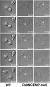
DdINCENP null cells have a late cytokinesis defect. Time-lapse video microscopy of a wild-type cell and a DdINCENP null cell undergoing cytokinesis. Cytokinesis of the wild-type cell was completed in ∼3 min. In contrast, DdINCENP null cell failed in cytokinesis because of the inability to break the thin cytoplasmic bridge connecting the daughter cells (arrowhead). The times are indicated in minutes:seconds. Bar, 5 μm.
Figure 9.
Figure 9.
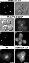
DdINCENP is not essential for the formation of equatorial or ectopic cleavage furrows during cytokinesis. (A and A′) Microscopy images of a DdINCENP null cell forming Rappaport (ectopic) furrows during four-way cytokinesis. As shown, DdINCENP null cells are able to make normal and ectopic cleavage furrows. (B, B′, C, and C′) GFP-DdINCENP localized only to one pair of cleavage furrows in a four-way cell division. Arrows indicate the position of the presumptive equatorial cleavage furrows and arrowheads indicate the position of presumptive ectopic furrows. (B and B′) Live DIC and fluorescent images of an AX2 cell expressing GFP-DdINCENP ongoing a four-way division. (C and C′) Fluorescent image of a wild-type cells expressing GFP-DdINCENP. The cell was stained by DAPI to better illustrate the four-way cytokinesis. (D) Live fluorescent image of an AX2 cell expressing GFP-MHC (myosin II heavy chain) during a four-way cytokinesis. Myosin II localized to both normal and ectopic furrows (arrows). (E) Myosin II localizes normally at the cleavage furrow of DdINCENP null cells. Fluorescent microscopy image of a DdINCENP null cell expressing GFP-Myosin II heavy chain. The absence of DdINCENP does not affect the localization of myosin II at the cleavage furrow (arrows). Bar, 5 μm.
Figure 10.
Figure 10.
The distribution of DdINCENP in the cleavage furrow area is different in the absence of myosin II. Time-lapse video microscopy was performed on a myosin heavy chain null cell expressing GFP-DdINCENP. The live images show a mutant cell attached to a substrate undergoing myosin II-independent cytokinesis B (Zang et al., 1997). In contrast to the cortical distribution of GFP-DdINCENP in wild-type cells (see Figure 2), GFP-DdINCENP was found as a narrow band at the equatorial plane of the dividing cell. This band of GFP-DdINCENP decreased in size as the cleavage furrow ingressed. The times are indicated in minutes:seconds. Bar, 5 μm.

Similar articles

Cited by

References

    1. Adams, R. R., Carmena, M., and Earnshaw, W. C. (2001a). Chromosomal passengers and the (aurora) ABCs of mitosis. Trends Cell Biol. 11, 49-54. - PubMed
    1. Adams, R. R., Maiato, H., Earnshaw, W. C., and Carmena, M. (2001b). Essential roles of Drosophila inner centromere protein (INCENP) and aurora B in histone H3 phosphorylation, metaphase chromosome alignment, kinetochore disjunction, and chromosome segregation. J. Cell Biol. 153, 865-880. - PMC - PubMed
    1. Ainsztein, A. M., Kandels-Lewis, S. E., Mackay, A. M., and Earnshaw, W. C. (1998). INCENP centromere and spindle targeting: identification of essential conserved motifs and involvement of heterochromatin protein HP1. J. Cell Biol. 143, 1763-1774. - PMC - PubMed
    1. Andrews, P. D., Ovechkina, Y., Morrice, N., Wagenbach, M., Duncan, K., Wordeman, L., and Swedlow, J. R. (2004). Aurora B regulates MCAK at the mitotic centromere. Dev. Cell 6, 253-268. - PubMed
    1. Bishop, J. D., and Schumacher, J. M. (2002). Phosphorylation of the carboxyl terminus of inner centromere protein (INCENP) by the Aurora B Kinase stimulates Aurora B kinase activity. J. Biol. Chem. 277, 27577-27580. - PMC - PubMed

Publication types

MeSH terms

Substances

LinkOut - more resources