. 2006 Feb;17(2):966-76.
doi: 10.1091/mbc.e05-07-0647. Epub 2005 Dec 7.

MAGI-1 is required for Rap1 activation upon cell-cell contact and for enhancement of vascular endothelial cadherin-mediated cell adhesion

Affiliations

MAGI-1 is required for Rap1 activation upon cell-cell contact and for enhancement of vascular endothelial cadherin-mediated cell adhesion

Atsuko Sakurai et al. Mol Biol Cell. 2006 Feb.

Abstract

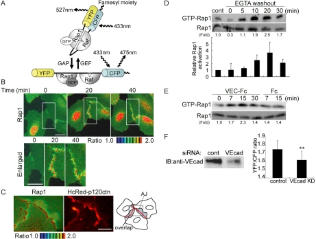
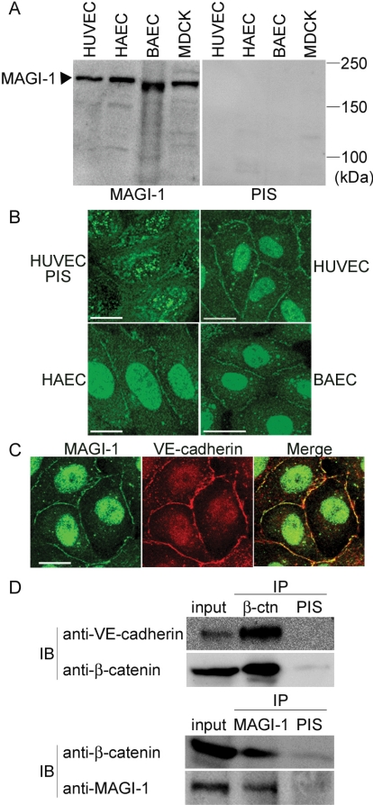
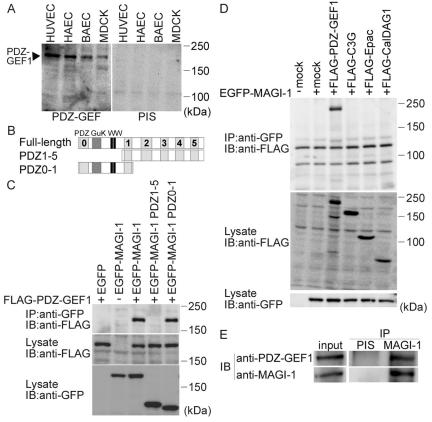
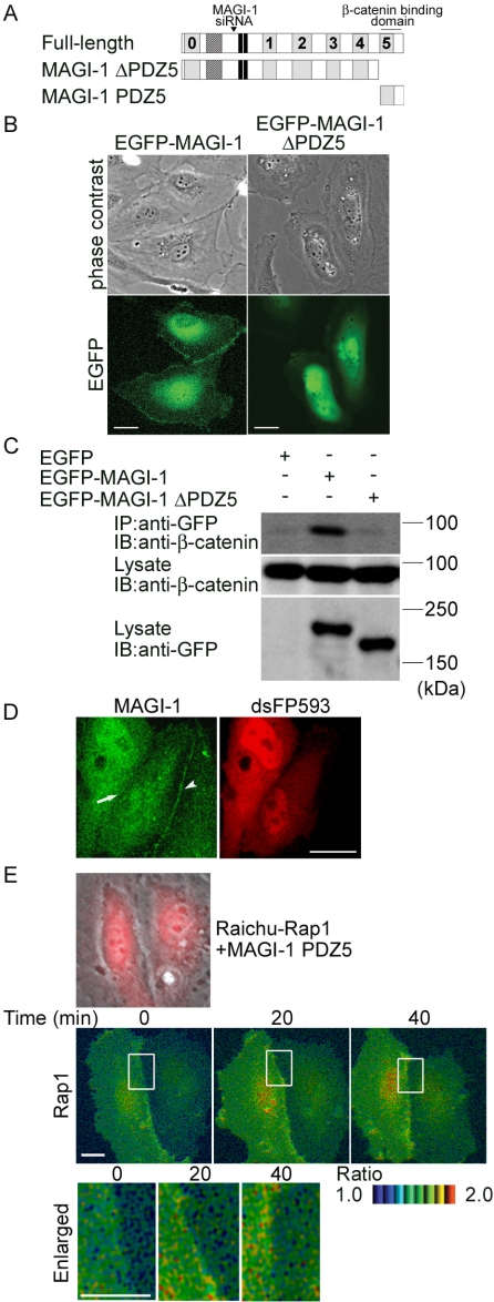
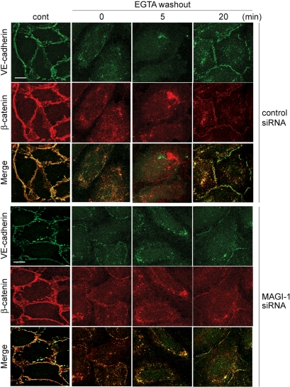
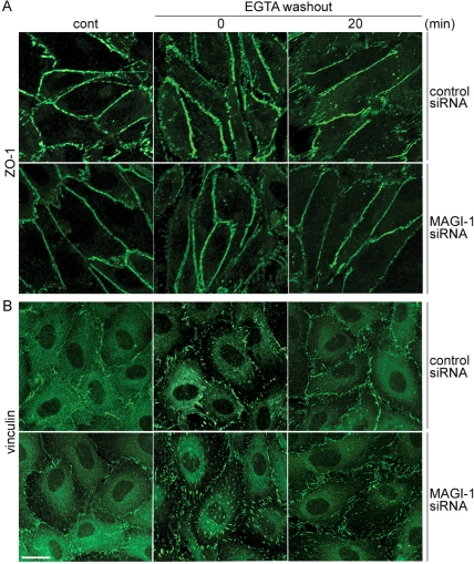
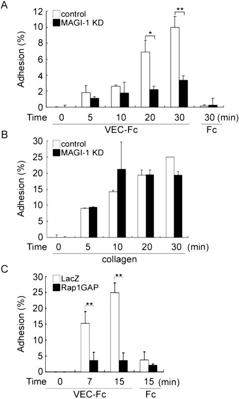
Rap1 is a small GTPase that regulates adherens junction maturation. It remains elusive how Rap1 is activated upon cell-cell contact. We demonstrate for the first time that Rap1 is activated upon homophilic engagement of vascular endothelial cadherin (VE-cadherin) at the cell-cell contacts in living cells and that MAGI-1 is required for VE-cadherin-dependent Rap1 activation. We found that MAGI-1 localized to cell-cell contacts presumably by associating with beta-catenin and that MAGI-1 bound to a guanine nucleotide exchange factor for Rap1, PDZ-GEF1. Depletion of MAGI-1 suppressed the cell-cell contact-induced Rap1 activation and the VE-cadherin-mediated cell-cell adhesion after Ca2+ switch. In addition, relocation of vinculin from cell-extracellular matrix contacts to cell-cell contacts after the Ca2+ switch was inhibited in MAGI-1-depleted cells. Furthermore, inactivation of Rap1 by overexpression of Rap1GAPII impaired the VE-cadherin-dependent cell adhesion. Collectively, MAGI-1 is important for VE-cadherin-dependent Rap1 activation upon cell-cell contact. In addition, once activated, Rap1 upon cell-cell contacts positively regulate the adherens junction formation by relocating vinculin that supports VE-cadherin-based cell adhesion.


Figures

Figure 1.
Figure 1.
Rap1 is activated by VE-cadherin-mediated cell adhesion upon cell-cell contact. (A) Schematic illustration of Raichu-Rap1. FRET efficiency depends on the guanine nucleotide binding state of Rap1. GDP-bound Raichu-Rap1 emits 475-nm fluorescence when excited at 433 nm, whereas GTP-bound Raichu-Rap1 emits 527-nm fluorescence due to FRET. Raf; Ras/Rap1 binding domain of Raf. (B) Motile HUVECs infected with Adeno-Raichu-Rap1 were monitored by FRET time-lapse imaging every 20 s. A ratio image of YFP to CFP reflects FRET efficiency. Ratio images are shown by the intensity modulated display, in which the upper and lower limits of the ratio (the intensity of YFP divided by that of CFP) are indicated by the red and blue hues, respectively, and the average intensity of YFP and CFP is used. Time since starting FRET imaging is indicated on the top (min). The boxed regions in the top panels were enlarged and are shown in the bottom panels. Bars, 20 μm. (C) HUVECs expressing both Raichu-Rap1 and HcRed-tagged p120 catenin were FRET-imaged and red-fluorescence-imaged. The real images of boxed region of the schema are shown as FRET image (left panel) and red-fluorescence image (center). Areas indicated by the gray are regions where protruding and overlapping regions of the contacting cells. Note that Rap1 is activated at the adherens junctions where p120 catenin localizes. Bar, 20 μm. (D) Confluent HUVECs cultured in medium 199 containing 1% BSA without serum were treated with EGTA for 30 min to disrupt Ca2+-dependent cell adhesion. Subsequently, the cells were treated with Ca2+-containing medium. Cell lysates at the time points indicated at the top were subjected to pulldown assay for detecting GTP-bound Rap1 as described in Materials and Methods. A representative results from three independent experiments is shown (top). Fold activation indicates the ratio of the GTP-Rap1 intensity of total Rap1 intensity to the control GTP-Rap1 intensity of total Rap1 intensity. The result from three independent experiments were shown (bottom). Control (cont) was prepared from cells in medium 199 before calcium switch. Cells treated with EGTA for 30 min (time 0). (E) HUVECs sparsely cultured on the dish were stimulated with 10 μg/ml VEC-Fc or Fc for the time indicated at the top. Rap1 activity was examined as described for D. Fold activation is analyzed similarly to D. (F) The effect of VE-cadherin siRNA on VE-cadherin expression was examined by immunoblotting (left). FRET at the cell-cell contacts were quantitatively analyzed in control siRNA-treated HUVECs and VE-cadherin-depleted HUVECs (right), as explained in Supplementary Figure 2. Mean values with SDs obtained by 30 cell-cell contact sites are shown as a representative result of three independent experiments. Statistical significance was analyzed by Student's t test; ** p < 0.01.
Figure 2.
Figure 2.
MAGI-1 localizes to cell-cell contacts and forms a complex with VE-cadherin and β-catenin. (A) Lysates from the cells indicated at the top were subjected to SDS-PAGE followed by immunoblotting with anti-MAGI-1 antibody (left) and with pre-immune serum (PIS, right). (B) Endothelial cells were immunostained with pre-immune serum (PIS, left top) and anti-MAGI-1 antibody. Immunoreaction was visualized by fluorescent microscopy. Bars, 20 μm. (C) HUVECs were immunostained with both anti-MAGI-1 antibody (green) and anti-VE-cadherin antibody (red). A merged image is shown in the right panel (Merge). Bar, 20 μm. (D) Cell lysates from HUVECs were subjected to either immunoprecipitation (IP) with antibodies as indicated at the top followed by immunoblotting (IB) with antibodies as indicated at the left. VE-cadherin was coimmunoprecipitated with β-catenin (top). β-catenin was coimmunoprecipitated with MAGI-1 (bottom)
Figure 3.
Figure 3.
MAGI-1 interacts with PDZ-GEF1 in vascular endothelial cells. (A) Cell lysates from HUVECs, HAECs, BAECs, and MDCK cells were subjected to SDS-PAGE and immunoblotting with anti-PDZ-GEF1 antibody (left) or pre-immune serum (PIS, right). (B) Schematic illustration of MAGI-1 (full length) and its deletion mutants. MAGI-1 consists of six PDZ domains (PDZ0-5, indicated by gray boxes), a guanylate kinase domain (GuK), and two WW domains. Deletion mutants, PDZ1-5 and PDZ0-1, consist of PDZ1 to PDZ5 and the amino-terminus to PDZ1, respectively. (C) 293T cells were transfected with the plasmids together with (+) or without (-) FLAG-tagged PDZ-GEF1 expressing vector as indicated at the top. Cell lysates were subjected to immunoprecipitation (IP) with anti-GFP antibody followed by immunoblotting (IB) or directly to immunoblotting using the antibodies as indicated. Note that FLAG-tagged PDZ-GEF1 is coimmunoprecipitated with GFP-tagged PDZ0-1. (D) Cells transfected with a panel of FALG-tagged Rap1 GEF-expressing plasmids together with (+) or without (-) EGFP-tagged MAGI-1-expressing plasmid. Cell lysates were subjected to immunoprecipitation (IP) followed by immunoblotting (IB) similarly to C. Note that only FLAG-tagged PDZ-GEF-1 among several Rap1 GEFs is coimmunoprecipitated with MAGI-1. (E) The lysate of HUVECs was incubated with either pre-immune serum (PIS) or anti-MAGI-1 antibody, followed by immunoblotting with anti-PDZ-GEF1 antibody. Note that MAGI-1 is coimmunoprecipitated with PDZ-GEF1.
Figure 4.
Figure 4.
MAGI-1 localizing to cell-cell contact via β-catenin is required for Rap1 activation. (A) Schematic illustration of MAGI-1 and its mutants. The corresponding region of siRNA for MAGI-1 used in Figure 5 is also indicated in this schema.(B) HUVECs were transfected with the plasmids indicated at the top and imaged on an Olympus IX-81 fluorescent microscope. Bars, 20 μm. Note the localization of EGFP-MAGI-1 at the cell-cell contact but not MAGI-1 lacking PDZ domain 5. (C) 293T cells were transfected with the plasmids as indicated at the top. Cell lysates were subjected to immunoprecipitation (IP) with anti-GFP followed by immunoblotting (IB) or directly to immunoblotting using the antibodies indicated at the left. Note that endogenous β-catenin is coimmunoprecipitated with EGFP-tagged MAGI-1, but not with that lacking PDZ5. (D) HUVECs expressing PDZ domain 5 of MAGI-1 was immunostained with anti-MAGI-1 antibody. No immunoreaction was detected at the cell-cell contact between PDZ domain 5-expressing cells (arrow), whereas immunoreaction was detected at the contact between PDZ domain 5-expressing cell and untransfected cell (arrow-head). (E) HUVECs transfected with Raichu-Rap1 and pIRM21-MAGI-1-PDZ5 were FRET-imaged (middle). Phase contrast image was overlaid onto the image for dsFP593 to distinguish HUVECs transfected with pIRM21-MAGI-1-PDZ5 from those transfected only with Raichu-Rap1 (top). Red and blue hues indicated by intensity modulated display reflect increased and decreased FRET, respectively. The boxed regions in the middle panels were enlarged (bottom). The upper and lower limits of the ratio range are shown at the bottom right. Bars, 20 μm.
Figure 5.
Figure 5.
Depletion of MAGI-1 inhibits Rap1 activation upon cell-cell contact. (A) HUVECs transfected with control siRNAs or MAGI-1 siRNAs were cultured for 48 h. The cells were lysed, subjected to SDS-PAGE, and immunoblotted with anti-MAGI-1 and anti-β-catenin. (B) HUVECs transfected with control siRNAs or MAGI-1 siRNAs were cultured for 48 h and immunostained with anti-MAGI-1. Bars, 20 μm. (C) MAGI-1-depleted HUVECs were infected with Adeno-Raichu-Rap1 and FRET-imaged. The ratio-images indicate Rap1 activation by red hue and Rap1 inactivation by blue hue (top). The boxed region between two neighboring cells is enlarged (bottom). Bars, 20 μm. (D) Quantitative FRET analysis at the cell-cell contacts were performed in cells treated with control siRNA-treated HUVECs (control) and with MAGI-1-depleted cells (MAGI-1 KD). Quantitative FRET analysis is explained in Supplementary Figure 2. Mean values with standard deviations obtained by 30 cell-cell contact sites are shown as a representative result of three independent experiments. Statistical significance was analyzed by Student's t test and is indicated as ** p < 0.01.
Figure 6.
Figure 6.
Depletion of MAGI-1 impairs AJ formation. HUVECs transfected with control siRNAs (top three columns) or MAGI-1 siRNAs (bottom three columns) were cultured for 48 h. Cells were replated onto the glass-base dishes for another 24 h to constitute the cell-cell contacts. The cells were treated with EGTA for 30 min to disrupt VE-cadherin-dependent junctions and kept in the replaced medium containing Ca2+ for the time indicated at the top. The cells were immunostained with anti-VE-cadherin antibody (green) and anti-β-catenin antibody (red). The merged images are shown in the bottom panels (Merge). Bars, 20 μm. VE-cadherin remarkably accumulated 20 min after Ca2+ restoration in control siRNA-treated HUVECs, whereas slight accumulation was observed in MAGI-1 siRNA-treated HUVECs.
Figure 7.
Figure 7.
MAGI-1 depletion affects an AJ-supporting molecule, vinculin, but not a TJ-supporting molecule, ZO-1. (A) Similarly to Figure 6, HUVECs treated with control siRNAs (top panels) and MAGI-1 siRNAs (bottom panels) were immunostained with anti-ZO-1 after calcium switch. Bar, 20 μm. Note that ZO-1-positive cells were affected neither by calcium switch nor MAGI-1 depletion. (B) Similarly to A, control siRNA-treated cells and MAGI-1-depleted cells were immunostained with anti-vinculin before and after Ca2+ switch. Ca2+ depletion from the culture medium resulted in displacement of vinculin from cell-cell contacts to cell-ECM contacts and Ca2+-restoration induced relocalization from cell-ECM contacts to cell-cell contacts in control HUVECs. In clear contrast, vinculin remained at the cell-ECM contacts even 20 min after Ca2+ restoration in MAGI-1-depleted cells.
Figure 8.
Figure 8.
Depletion of MAGI-1 and inactivation of Rap1 inhibits VE-cadherin-mediated cell adhesion. (A) HUVECs transfected with control siRNAs (white column) or MAGI-1 siRNAs (black column) were cultured for 48 h, suspended in 0.5% BSA-containing medium 199, and incubated for 30 min at 37°C. Cells, 2.0 × 10⁵, were plated onto either a VEC-Fc- or Fc-coated well for the time indicated at the bottom. Cell adhesion was quantified as described in Materials and Methods. The averages of triplicate (plus SDs) are presented. A representative result of three independent experiments is shown. Statistical significance was analyzed by Student's t test; * p < 0.05 and ** p < 0.01. Note that adhesion of HUVECs treated with MAGI-1 siRNAs to the VEC-Fc-coated dish was significantly reduced compared with mock-treated HUVECs. (B) Adhesion of MAGI-1-depleted cells to a collagen-coated dish was comparable to mock-treated HUVECs, as analyzed by the same method described in the legend for A. (C) HUVECs infected with either LacZ-expressing adenovirus (LacZ) or Rap1GAPII-expressing virus (Rap1GAP) were analyzed for adhesion to a VEC-Fc-coated dish similarly to A.

Similar articles

Cited by

References

    1. Adamsky, K., Arnold, K., Sabanay, H., and Peles, E. (2003). Junctional protein MAGI-3 interacts with receptor tyrosine phosphatase beta (RPTP beta) and tyrosine-phosphorylated proteins. J. Cell Sci. 116, 1279-1289. - PubMed
    1. Boettner, B., Govek, E. E., Cross, J., and Van Aelst, L. (2000). The junctional multidomain protein AF-6 is a binding partner of the Rap1A GTPase and associates with the actin cytoskeletal regulator profilin. Proc. Natl. Acad. Sci. USA 97, 9064-9069. - PMC - PubMed
    1. Boettner, B., Harjes, P., Ishimaru, S., Heke, M., Fan, H. Q., Qin, Y., Van Aelst, L., and Gaul, U. (2003). The AF-6 homolog canoe acts as a Rap1 effector during dorsal closure of the Drosophila embryo. Genetics 165, 159-169. - PMC - PubMed
    1. Bos, J. L. (2005). Linking Rap to cell adhesion. Curr. Opin. Cell Biol. 17, 123-128. - PubMed
    1. Bos, J. L., de Rooij, J., and Reedquist, K. A. (2001). Rap1 signalling: adhering to new models. Nat. Rev. Mol. Cell Biol. 2, 369-377. - PubMed

Publication types

MeSH terms

LinkOut - more resources