. 2006 Feb;17(2):907-16.
doi: 10.1091/mbc.e05-06-0542. Epub 2005 Dec 7.

Conventional kinesin mediates microtubule-microtubule interactions in vivo

Affiliations

Conventional kinesin mediates microtubule-microtubule interactions in vivo

Anne Straube et al. Mol Biol Cell. 2006 Feb.

Abstract

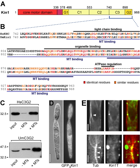
Conventional kinesin is a ubiquitous organelle transporter that moves cargo toward the plus-ends of microtubules. In addition, several in vitro studies indicated a role of conventional kinesin in cross-bridging and sliding microtubules, but in vivo evidence for such a role is missing. In this study, we show that conventional kinesin mediates microtubule-microtubule interactions in the model fungus Ustilago maydis. Live cell imaging and ultrastructural analysis of various mutants in Kin1 revealed that this kinesin-1 motor is required for efficient microtubule bundling and participates in microtubule bending in vivo. High levels of Kin1 led to increased microtubule bending, whereas a rigor-mutation in the motor head suppressed all microtubule motility and promoted strong microtubule bundling, indicating that kinesin can form cross-bridges between microtubules in living cells. This effect required a conserved region in the C terminus of Kin1, which was shown to bind microtubules in vitro. In addition, a fusion protein of yellow fluorescent protein and the Kin1tail localized to microtubule bundles, further supporting the idea that a conserved microtubule binding activity in the tail of conventional kinesins mediates microtubule-microtubule interactions in vivo.


Figures

Figure 1.
Figure 1.
A second microtubule binding site in conventional kinesin. (A) Kin1 is the conventional kinesin from U. maydis (Lehmler et al., 1997). It has an N-terminal motor core. The Kin1 tail is predicted to consist of a globular domain (G1), three coiled-coils (C1-C3), and a C-terminal globular domain (G2). Amino acids are given above the bar. (B) The C-terminal portion of conventional kinesin tails contains binding sites for light chains (Diefenbach et al., 1998) and organelles (Seiler et al., 2000) as well as a conserved motif that is implied in regulation of motor activity (Stock et al., 1999; Seiler et al., 2000). In addition, the C-terminal 196 amino acids of human kinesin heavy chain (HsKHC) localize to microtubules in vivo (Navone et al., 1992); region indicated by light blue bars). A microtubule binding activity was mapped to 55 amino acids between organelle binding and ATPase regulation sites (Hackney and Stock, 2000). Sequence similarity between human and fungal (UmKin1) kinesin is significantly higher within the functional regions. Note that fungal kinesins do not possess light chains. (C) Recombinant tail polypeptides from human kinesin-1 (HsC3G2) and U. maydis kinesin-1 (UmC3G2) cosedimented specifically with taxol-stabilized microtubules. Compare pellets after centrifugation with and without microtubules (+MT and -MT) from E. coli extracts (input). Note that microtubules were limiting in that experiment, thus similar amounts of both polypeptides were pulled down. (D) Expression of full-length Kin1 tagged with triple GFP resulted in an even cytoplasmic background. Bar, 5 μm. (E) A fusion protein of YFP and the Kin1 tail (amino acids 336-968) localized to microtubules (CFP-α-tubulin, red) in U. maydis Δkin1 cells. Note that colocalization occurs mainly within microtubule bundles (marked by arrowheads). Bars, 1 μm.
Figure 2.
Figure 2.
Kin1 in microtubule bending and bundling. A, In kin1-null mutants no Kin1 was found in Western analysis, whereas expression of Kin1 under control of the strong crg-promoter (Bottin et al., 1996) led to ∼100-fold overexpression. Note that a 1:50 dilution was loaded for the overexpressing strain as seen in tubulin loading control panel. (B) Control cells contain microtubules that are partially bundled (control). Deletion of kin1 results in less bundling and shorter microtubules (ΔKin1). In contrast, overexpression of Kin1 led to less organized microtubules that were longer and formed more intensively stained bundles (Kin1↑). Bar, 5 μm. (C) Ultrastructural analysis of U. maydis wild-type cells shows long bundles of microtubules. Cross-sections reveal that bundles consist of up to three microtubules. Bars, 100 nm. (D) In electron microscopy (EM) images, 25% of all cross-sectioned microtubules were found in bundles of two (light gray) or three (dark gray) in control cells. Bundling was threefold reduced in the Kin1 deletion mutant (ΔKin1), but EM analysis failed to detect increased bundling in the Kin1-overexpressing strain (Kin1↑). Note that significantly more bundling was found during analysis of GFP-fluorescence in this strain (Figure 3F). (E1) Repressed expression of GFP-Tub1, which was integrated into the genome as an additional copy (strain FB2rGFPTub1) led to decreased microtubule labeling. Movement of speckles (arrow) that serve as structural landmarks illustrate that bending results from sliding of a microtubule over another microtubule. Bar, 2 μm. (F1) Image series showing bending of GFP-labeled microtubules in a strain that also expresses a fusion of Peb1, the EB1-homologue of U. maydis (Straube et al., 2003) with monomeric red fluorescent protein (Campbell et al., 2002). A microtubule slides over a second microtubule (arrows) and forms a loop. RFP-Peb1 (arrowhead) indicates that the microtubule slides with plus-end trailing. Elapsed time is given in seconds. Bar, 2 μm. (F2) The cartoon illustrates how a plus-end-directed kinesin molecule could accomplish such a bending event. The motor (red) cross-bridges two microtubules in a bundle. Although it walks toward the plus-end of one microtubule, this microtubule is pushed backward and bent. (G) Microtubule polarity within microtubule bundles is determined using Peb1-RFP marked growing microtubule plus-ends within microtubule bundles (green, GFP-tubulin). An example for a parallel and an antiparallel microtubule bundle is shown. Bar, 5 μm. (H) In control cells, microtubule bending motility was observed with a frequency of one event per 40 s. Deletion of Kin1 reduced this frequency to below one event per minute, whereas high levels of Kin1 doubled the bending activity. See supplemental movies (Videos 2-4). (I) Time-lapse images illustrate the reduced motility of the microtubule cytoskeleton in the absence of Kin1. The false-colored image merges two images that span a time interval of 18 s (T, 0 in red; T, 18 s in green; stationary microtubules in yellow). Elapsed time is given in seconds. Bar, 3 μm. (K) Time-lapse images of a Kin1-overexpressing cell reveal an enhance microtubule bending activity. Bending events are marked with arrowheads. The false-colored image merges two images that span a time interval of 18 s (T, 0 in red; T, 18 s in green; stationary microtubules in yellow). Elapsed time is given in seconds. Bar, 3 μm.
Figure 3.
Figure 3.
Kinesin rigor mutants induce rigid microtubule cross-bridges. (A) A point mutation in the P-loop of kinesins interferes with ATP hydrolysis and confers “rigorous” binding to microtubules (Wedlich-Soldner et al., 2002). The fusion protein K3rigorK1T consists of the tail of Kin1 and the rigor mutated motor domain of Kin3, a Kif1a/Unc104 homologue of U. maydis. (B) Mutated Kin1 protein (Kin1rigor) but not Kin3rigor led to thicker microtubule bundles. Cells expressing the K3rigorK1T fusion protein contained long and very bright microtubule bundles (K3rigorK1T). Arrowheads mark thick microtubule bundles. Bar, 5 μm. (C) Both Kin1rigor and K3rigorK1T inhibited microtubule bending, whereas Kin3rigor was without effect. See supplemental movies (Videos 5 and 6). (D) In cells expressing Kin1rigor (D1) or K3rigorK1T (D2 and D3), microtubule bundles of up to nine microtubules were found that were surrounded by a fine matrix. Bars, 100 nm. (E) 69% and 86% of cross-sectioned microtubules were bundled in Kin1rigor- and K3rigorK1T-expressing cells, respectively. (F) Fluorescence intensity measurement of GFP-labeled microtubule bundles in various kinesin mutants demonstrated that bundles in control cells contain on average 3 microtubules. In kin1 deletion mutants, the number of microtubules in the remaining bundles is significantly reduced. Expression of Kin1 at a high level significantly enhances microtubule bundling (p values are indicated by asterisks). The rigor constructs Kin1rigor and Kin3rigorK1T raise the number per bundle to approximately 5 and approximately 7.5, respectively, whereas Kin3rigor was without effect. (G) The mean distance between neighboring microtubules was determined to be ∼10 nm for control cells, ∼41 nm for Kin1rigor, and ∼45 nm for K3rigorK1T.
Figure 4.
Figure 4.
The bundling activity in the Kin1 tail is located at its C terminus. (A) Constructs used to identify the region in the Kin1 tail that mediates heavy bundling and suppression of bending in K3rigorK1T. (B) Western analysis demonstrates comparable expression levels of the mutant proteins. (C) Deletion of the second coiled-coil (ΔC2) was without effect on microtubule bundling. However, deletion of the third coiled-coil alone or together with the C-terminal globular domain (ΔC3G2) eliminated the bundling activity of K3rigorK1T. Images for control and K3rigorK1T are included for comparison. To visualize single microtubules as well, images appear saturated where thick microtubule bundles occur. Asterisks mark the bud neck. Bars, 5 μm. (D) Fluorescence intensity measurement of GFP-labeled microtubule bundles in cells expressing Kin1 tail deletion constructs showed that the second coiled-coil domain is dispensable for the microtubule bundling activity in the Kin1 tail (Kin3rigorK1TΔC2). In contrast, deletion of the third coiled-coil and the C-terminal globular domain (ΔC3G2) from the Kin3rigorK1T chimera disabled microtubule bundling completely. (E) Deletion of C3 and G2 restored bending activity to the level of the Kin1 deletion mutant, whereas the mutant construct with deletion of C2 suppressed microtubule bending as effective as the full-length Kin3rigorK1T construct did. Data for control, ΔKin1 and K3rigorK1T are included for comparison. See supplemental movies (Videos 2, 6, and 7). (F) Time-lapse images illustrate the strongly reduced motility of the microtubule cytoskeleton in Kin3rigorK1T-expressing cells. The false-colored image merges two images that span a time interval of 18 s (T, 0 in red; T, 18 s in green; stationary microtubules in yellow). Elapsed time is given in seconds. Bar, 3 μm. (G) Time-lapse images illustrate the recovered motility of the microtubule cytoskeleton in cells expressing the deletion construct Kin3rigorK1TΔC3G2. The false-colored image merges two images that span a time interval of 18 s (T, 0 in red; T, 18 s in green; stationary microtubules in yellow). Elapsed time is given in seconds. Bar, 3 μm.

Similar articles

Cited by

References

    1. Adamikova, L., Straube, A., Schulz, I., and Steinberg, G. (2004). Calcium signaling is involved in dynein-dependent microtubule organization. Mol. Biol. Cell 15, 1969-1980. - PMC - PubMed
    1. Allan, V. (1995). Membrane traffic motors. FEBS Lett 369, 101-106. - PubMed
    1. Amos, L. A. (1987). Kinesin from pig brain studied by electron microscopy. J. Cell Sci. 87, 105-111. - PubMed
    1. Andrews, S. B., Gallant, P. E., Leapman, R. D., Schnapp, B. J., and Reese, T. S. (1993). Single kinesin molecules crossbridge microtubules in vitro. Proc. Natl. Acad. Sci. USA 90, 6503-6507. - PMC - PubMed
    1. Baas, P. W. (2002). Microtubule transport in the axon. Int. Rev. Cytol. 212, 41-62. - PubMed

Publication types

MeSH terms

LinkOut - more resources