. 2005 Dec 20;102(51):18467-72.
doi: 10.1073/pnas.0506187102. Epub 2005 Dec 8.

Depletion of minichromosome maintenance protein 5 in the zebrafish retina causes cell-cycle defect and apoptosis

Affiliations

Depletion of minichromosome maintenance protein 5 in the zebrafish retina causes cell-cycle defect and apoptosis

Soojin Ryu et al. Proc Natl Acad Sci U S A. .

Abstract

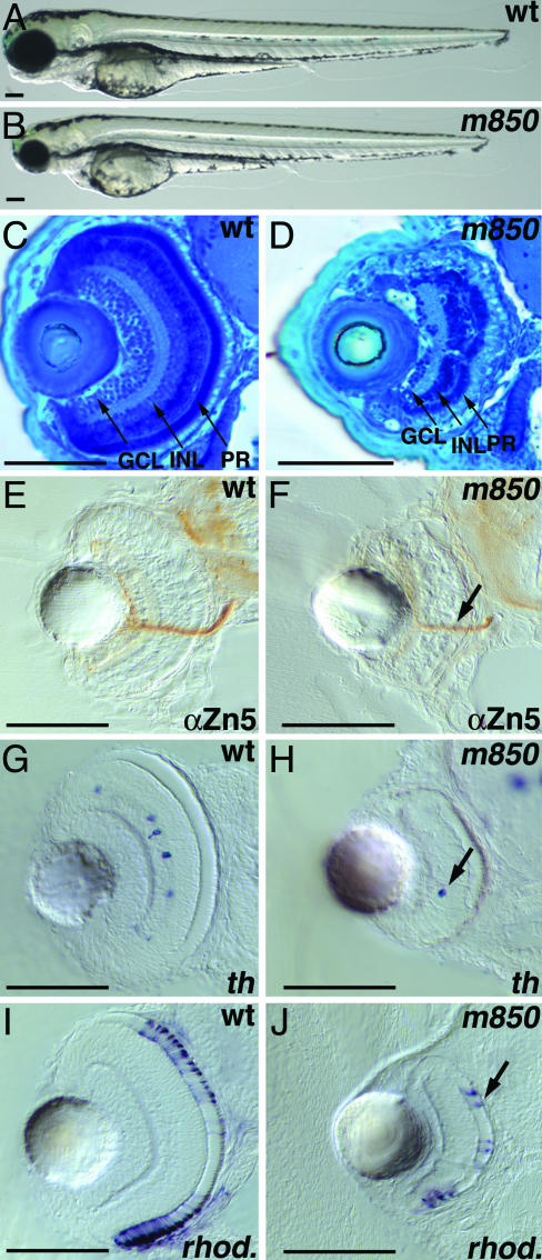
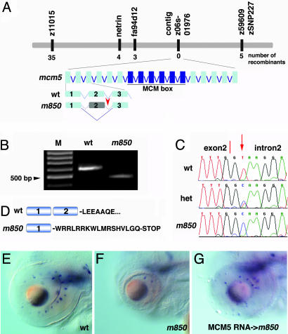
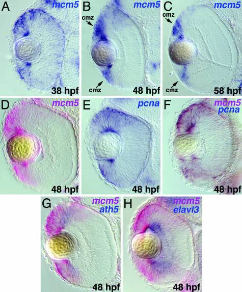
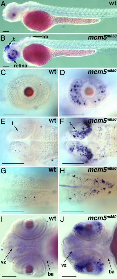
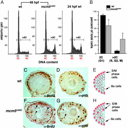
In multicellular organisms, the control of genome duplication and cell division must be tightly coordinated. Essential roles of the minichromosome maintenance (MCM) proteins for genome duplication have been well established. However, no genetic model has been available to address the function of MCM proteins in the context of vertebrate organogenesis. Here, we present positional cloning of a zebrafish mcm5 mutation and characterization of its retina phenotype. In the retina, mcm5 expression correlates closely with the pattern of cell proliferation. By the third day of development, mcm5 is down-regulated in differentiated cells but is maintained in regions containing retinal stem cells. We demonstrate that a gradual depletion of maternally derived MCM5 protein leads to a prolonged S phase, cell-cycle-exit failure, apoptosis, and reduction in cell number in mcm5(m850) mutant embryos. Interestingly, by the third day of development, increased apoptosis is detectable only in the retina, tectum, and hindbrain but not in other late-proliferating tissues, suggesting that different tissues may employ distinct cellular programs in responding to the depletion of MCM5.


Figures

Fig. 1.
Fig. 1.
m850 mutant embryos display reduction in cell number in the retina. Live wild-type (A) and m850 mutant (B) embryos at 3 dpf. A smaller head and smaller eyes characterize the m850 mutant. (C and D) Transverse sections through the eye at 3 dpf, stained with methylene blue. (C) The wild-type retina shows characteristic stratification in three nuclear layers (GCL, INL, and PR) and two plexiform layers. (D) In m850 mutant embryos, the stratification of retinal layers occurs, although the number of cells across all three retinal nuclear layers is severely reduced. (E and F) Transverse sections of 3-dpf embryos stained with anti-Zn5 (α-Zn5) antibody exhibit the presence of optic nerves in wild-type (E) and m850 mutant (F) embryos, albeit here with reduced intensity. (G and H) Transverse sections through the retinae of wild-type embryos (G) display dopaminergic amacrine cells, as detected by the expression of tyrosine hydroxylase (th) at 3 dpf. Dopaminergic amacrine cells can also be detected in mutant embryos (H). (I and J) Transverse sections through the retinae of 3-dpf embryos showing rod photoreceptors, as detected by the expression of rhodopsin (rhod.). The presence of differentiated rod photoreceptors is clearly visible in mutants (I). (A and B) Lateral views, anterior left, dorsal up; (CJ) transversal sections, dorsal up. (Scale bars, 100 μm.) Black arrows indicate the optic nerve (F), a dopaminergic amacrine cell (H), or a photoreceptor cell (J). GCL, ganglion cell layer; INL, inner nuclear layer; PR, photoreceptor layer.
Fig. 2.
Fig. 2.
The zebrafish mcm5 gene is disrupted in m850. (A) Schematic representation of the genetic map of linkage group 3 (LG 3) and genomic organization of the mcm5 locus showing the position of the mutation. Some of the closest simple-sequence-polymorphism and SNP markers are listed, and their genetic distances relative to the mutation are indicated as the number of recombinants in a total of 1,786 meioses. (B) RT-PCR from wild-type and mutant RNA using primers from exons 1 and 3 reveals that the mutant transcript is shorter. M, 100-bp DNA ladder. (C) Chromatogram of genomic sequences from the 3′ end of exon 2 from wild-type, heterozygous, and mutant embryos. The T-to-C base change in the mutant embryo affects the first of the two core bases, highly conserved in eukaryotic splice donor sites. (D) Skipping exon 2 produces a frame shift, which introduces a stop codon at amino acid residue 74 in mutant embryos. (EG) RNA rescue of the m850 mutant phenotype. The mutant phenotype was judged by the eye and head size and the presence of dopaminergic amacrine cells, as detected by th whole-mount in situ hybridization at 3 dpf. All pictures show lateral views, dorsal up. (E) Wild-type retina. (F) m850 mutant retina. (G) Injection of 200 pg of mcm5 RNA into m850 mutant embryos rescued the mutant phenotype (n = 19 of 26).
Fig. 3.
Fig. 3.
In the retina, mcm5 expression is down-regulated in regions containing differentiated cells but is maintained in the region containing retinal stem cells. (AC) Transverse sections of the retina of wild-type embryos after whole-mount in situ hybridization for mcm5. (A) At 38 hpf, whereas mcm5 expression in the retina is broad, it is stronger in the marginal zone. At 48 (B) and 58 (C) hpf, strong mcm5 expression is maintained in the ciliary marginal zone (cmz) but is down-regulated in other areas of the retina. (DF) Expression of pcna (blue) (E), marking noncommitted proliferating cells, overlaps exactly with that of mcm5 (red) (D), resulting in brown staining (F). (G) ath5 (blue), marking neurons before full differentiation, overlaps with the lateral extent of mcm5 (red). (H) elavl3 (blue), marking fully differentiated neurons, shows no overlap with mcm5 (red). Anterior is up.
Fig. 4.
Fig. 4.
Apoptosis is activated in CNS tissues in m850 mutant embryos. (AJ) TUNEL assay for apoptosis in whole-mount wild-type and mcm5m850 mutant embryos at 2 dpf. (A and B) Apoptosis is activated in retina, tectum, and hindbrain in the mutant embryos. (CH) mcm5m850 mutant embryos show more apoptotic cells in the retina (C and D), the tectum (E and F), and the hindbrain (G and H) but not in the branchial arches and the brain ventricular zone (I and J), compared with wild-type embryos. (AD) Lateral view; (EH) dorsal views; (I and J) ventral views. Anterior toward the left. (Scale bars, 100 μm.) ba, branchial arches; hb, hindbrain; t, tectum; vz, ventricular zone.
Fig. 5.
Fig. 5.
S-phase prolongation and cell-cycle-progression defects can be detected in the retinae of mcm5m850 mutant embryos. (A) DNA content of dissociated retinal cells at 48–50 hpf from wild-type (wt) (Left) and mcm5m850 mutant (Center) embryos and at 24 hpf from wild-type embryos, as measured by quantitative FACS analysis of propidium-iodide-labeled cells. Rapidly proliferating retina cells at 24 hpf allow determination of the location of the 4C peak (Right). In contrast, at 48–50 hpf, retinae from wild-type embryos contain mostly cells with 2C DNA content (1C = one haploid genome equivalent). The mutant retinae contained an increased portion of cells with DNA content >2C. (B) Bar graph summarizing results from four independent experiments. Retinae from mcm5m850mutant embryos contained fewer cells in G1 phase and more cells in S, G2, or M phase. The error bars represent the standard deviation. (C, D, F, and G) Lateral sections through the eye of paraffin-embedded wild-type or mutant embryos, probed with antibodies against Br-dUrd or phosphorylated histone H3 (pH3) to label S or M phase cells, respectively. At 48 hpf, in wild-type retinae, the great majority of cells in the central region have already exited the cell cycle. In contrast, mcm5m850 mutant retinae contain cells in S or M phase in the central region, which are thus either delayed or arrested in S or M phase (black arrowheads). (E and H) Summary diagrams representing the results from C and D and F and G, respectively.

References

    1. Blow, J. J. (2001) EMBO J. 20, 3293–3297. - PMC - PubMed
    1. Donaldson, A. D. & Blow, J. J. (1999) Curr. Opin. Genet. Dev. 9, 62–68. - PubMed
    1. Diffley, J. F. X. & Labib, K. (2002) J. Cell. Sci. 115, 869–872. - PubMed
    1. Tye, B. K. (1999) Annu. Rev. Biochem. 68, 649–686. - PubMed
    1. Davey, M. J., Jeruzalmi, D., Kuriyan, J. & O'Donnell, M. (2002) Nat. Rev. Mol. Cell Biol. 3, 826–835. - PubMed

Publication types

MeSH terms

Substances