Comparative Study
. 2005 Dec;15(12):1831-7.
doi: 10.1101/gr.3825705.

Construction of a 2-Mb resolution BAC microarray for CGH analysis of canine tumors

Affiliations
Comparative Study

Construction of a 2-Mb resolution BAC microarray for CGH analysis of canine tumors

Rachael Thomas et al. Genome Res. 2005 Dec.

Abstract

Recognition of the domestic dog as a model for the comparative study of human genetic traits has led to major advances in canine genomics. The pathophysiological similarities shared between many human and dog diseases extend to a range of cancers. Human tumors frequently display recurrent chromosome aberrations, many of which are hallmarks of particular tumor subtypes. Using a range of molecular cytogenetic techniques we have generated evidence indicating that this is also true of canine tumors. Detailed knowledge of these genomic abnormalities has the potential to aid diagnosis, prognosis, and the selection of appropriate therapy in both species. We recently improved the efficiency and resolution of canine cancer cytogenetics studies by developing a small-scale genomic microarray comprising a panel of canine BAC clones representing subgenomic regions of particular interest. We have now extended these studies to generate a comprehensive canine comparative genomic hybridization (CGH) array that comprises 1158 canine BAC clones ordered throughout the genome with an average interval of 2 Mb. Most of the clones (84.3%) have been assigned to a precise cytogenetic location by fluorescence in situ hybridization (FISH), and 98.5% are also directly anchored within the current canine genome assembly, permitting direct translation from cytogenetic aberration to DNA sequence. We are now using this resource routinely for high-throughput array CGH and single-locus probe analysis of a range of canine cancers. Here we provide examples of the varied applications of this resource to tumor cytogenetics, in combination with other molecular cytogenetic techniques.


Figures

Figure 1.
Figure 1.
(A) DAPI-banded metaphase spread from a clinically normal male dog. Note that the karyotype (2n = 78) comprises 38 pairs of acrocentric autosomes, a large submetacentric X chromosome, and a small metacentric Y chromosome. (B) Typical DAPI-banded metaphase preparation of a cell derived from canine osteosarcoma case OS-a. This tumor has a consistent chromosome number of 34 (30/30 cells) and presented with a complex karyotype comprising multiple metacentric chromosomes.
Figure 2.
Figure 2.
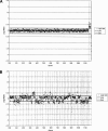
(A) Array CGH profile of a sex-mismatched hybridization performed with normal reference genomic DNA. Data are plotted as the mean, normalized, and background-subtracted log2 ratio of the replicate spots for each clone. Clones are plotted in genomic order from CFA 1qcen to CFA Yqtel. Clones derived from CFA X and CFA Y are shown with vertical broken lines on the right side of the profile. Log2 ratios representing genomic gain and loss are indicated by horizontal bars above and below the midline representing normal copy number. (B) Array CGH profile for case OS-a. Clones representing each chromosome are delineated by vertical bars. The profile demonstrates the range of genomic gains and losses present in the tumor, which are summarized in the text.
Figure 3.
Figure 3.
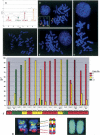
(a) Enlarged partial aCGH profile from Fig. 2B, showing 36 BAC clones distributed along the length of CFA 14. The pattern of fluorescence ratios indicates a copy number loss of the proximal region of CFA 14, interrupted by a small region with a normal copy number. A copy number gain is observed for clones within CFA 14qdist. (b) SLP analysis of four clones from CFA 14 supports these findings. Two proximal clones, 372-e16 (14q12, labeled red) and 375-a14 (14q14, green), are both present in a single copy on one arm of a novel metacentric chromosome in >90% of cells analyzed (shown as “i” in the inset). Two distal clones on CFA 14, 381-m15 (14q15-q21.1, labeled purple) and 375-k10 (14q22, yellow), are also present on the same derivative chromosome arm, suggesting a centric fusion event involving a grossly intact copy of CFA 14. Both distal clones are also present within a second novel chromosome structure (shown as “ii” in the inset). Clone 381-m15 has a normal copy number in >95% of cells, whereas chromosome duplication and translocation have generated a third copy of clone 375-k10 in >96% of cells (shown as “iii” in the inset). Inset: the three derivative chromosomes from this spread that contain segments of CFA 14. (c) SLP analysis of two clones from each of CFA 8 (374-o17, labeled yellow; 381-014, green) and CFA 13 (373-i23, red; 265-l22, blue). This figure shows one of the 15%-25% of cells in this tumor with two copies of both CFA 8 clones. Although all four probes show a normal copy number, there is clear indication of structural rearrangements involving both CFA 8 and CFA 13. (d) SLP analysis of clones from CFA 3 (191-f03, red), CFA 18 (245-k04, green) and CFA 37 (374-e01, purple) demonstrated a single copy number, correlating with observations from aCGH. A fourth probe, clone 169-o10 (CFA 26, labeled yellow) indicated a homozygous deletion (i.e., no visible signal) in > 95% of cells studied, which is also apparent from aCGH analysis. (e) SLP analysis of clones from CFA 11 (376-h09, red), CFA 14 (375-e19, purple), and CFA 26 (373-e12, green) demonstrated a single copy number. Clone 372-i12 (CFA 9q26, yellow) is present as two copies, correlating with the normal aCGH profile; however, it is clear from the position of the probe signals that CFA 9 has undergone structural rearrangement. (f) Combined summary of the aCGH and SLP analyses for these 16 loci. The frequency of probe signals (n = 0, 1, 2, or >2) in the cell population is plotted against the corresponding locus. Beneath each BAC address the aCGH ratio of each locus is indicated and color-coded according to the threshold of copy number: loss (red), normal (yellow), gain (green). In each instance there is agreement between the copy number data derived from both approaches. (g) Application of a chromosome-specific tiling panel in FISH analysis. The central feature shows a CFA 38 ideogram and the cytogenetic location of 10 BAC clones that have been mapped to this chromosome. When all 10 of these clones are labeled with the same fluorochrome, cohybridization results in painting of the chromosome as shown on the right. Conversely, if individual clones are labeled with different fluorochromes, the pool of fluorescently labeled BAC clones (in this instance, five clones) may be used to generate a multicolored chromosome-specific “tiling-set” for the study structural rearrangements (left).

Similar articles

Cited by

References

    1. Altschul, S.F., Madden, T.L., Schaffer, A.A., Zhang, J., Zhang, Z., Miller, W., and Lipman, D.J. 1997. Gapped BLAST and PSI-BLAST: A new generation of protein database search programs. Nucleic Acids Res. 25: 3389-3402. - PMC - PubMed
    1. Bannasch, D.L., Bannasch, M.J., Ryun, J.R., Famula, T.R., and Pedersen, N.C. 2005. Y chromosome haplotype analysis in purebred dogs. Mamm. Genome 16: 273-280. - PubMed
    1. Breen, M., Thomas, R., Faircloth, S., Mahoney, J., Pettengill, M., Scott, A., Thomson, S., Hudson, R., Bianco, S., Fosmire, S., et al. 2003. Molecular cytogenetics of canine lymphoma and leukaemia. In Genes, dogs and cancer: 3rd Annual Canine Cancer Conference. (ed. J. Modiano), pp. P3021.0903. International Veterinary Information Service, Ithaca NY (www.ivis.org), Seattle, WA.
    1. Breen, M., Hitte, C., Lorentzen, T.D., Thomas, R., Cadieu, E., Sabacan, L., Scott, A., Evanno, G., Parker, H.G., Kirkness, E.F., et al. 2004. An integrated 4249 marker FISH/RH map of the canine genome. BMC Genomics 5: 65-75. - PMC - PubMed
    1. Chung, Y.J., Jonkers, J., Kitson, H., Fiegler, H., Humphray, S., Scott, C., Hunt, S., Yu, Y., Nishijima, I., Velds, A., et al. 2004. A whole-genome mouse BAC microarray with 1-Mb resolution for analysis of DNA copy number changes by array comparative genomic hybridization. Genome Res. 14: 188-196. - PMC - PubMed

Web site references

    1. http://www.cvm.ncsu.edu/mbs/breen_matthew.htm; Primary authors' laboratory Web site, with details of all clones used on this array.
    1. http://cgap.nci.nih.gov/Chromosomes/Mitelman; Mitelman Database of Chromosome Aberrations in Cancer 2005. Mitelman, F., Johansson, B., and Mertens, F. (eds).

Publication types

LinkOut - more resources