. 2005 Dec;4(12):2017-28.
doi: 10.1128/EC.4.12.2017-2028.2005.

The polybasic juxtamembrane region of Sso1p is required for SNARE function in vivo

Affiliations

The polybasic juxtamembrane region of Sso1p is required for SNARE function in vivo

Jeffrey S Van Komen et al. Eukaryot Cell. 2005 Dec.

Abstract

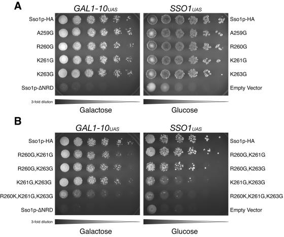
Exocytosis in Saccharomyces cerevisiae requires the specific interaction between the plasma membrane t-SNARE complex (Sso1/2p;Sec9p)and a vesicular v-SNARE (Snc1/2p). While SNARE proteins drive membrane fusion, many aspects of SNARE assembly and regulation are ill defined. Plasma membrane syntaxin homologs (including Sso1p) contain a highly charged juxtamembrane region between the transmembrane helix and the "SNARE domain" or core complex domain. We examined this region in vitro and in vivo by targeted sequence modification, including insertions and replacements. These modified Sso1 proteins were expressed as the sole copy of Sso in S. cerevisiae and examined for viability. We found that mutant Sso1 proteins with insertions or duplications show limited function, whereas replacement of as few as three amino acids preceding the transmembrane domain resulted in a nonfunctional SNARE in vivo. Viability is also maintained when two proline residues are inserted in the juxtamembrane of Sso1p, suggesting that helical continuity between the transmembrane domain and the core coiled-coil domain is not absolutely required. Analysis of these mutations in vitro utilizing a reconstituted fusion assay illustrates that the mutant Sso1 proteins are only moderately impaired in fusion. These results suggest that the sequence of the juxtamembrane region of Sso1p is vital for function in vivo, independent of the ability of these proteins to direct membrane fusion.


Figures

FIG. 3.
FIG. 3.
Effects of insertions in the Sso1p juxtamembrane in vivo. Constructs containing Sso1p insertion mutations described in Fig. 1 were assayed for in vivo function by plasmid shuffling. A. Growth on 5-FOA. Left: threefold serial dilutions of JMY303 (Sso1p-HA), JMY372 (Sso1p-KLGGP-HA), JMY371 (Sso1p-6AA insertion-HA), JMY369 (Sso1p-1x linker-HA), JMY370 (Sso1p-2x juxtamembrane-HA), JMY368 (Sso1p-3x linker-HA), or JMY305 (Sso1pΔNRD) were spotted onto synthetic complete media with 2% galactose containing 1 mg/ml 5-fluoroorotic acid and grown at 30°C for 72 h. Right: threefold serial dilutions of JMY384 (Sso1p-HA), JMY387 (Sso1p-KLGGP-HA), JMY386 (Sso1p-6AA insertion-HA), JMY388 (Sso1p-1x linker-HA), JMY389 (Sso1p-2x juxtamembrane-HA), JMY390 (Sso1p-3x linker-HA), or JMY402 (empty vector) were spotted onto synthetic complete media with 2% glucose containing 1 mg/ml 5-fluoroorotic acid and grown at 30°C for 72 h. B. Plasma membrane localization. Differential interference contrast (DIC) images and indirect immunofluorescence images are shown for JMY303 (Sso1p-HA), JMY371 (Sso1p-6AA insertion-HA), JMY369 (Sso1p-1x linker-HA), and JMY370 (Sso1p-2x juxtamembrane-HA). Localization was determined by staining with an anti-HA antibody. Scale bar equals 5 μm. C. Expression. Whole-cell extracts of JMY303 (Sso1p-HA), JMY372 (Sso1p-KLGGP-HA), JMY371 (Sso1p-6AA insertion-HA), JMY369 (Sso1p-1x linker-HA), JMY370 (Sso1p-2x juxtamembrane-HA), JMY368 (Sso1p-3x linker-HA), JMY384 (Sso1p-HA), JMY387 (Sso1p-KLGGP-HA), JMY386 (Sso1p-6AA insertion-HA), JMY388 (Sso1p-1x linker-HA), JMY389 (Sso1p-2x juxtamembrane-HA), and JMY390 (Sso1p-3x linker-HA) were resolved by SDS-PAGE on a 4 to 10% bis-Tris NuPAGE gel and blotted with an anti-HA antibody (∼16 μg total protein per lane or ∼83 μg total protein per lane). Five times the amount of extract was used for strains expressing Sso1p from the CEN plasmids. D. Relative Sso1p expression. A whole-cell extract of JMY384 (Sso1p-HA) was resolved by SDS-PAGE on a 4 to 10% bis-Tris NuPAGE gel and blotted with an anti-Sso antibody (∼30 μg total protein).
FIG. 4.
FIG. 4.
In vitro fusion reactions with recombinant Sso1p juxtamembrane insertions. A. Kinetic fusion assay comparing different acceptor t-SNARE liposomes containing t-SNARE complexes composed of GST-Sec9c and the indicated Sso1 protein. Each t-SNARE liposome population (45 μl) was mixed with fluorescent donor v-SNARE liposomes containing Snc1p (5 μl) and NBD fluorescence monitored in a fluorescent plate reader for 2 h. B. Coomassie blue-stained gel of the liposomes used in panel A, indicating that very similar amounts of various Sso1p mutants were reconstituted.
FIG. 6.
FIG. 6.
In vivo effects of single, double, and triple point mutations in the Sso1p juxtamembrane region. A. Growth on 5-FOA with single point mutation. Left: threefold serial dilutions of JMY303 (Sso1p-HA), JMY384 (Sso1p-A259G-HA), JMY380 (Sso1p-R260G-HA), JMY381 (Sso1p-K261G-HA), JMY382 (Sso1p-K263G-HA), or JMY305 (Sso1pΔNRD) were spotted onto synthetic complete media with 2% galactose containing 1 mg/ml 5-fluoroorotic acid and grown at 30°C for 72 h. Right: threefold serial dilutions of JMY384 (Sso1p-HA), JMY394 (Sso1p-A259G-HA), JMY395 (Sso1p-R260G-HA), JMY396 (Sso1p-K261G-HA), JMY397 (Sso1p-K263G-HA), or JMY402 (vector) were spotted onto synthetic complete media with 2% glucose containing 1 mg/ml 5-fluoroorotic acid and grown at 30°C for 72 h. B. Growth on 5-FOA with double and triple point mutations. Left: threefold serial dilutions of JMY303 (Sso1p-HA), JMY376 (Sso1p-R260G, K261G-HA), JMY377 (Sso1p-R260G, K263GHA), JMY378 (Sso1p-K261G, K263G-HA), JMY379 (Sso1p-R260G, K261G, K263G-HA), or JMY305 (Sso1pΔNRD) were spotted onto synthetic complete media with 2% galactose containing 1 mg/ml 5-fluoroorotic acid and grown at 30°C for 72 h. Right: threefold serial dilutions of JMY384 (Sso1p-HA), JMY398 (Sso1p-R260G, K261G-HA), JMY399 (Sso1p-R260G, K263G-HA), JMY400 (Sso1p-K261G, K263G-HA), JMY401 (Sso1p-R260G, K261G, K263G-HA), or JMY402 (vector) were spotted onto synthetic complete media with 2% glucose containing 1 mg/ml 5-fluoroorotic acid and grown at 30°C for 72 h.
FIG. 1.
FIG. 1.
Domain structure and sequence of Sso1p, Sso1p juxtamembrane mutations, and other plasma membrane syntaxins. A. The general domain structure of Sso1p is depicted. HA, HB, and HC refer to predicted coiled-coil domains that form an autonomously folding N-terminal regulatory domain. Mutations were introduced into the primary sequence of Sso1p by engineering a parent construct that contains a unique restriction site for insertions by double-stranded oligonucleotides (1x linker and 3x linker) or overlap PCR (6AA replaced, 6AA insertion, and 2x juxtamembrane). The linker constructs (6AA replaced, 6AA insertion, 1x linker, and 3x linker Sso1p) contain glycine-glycine-serine repeats. The 2x juxtamembrane construct duplicates 12 endogenous amino acids in the juxtamembrane region. Single, double, and triple point mutations were introduced by overlap PCR at the indicated residues. B. Sequence alignment of the juxtamembrane region of syntaxins from various species. Sc, Saccharomyces cerevisiae; Cn, Cryptococcus neoformans; Um, Ustilago maydis; Ag, Ashbya gossypii; Ca, Candida albicans; Sp, Schizosaccharomyces pombe; Ce, Caenorhabditis elegans; Hs, Homo sapiens; Ma, Macaca mulatta; Bt, Bos tarus; Rn, Rattus norvegicus; Mm, Mus musculus; Lo, Loligo pealei; Lp, Limulus polyphemus; Ls, Lymnaea stagnalis; St, Strongylocentrotus purpuratus. Numbering is relative to the S. cerevisiae Sso1p sequence.
FIG. 2.
FIG. 2.
Conversion of R265 and C266 to KL does not affect Sso1p function. A. Growth on 5-FOA. Left: threefold serial dilutions of JMY303 (Sso1p-HA) or JMY367 (Sso1p-KL-HA) were spotted onto synthetic complete media with 2% galactose containing 1 mg/ml 5-fluoroorotic acid and grown at 30°C for 72 h. Right: threefold serial dilutions of JMY384 (Sso1p-HA) or JMY385 (Sso1p-KL-HA) were spotted onto synthetic complete media with 2% glucose containing 1 mg/ml 5-fluoroorotic acid and grown at 30°C for 72 h. B. Kinetic fusion assay comparing different acceptor t-SNARE liposomes containing t-SNARE complexes composed of GST-Sec9c and the indicated Sso1 protein, H8-Sso1p (pJM88) or H8-Sso1p-KL-HA (pJM286). t-SNARE liposomes (45 μl) were mixed with fluorescent donor v-SNARE liposomes containing Snc1p (5 μl), and NBD fluorescence was monitored in a fluorescent plate reader for 2 h. C. Coomassie blue-stained gel of the liposomes used in panel B, indicating that very similar amounts of various Sso1p mutants were reconstituted.
FIG. 5.
FIG. 5.
In vivo effects of Sso1p juxtamembrane amino acid replacements. A. Constructs containing Sso1p replacement mutations described in Fig. 1 were assayed for in vivo function by plasmid shuffling. Growth on 5-FOA. Left: threefold serial dilutions of JMY303 (Sso1p-HA), JMY373 (N262G, K263G, I264S-Sso1p-A-HA), JMY374 (A259G, R260G, K261S-Sso1p-HA), JMY375 (Sso1p-6AA replaced-HA), or JMY305 (Sso1pΔNRD) were spotted onto synthetic complete media with 2% galactose containing 1 mg/ml 5-fluoroorotic acid and grown at 30°C for 72 h. Right: threefold serial dilutions of JMY384 (Sso1p-HA), JMY391 (Sso1p-N262G, K263G, I264S-HA), JMY392 (Sso1p-A259G, R260G, K261S-HA), JMY393 (Sso1p-6AA replaced-HA), or JMY402 (vector) were spotted onto synthetic complete media with 2% glucose containing 1 mg/ml 5-fluoroorotic acid and grown at 30°C for 72 h. B. Expression. Whole-cell extracts of JMY303 (Sso1p-HA), JMY373 (Sso1p-N262G, K263G, I264S-HA), JMY374 (Sso1p-A259G, R260G, K261S-HA), JMY375 (Sso1p-6AA replaced-HA), JMY384 (Sso1p-HA), JMY391 (Sso1p-N262G, K263G, I264S-HA), JMY392 (Sso1p-A259G, R260G, K261S-HA), or JMY393 (Sso1p-6AA replaced-HA) were resolved by SDS-PAGE on a 4 to 10% bis-Tris NuPAGE gel and blotted with an anti-HA antibody (∼29 μg total protein per lane or ∼137 μg total protein per lane). Five times the amount of extract was used for strains expressing Sso1p from the CEN plasmids. C. Plasma membrane localization. Differential interference contrast (DIC) image and indirect immunofluorescence image of JMY375 (Sso1p-6AA replaced-HA). Localization was determined by staining with an anti-HA antibody. Scale bar equals 5 μm. D. Kinetic fusion assay comparing different acceptor t-SNARE liposomes containing t-SNARE complexes composed of GST-Sec9c and the indicated Sso1 protein. t-SNARE liposomes (45 μl) were mixed with fluorescent donor v-SNARE liposomes containing Snc1p (5 μl) and NBD fluorescence monitored in a fluorescent plate reader for 2 h. E. Coomassie blue-stained gel of the liposomes used in panel D, indicating that very similar amounts of various Sso1p mutants were reconstituted.

Similar articles

Cited by

References

    1. Aalto, M. K., H. Ronne, and S. Keranen. 1993. Yeast syntaxins Sso1p and Sso2p belong to a family of related membrane proteins that function in vesicular transport. EMBO J. 12:4095-4104. - PMC - PubMed
    1. Bagnat, M., and K. Simons. 2002. Lipid rafts in protein sorting and cell polarity in budding yeast Saccharomyces cerevisiae. Biol. Chem. 383:1475-1480. - PubMed
    1. Bennett, M. K., N. Calakos, and R. H. Scheller. 1992. Syntaxin: a synaptic protein implicated in docking of synaptic vesicles at presynaptic active zones. Science 257:255-259. - PubMed
    1. Blumenthal, R., M. J. Clague, S. R. Durell, and R. M. Epand. 2003. Membrane fusion. Chem. Rev. 103:53-69. - PubMed
    1. Boeke, J. D., J. Trueheart, G. Natsoulis, and G. R. Fink. 1987. 5-Fluoroorotic acid as a selective agent in yeast molecular genetics. Methods Enzymol. 154:164-175. - PubMed

Publication types

MeSH terms