Comparative Study
. 2005 Dec;4(12):2044-56.
doi: 10.1128/EC.4.12.2044-2056.2005.

An Ustilago maydis septin is required for filamentous growth in culture and for full symptom development on maize

Affiliations
Comparative Study

An Ustilago maydis septin is required for filamentous growth in culture and for full symptom development on maize

Kylie J Boyce et al. Eukaryot Cell. 2005 Dec.

Abstract

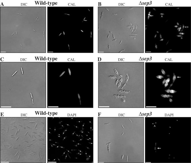
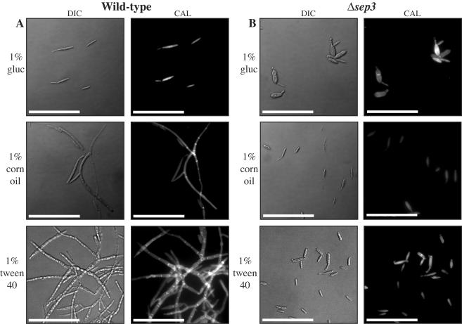
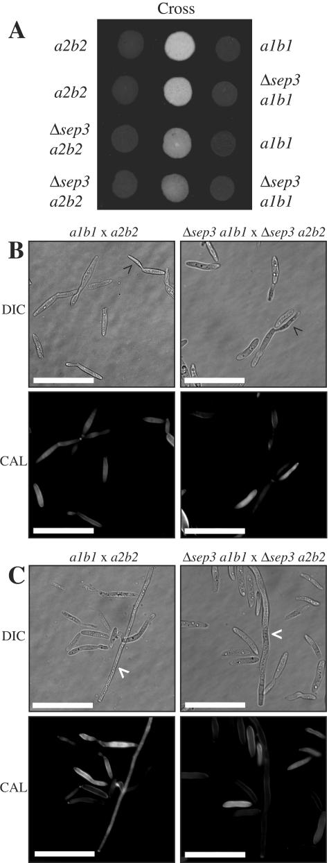
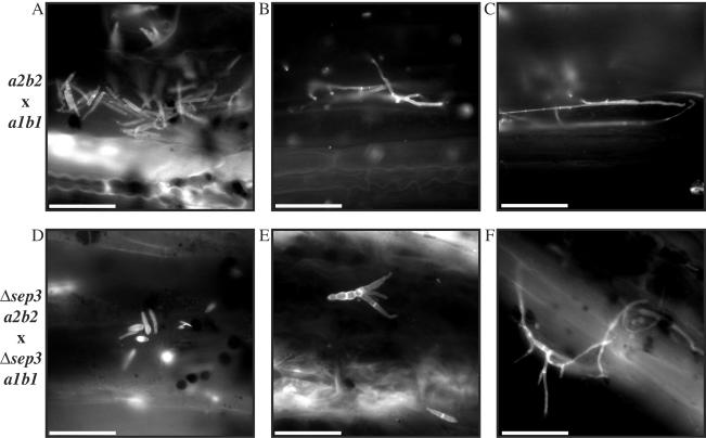
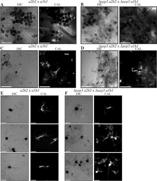
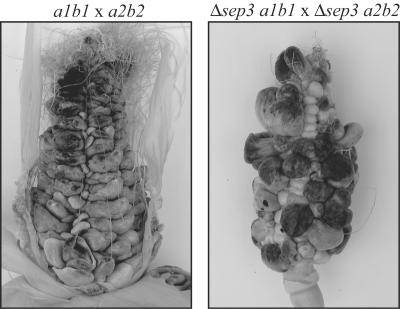
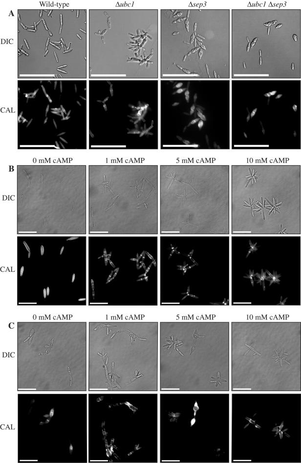
During maize infection, the fungal pathogen Ustilago maydis undergoes a dimorphic transition from budding, yeast-like cells to a filamentous dikaryon that proliferates in the host. This transition is regulated by mating and environmental signals. Septation is likely to be important in the growth of the infectious dikaryon because of the need to maintain specific cellular compartments during dikaryotic growth. Recently, we found that the transcript level for a septin gene was influenced by the conserved cyclic AMP (cAMP)/protein kinase A signaling pathway that participates in regulating dimorphism in U. maydis. In this study, we describe the detailed analysis of the function of this septin gene, designated sep3, in the growth, development, and pathogenesis of U. maydis. We show that sep3 is required for normal cellular morphology and the division of budding haploid cells. The gene is also required for lipid-induced filamentous growth in culture but not during the formation of mating filaments on agar medium or in planta. Strains with a deletion in sep3 have a reduction in symptom development in maize, with filamentous cells in planta displaying morphological defects. In addition, sep3 influences the differentiation of hyphae into teliospores and the germination of these teliospores to produce the meiotic haploid progeny that complete the disease life cycle. Finally, the deletion of sep3 was found to influence the multiple budding phenotype of a mutant with a defect in the regulatory subunit of protein kinase A. This result is consistent with a link between sep3 and the control of morphogenesis by cAMP signaling. Overall, this study highlights the importance of regulating septation and changes in morphology during phytopathogenesis.


Figures

FIG. 1.
FIG. 1.
Morphology of Δsep3 mutants. Wild-type 521 (a1b1) (A, C, and E) and Δsep3 (in a strain 521 a1b1 background) (B, D, and F) haploid strains were grown overnight in PDB medium and stained to visualize cell walls (CAL) or nuclei (DAPI). (A) Cell wall staining of wild-type cells showing septation at the mother-daughter bud junction at the distal end of the mother cell. The cells display even chitin distribution. (B) Cells of the Δsep3 mutant have an abnormal morphology resulting from aberrant division (white arrowhead) or multiple aberrant division sites (white arrow). Cells also often appear swollen and misshapen (white arrowhead with asterisk). Some cells also display an increase in chitin staining and uneven chitin distribution (white arrow). (C) Higher magnification of wild-type cells in panel A. (D) Higher magnification of Δsep3 cells in panel B. (E) Uninucleate cells of the wild-type strain 521 (a1b1). (F) Cells of the Δsep3 mutant are often multinucleate (white arrowhead and arrow). Cells with two nuclei appear morphologically similar to those in panel B, which result from one aberrant division (white arrowhead), while those with four nuclei appear morphologically similar to those in panel B, which result from three aberrant division sites (white arrow). Images were captured using DIC or epifluorescence to observe calcofluor-stained cell walls (CAL) or nuclei (DAPI). Scale bars = 50 μm.
FIG. 2.
FIG. 2.
The Δsep3 mutants fail to respond morphologically to lipids. Wild-type 521 (a1b1) (A) and Δsep3 (in a strain 521 a1b1 background) (B) haploid strains were grown for 5 days in minimal medium plus 1% glucose (gluc), 1% corn oil, or 1% Tween 40 and stained to visualize cell walls (CAL). (A) Wild-type haploid cells grow with a budding yeast cellular morphology in minimal medium plus 1% glucose. In minimal medium plus either 1% corn oil or 1% Tween 40, wild-type cells switch morphology to grow as septate, branched hyphae. (B) In minimal medium plus 1% glucose, the Δsep3 mutants appear swollen and have abnormal septation sites and morphology. In contrast to wild-type cells, Δsep3 cells have a budding yeast morphology in minimal medium plus 1% corn oil or 1% Tween 40. In addition, cells of the Δsep3 strains grown in lipids do not display morphology defects seen with mutant cells grown in 1% glucose. Images were captured using DIC or epifluorescence to observe calcofluor-stained cell walls (CAL). Scale bars = 50 μm.
FIG. 3.
FIG. 3.
Mating is unaffected in Δsep3 mutants. (A) Approximately 1 × 10⁵ cells were dropped onto DCM-plus-charcoal plates and incubated for 2 days at 30°C. Drops of single wild-type and mutant strains were included as controls along with combinations of wild-type and mutant strains to test for mating. Deletion of sep3 does not reduce the ability to mate. (B and C) To examine mating filaments, cells were scraped from DCM-plus-charcoal plates, resuspended in single-distillation H2O, and stained with CAL. Wild-type cells are present in a number of different morphological stages: yeast cells (B and C), yeast cells that have extended a conjugation tube (white arrowhead) (B), and mating filaments (black arrowhead) (C). Likewise, cells of the Δsep3 strains also are visible as yeast cells (B and C), yeast cells extending conjugation tubes (white arrowhead) (B), or hyphal filaments (black arrowhead) (C). Images were captured using DIC or epifluorescence to observe calcofluor-stained cell walls (CAL). Scale bar = 20 μm.
FIG. 4.
FIG. 4.
Cellular morphology in planta. Epidermal peels of leaves of infected maize plants were examined 5 days postinoculation. Maize seedlings were inoculated with crosses of wild-type strains 518 (a2b2) and 521 (a1b1) (A, B, and C) or Δsep3 (a2b2) and Δsep3 (a1b1) (D, E, and F). Epidermal peels were stained with calcofluor to reveal cell wall chitin and observed under epifluorescence. (A) Yeast cells of the wild-type strain 521 in infected leaves. (B) Filamentous growth of the wild-type cells, possibly towards mating partners. (C) Wild-type cells in a hyphal network around surrounding plant epidermal cells. Hyphal cells branch and have visible septa. (D) The Δsep3 yeast cells in infected leaves display morphological defects, including aberrant cell shape and swelling. (E) Yeast cells of the Δsep3 strain grow filamentously, possibly towards cells of the opposite mating partner. The cells display morphological defects, such as aberrant septation sites and abnormal cell shape. (F) Δsep3 hyphal cells on plant epidermal cells. Hyphal cells of the Δsep3 cells branch and have visible septa but have altered morphology, with hyphae appearing slightly curved. Images were captured using epifluorescence to observe calcofluor-stained cell walls. Scale bar = 50 μm.
FIG. 5.
FIG. 5.
Teliospore production and germination. Sections of tumors isolated from maize plants 14 days postinoculation (A and B), isolated teliospores harvested from maize plants 14 days postinoculation (C and D), and harvested teliospores germinated on potato dextrose agar slides for 24 h (E and F). Maize plants were inoculated with crosses of wild-type strains 518 (a2b2) and 521 (a1b1) (A, C, and E) or the Δsep3 a2b2 and Δsep3 a1b1 strains (B, D, and F). (A) Numerous mature teliospores (black with a rough surface coat which does not stain with calcofluor) are visible in sections from plants infected with wild-type strains. Immature teliospores could also be visualized in tumor sections when stained with the calcofluor cell wall stain. (B) Mature and immature teliospores are visible in tumor sections from plants infected with a cross of the Δsep3 a2b2 and Δsep3 a1b1 strains. However, the number of mature teliospores is reduced in these plants, and the number of immature teliospores is increased. (C) Teliospore preparations from plants infected with a wild-type cross. Ungerminated and germinating teliospores and some plant material are visible, but there are very few immature teliospores. (D) Teliospore preparations from plants infected with a Δsep3 a2b2 × Δsep3 a1b1 strain cross contain ungerminated and germinating teliospores and some plant material in addition to a large number of immature teliospores (white arrow) and many small, abnormal teliospores (black arrow). (E) Germinating teliospores from a wild-type cross. The teliospores extend a promycelium, from which haploid progeny are generated. (F) Germinating teliospores from a Δsep3 a2b2 × Δsep3 a1b1 strain cross show a variety of morphological defects. The promycelium and haploid progeny can be misshapen and swollen (black arrows). Images were captured under DIC or epifluorescence to reveal calcofluor-stained cell walls (CAL). Scale bars = 25 μm (A and B) and 50 μm (C, D, E, and F).
FIG. 6.
FIG. 6.
Pathogenicity of sep3 strains. Three-month-old maize plants were inoculated via injection into the silk channels of developing cobs. Plants were inoculated with crosses of wild-type strains 518 (a2b2) and 521 (a1b1) or the Δsep3 a1b1 and Δsep3 a2b2 mutant strains. Tumor formation and teliospore production were assessed 3 weeks postinoculation. Representative cobs of a 518 (a2b2) × 521 (a1b1) cross and a Δsep3 a1b1 × Δsep3 a2b2 strain cross are shown. A wild-type U. maydis infection results in the formation of large tumors filled with black teliospores. The deletion of sep3 does not inhibit the production of tumors or teliospores in infected plants.
FIG.7.
FIG.7.
Morphology of Δubc1 sep3 mutants and growth of Δsep3 strains in cAMP. (A) Wild-type 518 (a2b2) and Δubc1 a2b2, Δsep3 a2b2, and Δubc1 Δsep3 a2b2 haploid strains were grown overnight in PDB media and stained to visualize cell walls (CAL). The Δubc1 mutant exhibits a multibudded phenotype. The Δsep3 mutant cells are swollen, with abnormal morphology and septation sites. The Δubc1 sep3 double mutant strains have a cellular morphology similar to that of the Δsep3 single mutant strains. The cells are not multibudded like those of the Δubc1 mutant and have a swollen morphology with inappropriate septation sites. (B and C) Wild-type 518 (a2b2) (B) and Δsep3 a2b2 (C) haploid strains were grown overnight in minimal medium plus 1% glucose and either 0, 1, 5, or 10 mM cAMP and stained to visualize cell walls (CAL). (B) In 1 mM cAMP, wild-type cells exhibit an increase in budding and a greatly increased level of septation. Cells grown in 5 mM cAMP display less septation and more budding than cells grown in 1 mM cAMP. Wild-type cells grown in 10 mM cAMP exhibit little septation and greatly increased budding, such that cells appear as rosettes. (C) In 0 mM cAMP, cells of the Δsep3 strains appear misshapen and swollen and exhibit inappropriately localized septa. This phenotype is increased in severity in 1 mM cAMP, with swollen cells forming misshapen chains of septate cells. An increase in budding is evident in 5 mM cAMP and 10 mM cAMP. Like wild-type cells, Δsep3 cells grown in 10 mM cAMP form rosettes. The morphological defects of these strains at no or low levels of cAMP are reduced at high levels of cAMP. Images were captured under DIC or epifluorescence to reveal calcofluor stained cell walls (CAL). Scale bars = 50 μm (A) and 20 μm (B and C).

Similar articles

Cited by

References

    1. Andrews, D. L., J. D. Egan, M. E. Mayorga, and S. E. Gold. 2000. The Ustilago maydis ubc4 and ubc5 genes encode members of a MAP kinase cascade required for filamentous growth. Mol. Plant-Microbe Interact. 13:781-786. - PubMed
    1. Banuett, F., and I. Herskowitz. 1996. Discrete developmental stages during teliospore formation in the corn smut fungus, Ustilago maydis. Development 122:2965-2976. - PubMed
    1. Banuett, F., and I. Herskowitz. 1994. Identification of fuz7, a Ustilago maydis MEK/MAPKK homolog required for a-locus-dependent and -independent steps in the fungal life cycle. Genes Dev. 8:1367-1378. - PubMed
    1. Barrett, K. J., S. E. Gold, and J. W. Kronstad. 1993. Identification and complementation of a mutation to constitutive filamentous growth in Ustilago maydis. Mol. Plant-Microbe Interact. 6:274-283. - PubMed
    1. Bi, E., P. Maddox, D. J. Lew, E. D. Salmon, J. N. McMillan, E. Yeh, and J. R. Pringle. 1998. Involvement of an actomyosin contractile ring in Saccharomyces cerevisiae cytokinesis. J. Cell Biol. 142:1301-1312. - PMC - PubMed

Publication types

MeSH terms

LinkOut - more resources