Review
. 2005 Dec;69(4):544-64.
doi: 10.1128/MMBR.69.4.544-564.2005.

Molecules into cells: specifying spatial architecture

Affiliations
Review

Molecules into cells: specifying spatial architecture

Franklin M Harold. Microbiol Mol Biol Rev. 2005 Dec.

Abstract

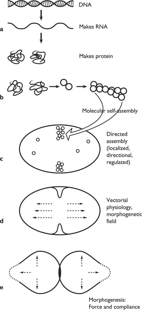
A living cell is not an aggregate of molecules but an organized pattern, structured in space and in time. This article addresses some conceptual issues in the genesis of spatial architecture, including how molecules find their proper location in cell space, the origins of supramolecular order, the role of the genes, cell morphology, the continuity of cells, and the inheritance of order. The discussion is framed around a hierarchy of physiological processes that bridge the gap between nanometer-sized molecules and cells three to six orders of magnitude larger. Stepping stones include molecular self-organization, directional physiology, spatial markers, gradients, fields, and physical forces. The knowledge at hand leads to an unconventional interpretation of biological order. I have come to think of cells as self-organized systems composed of genetically specified elements plus heritable structures. The smallest self that can be fairly said to organize itself is the whole cell. If structure, form, and function are ever to be computed from data at a lower level, the starting point will be not the genome, but a spatially organized system of molecules. This conclusion invites us to reconsider our understanding of what genes do, what organisms are, and how living systems could have arisen on the early Earth.


Figures

FIG. 1.
FIG. 1.
Hierarchy of biological order. The spatial and functional organization of cells is produced by a nested succession of processes that bridge the gap between the molecular scale and the cellular one. (a) DNA sequences are transcribed into RNA and then translated into amino acid chains; the latter fold spontaneously into functional proteins. (b) Ribosomes and other supramolecular complexes arise by self-assembly of their molecular constituents. (c) Cellular structures arise in a controlled manner at a particular time and place. (d) Many physiological processes have a direction in cell space. (e) Morphogenesis results from local compliance with applied forces, such as turgor pressure. (Modified from Harold [61].)
FIG. 2.
FIG. 2.
Self-organized microtubule patterns. Left, forms. (a) Multimeric kinesin only. (Adapted from Surrey et al. [152] with permission of the publisher.) (b) Multimeric kinesin plus a multimeric Drosophila motor protein. (Adapted from Surrey et al. [152] with permission of the publisher.) (c) Modified kinesin in a toroidal chamber. (Adapted from Nèdèlec et al. [118] with permission of the publisher.) Right, mechanisms. (d) A minus-directed motor (dynein) points the plus ends outward. (Adapted from Karsenti and Vernos [81] with permission of the publisher.) (e) A plus-directed motor (kinesin) points the minus ends outward. (Adapted from Karsenti and Vernos [81] with permission of the publisher.)
FIG. 3.
FIG. 3.
Direction and location. (a) Vectorial metabolism. Protons are pumped outward by the respiratory chain and return via the ATP synthase. (b) A polarized cell, with much of its physiology directed towards the transport of secretory vesicles to the tip. n, nucleus; v, vacuole; m, mitochondrion. Based on an electron micrograph of a shmoo of S. cerevisiae by Baba et al. (6). (c) Three patterns of localized wall deposition in growing bacteria: zonal, dispersed, and apical.
FIG. 4.
FIG. 4.
Duplicating the rod: some localized and oriented processes. (a) Actin-like cytoskeleton and dispersed synthesis of sidewall. Stippling indicates intensity of wall synthesis. (b) Segregation of nucleoids to the poles by an active mechanism; sidewalls elongate. (c) Cell finds its midpoint by the oscillation of Min proteins (nucleoid occlusion not shown). (d) Construction of divisome at the midpoint, wall synthesis focused there. (e) Construction of the septum, sidewall elongation ceases. Note that the cytoskeleton presumably undergoes rearrangements during the cell cycle which remain to be described.
FIG. 5.
FIG. 5.
The life cycle of Caulobacter crescentus. (a) Motile swarmer cell. (b) The swarmer cell settles down, loses its flagellum and pili and forms a stalk in their place. (c) As the cell grows, it begins to replicate DNA and assembles flagellar precursors at the distal pole. (d) Prior to division, the distal pole sprouts a flagellum and motility is activated. (e) The progeny stalked cell initiates a new round of replication, while the progeny swarmer cell swims off. (After Ausmees and Jacobs-Wagner [4], with permission of the publisher.)
FIG. 6.
FIG. 6.
Localization of some signal transduction proteins during the cell cycle of Caulobacter crescentus. See text for the functions of the kinases. (Adapted from Ausmees and Jacobs-Wagner [4] with permission of the publisher.)
FIG. 7.
FIG. 7.
Targeting secretory vesicles: some current ideas. a. Saccharomyces cerevisiae: vesicles delivered to marked site on actin cables. (After Pruyne and Bretsher [131], with permission of the publisher.) b. Schizosaccharomyces pombe: vesicles delivered on actin cables to apical sites, marked by proteins carried on microtubules. c. Fungal hyphae: vesicles delivered on microtubules to a Spitzenkörper, from which they proceed to the apex, perhaps via actin filaments. d. Silvetia compressa (previously known as Pelvetia compressa): a current of calcium ions localizes the site of outgrowth, to which secretory vesicles travel on actin cables. e. Lily pollen tube: currents of calcium ions and protons into the tip localize the site of exocytosis; actin involved in this and in vesicle transport.
FIG. 8.
FIG. 8.
Cortical landmark localizes the site of the next bud in diploid yeast cells. Diploid cells bud from the poles, either the pole that bears the birth scar (a) or its opposite (b). Localization depends on cortical landmarks (*) laid down during budding and transmitted structurally. Internal arrows indicate the axis of polarization. (Based on studies by Chant [26] and others.)

Similar articles

Cited by

References

    1. Adams, I. R., and J. V. Kilmartin. 2000. Spindle pole body duplication: a model for centrosome duplication? Trends Cell Biol. 10:329-334. - PubMed
    1. Albrecht-Buehler, G. 1990. In defense of “non-molecular” cell biology. Internat. Rev. Cytol. 120:191-241. - PubMed
    1. Arnone, M., and E. H. Davidson. 1997. The hardwiring of development: organization and function of genomic regulatory systems. Development 124:1851-1864. - PubMed
    1. Ausmees, N., and C. Jacobs-Wagner. 2003. Spatial and temporal control of differentiation and cell cycle progression in Caulobacter crescentus. Annu. Rev. Microbiol. 57:225-247. - PubMed
    1. Ausmees, N., J. R. Kuhn, and C. Jacobs-Wagner. 2003. The bacterial cytoskeleton: an intermediate filament-like function in cell shape. Cell 115:705-713. - PubMed