Review
. 2005 Dec;69(4):665-95.
doi: 10.1128/MMBR.69.4.665-695.2005.

The 2-hydroxycarboxylate transporter family: physiology, structure, and mechanism

Affiliations
Review

The 2-hydroxycarboxylate transporter family: physiology, structure, and mechanism

Iwona Sobczak et al. Microbiol Mol Biol Rev. 2005 Dec.

Abstract

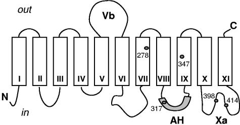
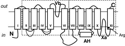
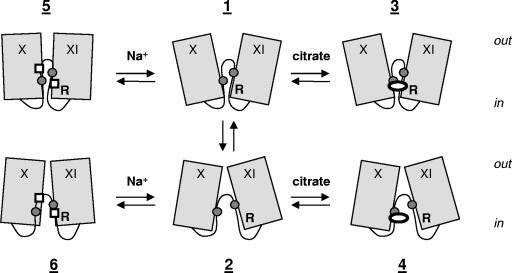
The 2-hydroxycarboxylate transporter family is a family of secondary transporters found exclusively in the bacterial kingdom. They function in the metabolism of the di- and tricarboxylates malate and citrate, mostly in fermentative pathways involving decarboxylation of malate or oxaloacetate. These pathways are found in the class Bacillales of the low-CG gram-positive bacteria and in the gamma subdivision of the Proteobacteria. The pathways have evolved into a remarkable diversity in terms of the combinations of enzymes and transporters that built the pathways and of energy conservation mechanisms. The transporter family includes H+ and Na+ symporters and precursor/product exchangers. The proteins consist of a bundle of 11 transmembrane helices formed from two homologous domains containing five transmembrane segments each, plus one additional segment at the N terminus. The two domains have opposite orientations in the membrane and contain a pore-loop or reentrant loop structure between the fourth and fifth transmembrane segments. The two pore-loops enter the membrane from opposite sides and are believed to be part of the translocation site. The binding site is located asymmetrically in the membrane, close to the interface of membrane and cytoplasm. The binding site in the translocation pore is believed to be alternatively exposed to the internal and external media. The proposed structure of the 2HCT transporters is different from any known structure of a membrane protein and represents a new structural class of secondary transporters.


Figures

FIG. 1.
FIG. 1.
Unrooted phylogenetic tree of members of the 2HCT family. Phylogenetic relationships were analyzed with the CLUSTAL W program using the default settings (141). The tree was generated with the DRAWTREE program in the Phylip package (J. Felsenstein, PHYLIP (Phylogeny Inference Package), version 3.6.a3, Department of Genome Sciences, University of Washington, Seattle, 2002). The tree is based on the C-terminal part of the multiple-sequence alignment, which contains the fewest gaps (positions 300 to 500 in Fig. 5A). Sequences included in the alignment correspond to the “typical” sequences in the “2HCT” column of Table 1. All other members of the 2HCT transporter family share over 60% sequence identity with one of the “typical” sequences. The six clusters in the tree are indicated by I to VI.
FIG. 2.
FIG. 2.
Schematic representation of physiological pathways for citrate and malate degradation involving 2HCT family members. (A) Malolactic fermentation; (B) citrolactic fermentation; (C) oxidative malate decarboxylation pathway; (D) citrate fermentation in gram-negative bacteria. Shaded circles represent the 2HCT transporter proteins. The stoichiometry of the transporters and the pyruvate lyase step are omitted in panel D. Abbreviations: cit, citrate; mal, malate; lac, lactate; oxace, oxaloacetate; ace, acetate; pyr, pyruvate; ME, malic enzyme; CL, citrate lyase; OAD, oxaloacetate decarboxylase; LDH, lactate dehydrogenase; AK, acetate kinase.
FIG. 3.
FIG. 3.
Mechanism of enzymes involved in citrate and malate metabolic pathways. (A) Malic enzymes. Malic enzyme homologs catalyze the conversion of malate to oxaloacetate, oxaloacetate to pyruvate, and pyruvate to lactate, while the substrate remains bound to the enzyme. Different types of malic enzymes (MleS, MalS, and CitM) catalyze different parts of the sequence as indicated at the top. (B) Oxaloacetate decarboxylase. Left, the OAD complex of K. pneumoniae, showing the domain, subunit composition, and the names of the corresponding structural genes. Right, mechanism of catalysis. The α domain on the αδ subunit transfers the carboxyl group of oxaloacetate to the biotin group attached to the δ domain, after which the decarboxylation of the carboxy-biotin group is coupled to the pumping of Na+ ions across the membrane. (C) Citrate lyase. Left, composition of the CL complex and the accessory enzymes necessary for the incorporation and activation of the modified CoA prosthetic group R-SH (2′-(5"-phosphoribosyl)-3′-dephospho-CoA) on the gamma subunit. The corresponding structural genes are indicated as well. Right, mechanism of catalysis. The gamma subunit is an intermediate acyl carrier protein that cycles between the citryl- and acetyl-loaded state during turnover. Abbreviations: Cit, citrate; OxAce, oxaloacetate; Ace, acetate; Pyr, pyruvate.
FIG. 4.
FIG. 4.
Energy coupling to oxaloacetate decarboxylation in bacteria. (A and C) Primordial oxaloacetate decarboxylases of the ME type (A) and OAD type (C) do not conserve the free energy released in the reaction. (B) Metabolic coupling. In citrolactic fermentation, the malic enzyme activity drives the exchange of external citrate for internal lactate catalyzed by the 2HCT exchanger CitP, thereby generating membrane potential. (F) Direct coupling. The OAD complex couples the decarboxylation reaction directly to the pumping of Na+ across the membrane. (D and E) Intermediate evolutionary states of energy coupling by OAD type of decarboxylases. The organisms in which the pathways are found and the transporter involved in the uptake of citrate into the cell are indicated at the bottom. For clusters of the 2HCT family, see Fig. 1. Abbreviations: Cit, citrate; OxaCe, oxaloacetate; Lac, lactate; Pyr, pyruvate. See the text for further explanation.
FIG. 5.
FIG. 5.
Profile analysis of the 2HCT family of transporters. The profiles were calculated based upon a multiple-sequence alignment of the “typical” sequences listed in Table 1, using a window of 20 residues (see also the legend to Fig. 1). Bars in the tops of the panels indicate the positions of gaps in the sequences. (A) Hydropathy profile using the Eisenberg scale (38). (B) Pairwise sequence identity (PSI) profile. The fraction of identical pairs at each position averaged over the window is plotted. (C) Hydrophobic moment profile. The hydrophobic moment was calculated at a periodicity of 100o (α-helix). (D) Frequency distribution of the glycine, alanine, and serine residues.
FIG. 6.
FIG. 6.
Multiple-sequence alignment of N- and C-terminal domains of selected members of the 2HCT family. N-terminal halves of sequences that produced hits with C-terminal halves, and vice versa, were selected, and a multiple sequence alignment was produced with CLUSTAL W (141). The overall sequence similarity between the two groups of five N-terminal (top) and C-terminal (bottom) halves is indicated with an asterisk for identical residues and with a tilde for similar residues. The positions of the putative TMSs in the N- and C-terminal halves are indicated by solid lines above and below the sequences, respectively. Similarly, the positions of the conserved regions, Vb and Xa, are indicated by dotted lines.
FIG. 7.
FIG. 7.
Sequence logos of the region around the sequence motif GGxG (positions 5, 6, and 8) in the N-terminal (top) and C-terminal (bottom) domains of the 2HCT transporters. The multiple-sequence alignment included all typical sequences (see Fig. 1 and Table 1). The logos were generated using WebLogo, version 2.8.1 (http://www.bio.cam.ac.uk/cgi-bin/seqlogo/logo.cgi).
FIG. 8.
FIG. 8.
Topology model of the members of the 2HCT family, based on experimental data for CitS of K. pneumoniae. Boxes represent putative transmembrane segments. The lengths of the loops correlate with the numbers of residues present in the loops. Segment Vb corresponds to a hydrophobic region that was predicted to be transmembrane. Loop Xa is one of the best-conserved regions in the 2HCT family and folds as a pore-loop structure. The AH loop folds into an amphipathic surface helix. The positions of the five endogenous cysteine residues Cys 278, Cys317, Cys347, Cys398, and Cys414 in CitS of K. pneumoniae are indicated by dots.
FIG. 9.
FIG. 9.
New structural model for the members of 2HCT transporter family. Two homologous domains containing five TMSs each, with an inverted topology in the membrane, are surrounded by dashed boxes. The two domains contain a pore-loop structure and enter the membrane-embedded part of the protein from the periplasmic or cytoplasmic side of the membrane, respectively (Vb and Xa). AH represents an amphipathic surface helix. The position of the conserved arginine residue in TMS XI is indicated by a black dot.
FIG. 10.
FIG. 10.
Kinetic schemes representing mechanisms of a symporter (A), a symporter in the exchange mode (B), and an antiporter (C). E represents the transporter. The subscript i and o indicate the orientation of the binding sites toward the cytoplasm and periplasm, respectively. The antiport mechanism in panel C assumes a dimer of two transporter molecules with one binding site oriented to the periplasm and the other to the cytoplasm. H, proton; S, substrate; P, product; A and B, two substrates at different sides of the membrane.
FIG. 11.
FIG. 11.
Schematic representation of the alternate access model for substrate translocation in the symport mode of transport, based on CitS of K. pneumoniae. The protein can reorient the binding sites in the “unloaded” (states 1 and 2) and “fully loaded” (states 5 and 6) states. Reorientation is blocked in the citrate-bound (states 7 and 8) and Na+-bound (states 3 and 4) states. Symbols: ovals, citrate; squares, Na+.
FIG. 12.
FIG. 12.
Models for binding of tri-, di-, and monocarboxylates to 2HCT transporters. Open arrows point to the carboxylate and hydroxy groups of the 2-hydroxycarboxylate motif that is common to all substrates. The arrows represent essential interactions between the substrate molecules and binding site residues on the protein. Arg+ represents the conserved arginine residue in TMS XI that interacts with a second carboxylate of citrate (top panel) and (S)-malate (middle panel), resulting in high affinity binding. Citrate binds in the divalent anionic state, and the protonated carboxylate group points away from the Arg residue. In the monocarboxylate 2-hydroxyisobutyrate, the interaction is not favorable, resulting in low-affinity binding (bottom panel).
FIG. 13.
FIG. 13.
Mechanistic model for the transporters of the 2HCT family. The scheme shows TMSs X and XI and pore-loop Xa in the C-terminal domain. States 1, 3, and 5 represent conformations of the protein with the binding site exposed to the external medium. States 2, 4, and 6 represent conformations with the binding site facing the cytoplasm. Isomerization of the two conformations is allowed only between states 1 and 2 and between the states that have bound both citrate and Na+ (not shown in the model). See text for further explanation. Symbols: ovals, citrate; squares, Na+ ions; circles, Cys residues; R, conserved arginine residue.

Similar articles

Cited by

References

    1. Abe, K., Z. S. Ruan, and P. C. Maloney. 1996. Cloning, sequencing, and expression in Escherichia coli of OxlT, the oxalate:formate exchange protein of Oxalobacter formigens. J. Biol. Chem. 271:6789-6793. - PubMed
    1. Abramson, J., I. Smirnova, V. Kasho, G. Verner, H. R. Kaback, and S. Iwata. 2003. Structure and mechanism of the lactose permease of Escherichia coli. Science 301:610-615. - PubMed
    1. Accardi, A., and C. Miller. 2004. Secondary active transport mediated by a prokaryotic homologue of ClC Cl− channels. Nature 427:803-807. - PubMed
    1. Altschul, S. F., T. L. Madden, A. A. Schaffer, J. Zhang, Z. Zhang, W. Miller, and D. J. Lipman. 1997. Gapped BLAST and PSI-BLAST: a new generation of protein database search. Nucleic Acids Res. 25:3389-3402. - PMC - PubMed
    1. Anantharam, V., M. J. Allison, and P. C. Maloney. 1989. Oxalate:formate exchange. The basis for energy coupling in Oxalobacter. J. Biol. Chem. 264:7244-7250. - PubMed

Publication types

MeSH terms

LinkOut - more resources