. 2006 Feb 10;281(6):3458-62.
doi: 10.1074/jbc.M512148200. Epub 2005 Dec 8.

The differential engagement of arrestin surface charges by the various functional forms of the receptor

Affiliations

The differential engagement of arrestin surface charges by the various functional forms of the receptor

Susan M Hanson et al. J Biol Chem. .

Abstract

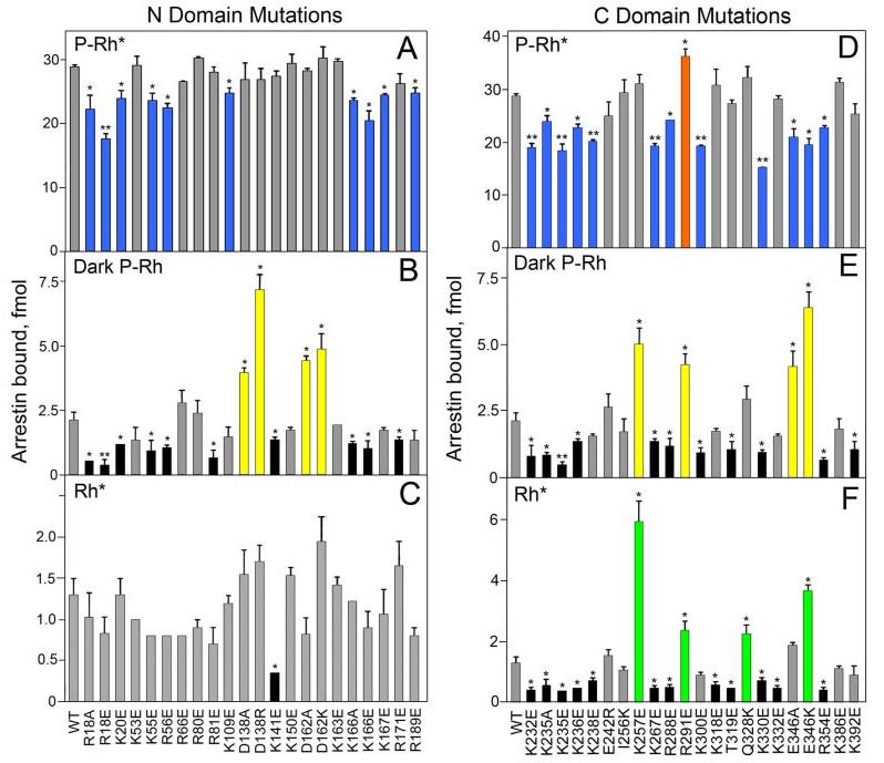
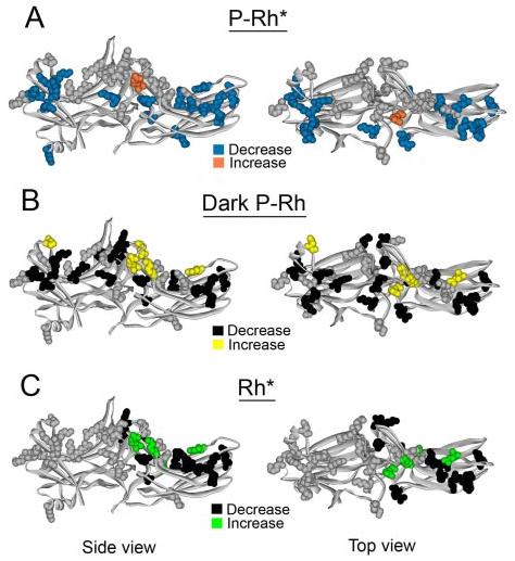
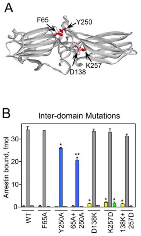
G-protein-coupled receptor signaling is terminated by arrestin proteins that preferentially bind to the activated phosphorylated form of the receptor. Arrestins also bind active unphosphorylated and inactive phosphorylated receptors. Binding to the non-preferred forms of the receptor is important for visual arrestin translocation in rod photoreceptors and the regulation of receptor signaling and trafficking by non-visual arrestins. Given the importance of arrestin interactions with the various functional forms of the receptor, we performed an extensive analysis of the receptor-binding surface of arrestin using site-directed mutagenesis. The data indicated that a large number of surface charges are important for arrestin interaction with all forms of the receptor. Arrestin elements involved in receptor binding are differentially engaged by the various functional forms of the receptor, each requiring a unique subset of arrestin residues in a specific spatial configuration. We identified several additional phosphate-binding elements in the N-domain and demonstrated for the first time that the active receptor preferentially engages the arrestin C-domain. We also found that the interdomain contact surface is important for arrestin interaction with the non-preferred forms of the receptor and that residues in this region play a role in arrestin transition into its high affinity receptor binding state.


Figures

FIGURE 1
FIGURE 1. Identified receptor-binding elements and regulatory intramolecular interactions in arrestin
Shown is the visual arrestin crystal structure (6), highlighting receptor-binding elements as follows: blue, phosphate-binding residues (6, 8, 9, 12-15); green, β-strands and loops implicated in receptor binding by peptide competition (25, 26) and element swapping (15, 19). Residues participating in regulatory intramolecular interactions are indicated as follows: pink, polar core (9) (except phosphate-binding Arg-175, indicated in dark blue); orange, three-element interaction (8). Top panel, side view. Lower panel, top view (from the receptor-binding side).
FIGURE 2
FIGURE 2. The exposed charged residues in arrestin implicated in receptor binding
The binding of radiolabeled arrestins produced in cell-free translation to P-Rh* (A and D), dark P-Rh (B and E), and Rh* (C and F) was measured as described under “Experimental Procedures.” Binding to dark Rh was negligible in all cases (data not shown). Mutations in the N-domain (A–C) and C-domain (D–F) are color-coded as follows: blue, those that significantly reduce P-Rh* binding; orange, those that enhance P-Rh* binding (R291E); yellow, those that enhance dark P-Rh binding; green, those that enhance Rh* binding; black, those that reduce dark P-Rh and/or Rh* binding; gray, those that do not significantly affect binding. Means ± S.D. from two experiments performed in duplicate are shown. *, p < 0.05, **, p < 0.01, as compared with wild-type (WT) arrestin.
FIGURE 3
FIGURE 3. Map of exposed charged residues implicated in receptor binding
A–C, visual arrestin crystal structure highlighting mutations of exposed charges as follows. A, blue, those that significantly reduce P-Rh* binding; orange, those that enhance P-Rh* binding (R291E); gray, that do not affect P-Rh* binding. B, yellow, those that enhance dark P-Rh binding; black, that reduce dark P-Rh binding; gray, that do not affect dark P-Rh binding. C, green, those that enhance Rh* binding; black, those that reduce Rh* binding; gray, those that do not affect Rh* binding. (This figure was based on data in Fig. 2). Left panel, side view. Right panel, top view.
FIGURE 4
FIGURE 4. The effects of mutations in the interdomain region
A, top view of the visual arrestin crystal structure highlighting residues between the two domains that were mutated. B, the binding of each radiolabeled arrestin produced in cell-free translation to dark P-Rh (left), P-Rh* (center), and Rh* (right) is shown. Results are color-coded as follows: blue, mutations that significantly reduce P-Rh* binding; yellow, mutations that enhance dark P-Rh binding; green, mutations that enhance Rh* binding; gray, mutations that do not significantly affect binding. Means ± S.D. from two experiments performed in duplicate are shown. *, p < 0.05, **, p < 0.01, as compared with wild-type (WT) arrestin.

References

    1. Ferguson SS, Downey WE, Colapietro AM, Barak LS, Menard L, Caron MG. Science. 1996;271:363–366. - PubMed
    1. Perry SJ, Lefkowitz RJ. Trends Cell Biol. 2002;12:130–138. - PubMed
    1. Gurevich VV, Gurevich EV. Structure (Camb.) 2003;11:1037–1042. - PubMed
    1. Schleicher A, Kuhn H, Hofmann KP. Biochemistry. 1989;28:1770–1775. - PubMed
    1. Palczewski K, Pulvermuller A, Buczylko J, Hofmann KP. J. Biol. Chem. 1991;266:18649–18654. - PubMed

Publication types

MeSH terms

LinkOut - more resources