. 2005 Dec 27;102(52):18848-53.
doi: 10.1073/pnas.0505833102. Epub 2005 Dec 9.

A papain-like enzyme at work: native and acyl-enzyme intermediate structures in phytochelatin synthesis

Affiliations

A papain-like enzyme at work: native and acyl-enzyme intermediate structures in phytochelatin synthesis

Denis Vivares et al. Proc Natl Acad Sci U S A. .

Abstract

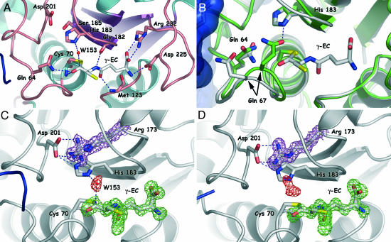
Phytochelatin synthase (PCS) is a key enzyme for heavy-metal detoxification in plants. PCS catalyzes the production of glutathione (GSH)-derived peptides (called phytochelatins or PCs) that bind heavy-metal ions before vacuolar sequestration. The enzyme can also hydrolyze GSH and GS-conjugated xenobiotics. In the cyanobacterium Nostoc, the enzyme (NsPCS) contains only the catalytic domain of the eukaryotic synthase and can act as a GSH hydrolase and weakly as a peptide ligase. The crystal structure of NsPCS in its native form solved at a 2.0-A resolution shows that NsPCS is a dimer that belongs to the papain superfamily of cysteine proteases, with a conserved catalytic machinery. Moreover, the structure of the protein solved as a complex with GSH at a 1.4-A resolution reveals a gamma-glutamyl cysteine acyl-enzyme intermediate stabilized in a cavity of the protein adjacent to a second putative GSH binding site. GSH hydrolase and PCS activities of the enzyme are discussed in the light of both structures.


Figures

Fig. 1.
Fig. 1.
Overall structure of the NsPCS dimer and its relationship with the papain family. (A) Stereo view of the overall NsPCS dimer. The ribbon is colored in gray for one monomer and in green for the other one. A comparison with the papain fold shows additional secondary structures drawn here in yellow or red depending on the monomer to which they belong. (B) Stereo view of the overlap of the NsPCS (green) and papain (white) active-site residues.
Fig. 2.
Fig. 2.
Residues involved in the binding of γ-EC and structural changes upon acylation. (A) View of selected residues in molecule A around the γ-EC binding site. Hydrogen bonds between the protein and the γ-EC moiety are depicted as blue broken lines. (B) Superimposition of the native NsPCS structure (green) and the acyl–enzyme complex (white). Note that Oε1 of Gln-64 in the native structure occupies the position of W153 (not shown for clarity) in the acyl–enzyme intermediate. (C and D) Comparison of the electron-density map around the γ-EC moiety in molecule A (C) and molecule B (D). In both cases, the electron-density omit map of γ-EC (contoured at +2.5σ and colored in green) is superimposed with the 2m FoFc map around the position of the catalytic water and of the residue Arg-173 (contoured at +1σ and drawn in red and magenta, respectively). In molecule B of the acyl–enzyme intermediate, the electron density depicted in red is clearly not compatible with the presence of a water molecule (see text for details). In all of the figures, the second monomer is colored in blue.
Fig. 3.
Fig. 3.
Sequence alignment of NsPCS with selected eukaryotic PCS sequences. Red stars denote the position of the catalytic triad. Red squares and triangles correspond to the residues whose main chain or side chain, respectively, is hydrogen-bonded to the γ-EC in the acyl–enzyme complex. Shading along the secondary structure elements represents the residues at the dimeric interface. In the consensus sequence, red boxes are drawn when the residues are strictly conserved along the alignment, whereas a simple letter is indicated when the sequences are conserved in all eukaryotic PCSs but Nostoc. Nos, Nostoc; Sp, S. pombe; Ce, C. elegans; At, A. thaliana PCS; Ta, T. aestivum; Nt, Nicotiana tabacum; Cons, consensus sequence.
Fig. 4.
Fig. 4.
Analysis of the surface conservation among the PCS family. (A and B) Mapping of the sequence conservation in the native (A) and acyl–enzyme (B) structures. In each case the surface is colored according to the alignment found in Fig. 3. Red patches correspond to identical and homologous sequence conservation (red and gray shading in the alignment). Residues conserved in all PCSs but Nostoc are colored in yellow, except for cysteine residues falling in this category, which are colored in blue.

Comment in

References

    1. Grill, E., Winnacker, E. L. & Zenk, M. H. (1985) Science 230, 674–676. - PubMed
    1. Cobbett, C. S. (1999) Trends Plant Sci. 4, 335–337. - PubMed
    1. Cobbett, C. & Goldsbrough, P. (2002) Annu. Rev. Plant Biol. 53, 159–182. - PubMed
    1. Clemens, S., Kim, E. J., Neumann, D. & Schroeder, J. I. (1999) EMBO J. 18, 3325–3333. - PMC - PubMed
    1. Grill, E., Loffler, S., Winnacker, E.-L. & Zenk, M. H. (1989) Proc. Natl. Acad. Sci. USA 86, 6838–6842. - PMC - PubMed

Publication types

MeSH terms