. 2006 Apr;34(4):426-33.
doi: 10.1165/rcmb.2005-0298OC. Epub 2005 Dec 9.

Syk activation in dendritic cells is essential for airway hyperresponsiveness and inflammation

Affiliations

Syk activation in dendritic cells is essential for airway hyperresponsiveness and inflammation

Shigeki Matsubara et al. Am J Respir Cell Mol Biol. 2006 Apr.

Abstract

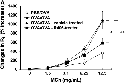
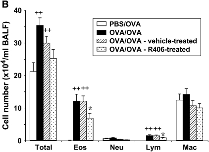
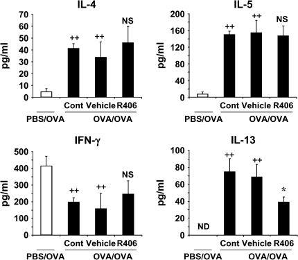
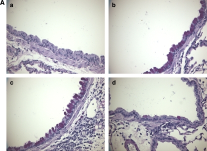
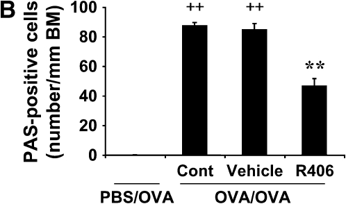
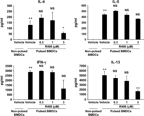
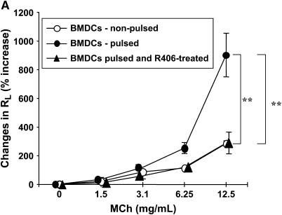
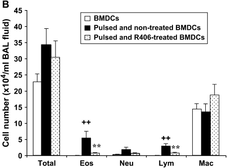
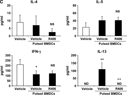
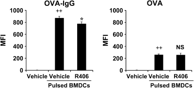
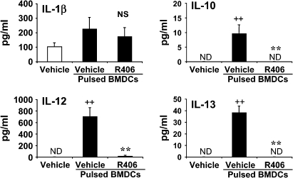
We evaluated the role of Syk, using an inhibitor, on allergen-induced airway hyperresponsiveness (AHR) and airway inflammation in a system shown to be B cell- and mast cell-independent. Sensitization of BALB/c mice with ovalbumin (OVA) and alum after three consecutive OVA challenges resulted in AHR to inhaled methacholine and airway inflammation. The Syk inhibitor R406 (30 mg/kg, administered orally, twice daily) prevented the development of AHR, increases in eosinophils and lymphocytes and IL-13 levels in bronchoalveolar lavage (BAL) fluid, and goblet cell metaplasia when administered after sensitization and before challenge with OVA. Levels of IL-4, IL-5, and IFN-gamma in BAL fluid and allergen-specific antibody levels in serum were not affected by treatment. Because many of these responses may be influenced by dendritic cell function, we investigated the effect of R406 on bone marrow-derived dendritic cell (BMDC) function. Co-culture of BMDC with immune complexes of OVA and IgG anti-OVA together with OVA-sensitized spleen mononuclear cells resulted in increases in IL-13 production. IL-13 production was inhibited if the BMDCs were pretreated with the Syk inhibitor. Intratracheal transfer of immune complex-pulsed BMDCs (but not nonpulsed BMDCs) to naive mice before airway allergen challenge induced the development of AHR and increases in BAL eosinophils and lymphocytes. All of these responses were inhibited if the transferred BMDCs were pretreated with R406. These results demonstrate that Syk inhibition prevents allergen-induced AHR and airway inflammation after systemic sensitization and challenge, at least in part through alteration of DC function.


Figures

<b>Figure 1.</b>
Figure 1.
Effects of R406 on development of AHR and airway inflammation. (A) R406 suppresses antigen-induced AHR. Sensitized mice received oral R406 (30 mg/kg,twice daily) or vehicle, administered from 1 d before the first OVA challenge to the day before measurement of AHR. Results are expressed as the percentage increase in airway resistance after MCh inhalation. (B) R406 suppresses allergen-induced inflammatory cell infiltration in the airways. PBS/OVA: nonsensitized and challenged; OVA/OVA: sensitized and challenged. Results represent mean ± SEM from three separate experiments (n = 12). ++P < 0.01 comparing sensitized and challenged to challenged alone. *P < 0.05 comparing vehicle-treated with R406-treated sensitized and challenged mice.
<b>Figure 2.</b>
Figure 2.
Cytokine levels in the BALF. Mice were the same as in the legend to Figure 1. ++P < 0.01 comparing PBS/OVA with OVA/OVA; *P < 0.05 comparing vehicle-treated and R406-treated groups. NS: nonsignificant comparing vehicle-treated with R406-treated group. ND: none detected.
<b>Figure 3.</b>
Figure 3.
R406 suppresses goblet cell metaplasia. Goblet cell metaplasia was detected by PAS staining 48 h after the last OVA challenge. (A) Airway epithelium stained with PAS. (a) PBS/OVA; (b) OVA/OVA; (c) OVA/OVA after vehicle treatment; (d) OVA/OVA after treatment with 30 mg/kg R406. (B) Quantification of goblet cell metaplasia. Goblet cell numbers were determined in lung tissue stained with PAS. Results are expressed as the number of PAS-positive cells per millimeter of basement membrane (BM). Each column represents the mean ± SEM of three separate experiments (n = 12). ++P < 0.01 comparing PBS/OVA with OVA/OVA; **P < 0.01 comparing vehicle-treated with R406-treated mice.
<b>Figure 4.</b>
Figure 4.
Decreased IL-13 release from OVA-sensitized spleen cells cultured with Syk inhibitor-treated BMDCs. BMDCs were preincubated with 0.3–3 μM of R406 for 1 h. The cells were pulsed with anti-OVA IgG/OVA for 1 h, thoroughly washed, and co-cultured with sensitized spleen mononuclear cells for 6 d. Cytokine levels in supernates were assayed by ELISA. Each column represents mean ± SEM from two experiments (n = 8). ++P < 0.01 comparing pulsed and nonpulsed BMDCs; **P < 0.01 and *P < 0.05 comparing vehicle-treated and R406 (3 μM)-treated and pulsed BMDCs. NS: nonsignificant comparing vehicle-treated and R406-treated groups.
<b>Figure 5.</b>
Figure 5.
Effects of intratracheal transfer of Syk inhibitor-treated BMDCs. Pulsed and nonpulsed BMDCs were prepared as described, and 5 × 10⁶ BMDCs were instilled intratracheally after sensitization but before OVA challenge. (A) AHR (n = 8 in each group). **P < 0.01 comparing recipients of nonpulsed and pulsed BMDC. **P < 0.01 comparing vehicle-treated and R406-treated BMDC recipients. (B) BAL inflammatory cell composition (n = 8 in each group). ++P < 0.01 comparing recipients of pulsed and nonpulsed BMDC; **P < 0.01 comparing vehicle-treated and R406 (3 μM)-treated and pulsed BMDCs. (C) BAL cytokine levels (n = 8 in each group). ++P < 0.01 and +P < 0.05 comparing recipients of pulsed and nonpulsed BMDC; **P < 0.01 comparing vehicle-treated and R406 (3 μM)-treated and pulsed BMDCs. NS: nonsignificant comparing vehicle-treated and R406-treated groups.
<b>Figure 6.</b>
Figure 6.
Effect of R406 on FITC-conjugated antigen uptake in BMDCs. BMDCs were incubated with R406 (3 μM) for 1 h before pulsing with FITC-OVA-IgG or FITC-OVA alone. Cells were washed 60 min after pulsing, and the amount of antigen uptake was measured by flow cytometry. The values were expressed as FITC-MFI calculated with Cell Quest Pro Software. Each column represents mean ± SEM from two experiments (n = 6). **P < 0.01 comparing pulsed and nonpulsed BMDCs; *P < 0.05 comparing vehicle-treated and R406-treated and pulsed BMDCs. NS: nonsignificant comparing vehicle-treated and R406-treated groups.
<b>Figure 7.</b>
Figure 7.
Effect of R406 on cytokine production in immune complex-pulsed BMDCs. BMDCs were incubated with R406 (3 μM) for 1 h before pulsing with OVA-IgG. The cells were cultured overnight, and cytokine levels in supernates were assayed by ELISA as described. Each column represents mean ± SEM from two experiments (n = 6). ++P < 0.01 comparing pulsed and nonpulsed BMDCs; **P < 0.01 comparing vehicle-treated and R406-treated and pulsed BMDCs. NS: nonsignificant comparing vehicle-treated and R406-treated groups.

Similar articles

Cited by

References

    1. Busse WW, Lemanske RF Jr. Asthma. N Engl J Med 2001;344:350–362. - PubMed
    1. Miyahara N, Swanson BJ, Takeda K, Taube C, Miyahara S, Kodama T, Dakhama A, Ott VL, Gelfand EW. Effector CD8+ T cells mediate inflammation and airway hyper-responsiveness. Nat Med 2004;10:865–869. - PubMed
    1. Robinson DS, Hamid Q, Ying S, Tsicopoulos A, Barkans J, Bentley AM, Corrigan C, Durham SR, Kay AB. Predominant TH2-like bronchoalveolar T-lymphocyte population in atopic asthma. N Engl J Med 1992;326:298–304. - PubMed
    1. Kuipers H, Lambrecht BN. The interplay of dendritic cells, Th2 cells and regulatory T cells in asthma. Curr Opin Immunol 2004;16:702–708. - PubMed
    1. van Rijt LS, Jung S, Kleinjan A, Vos N, Willart M, Duez C, Hoogsteden HC, Lambrecht BN. In vivo depletion of lung CD11c+ dendritic cells during allergen challenge abrogates the characteristic features of asthma. J Exp Med 2005;201:981–991. - PMC - PubMed

Publication types

MeSH terms