. 2005 Dec 8;438(7069):858-62.
doi: 10.1038/nature04175.

BMP inhibition-driven regulation of six-3 underlies induction of newt lens regeneration

Affiliations

BMP inhibition-driven regulation of six-3 underlies induction of newt lens regeneration

Matthew W Grogg et al. Nature. .

Abstract

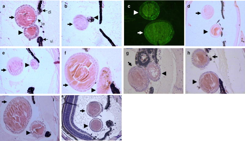
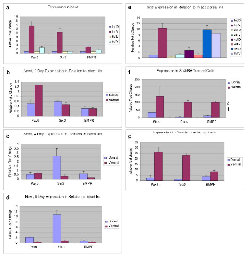
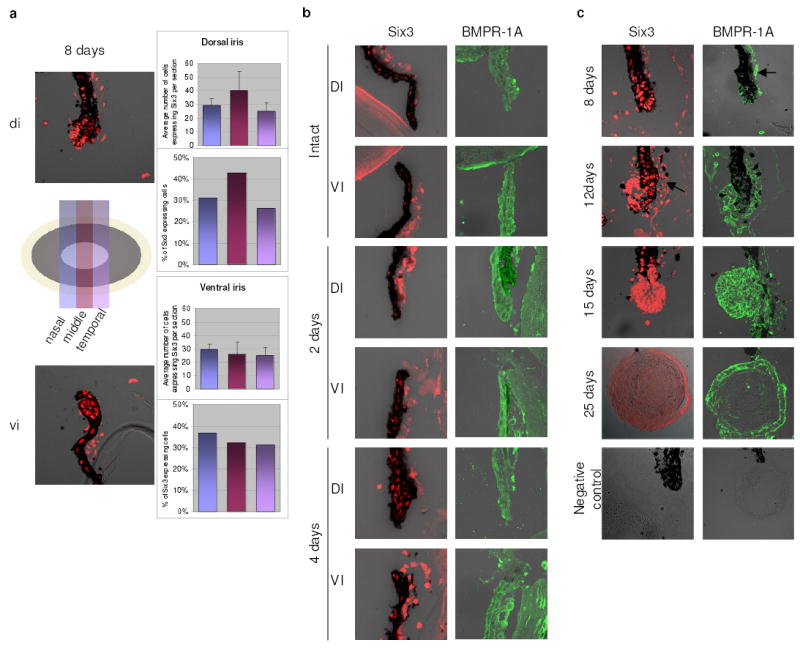
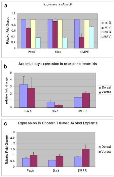
Lens regeneration in adult newts is a classic example of how cells can faithfully regenerate a complete organ through the process of transdifferentiation. After lens removal, the pigment epithelial cells of the dorsal, but not the ventral, iris dedifferentiate and then differentiate to form a new lens. Understanding how this process is regulated might provide clues about why lens regeneration does not occur in higher vertebrates. The genes six-3 and pax-6 are known to induce ectopic lenses during embryogenesis. Here we tested these genes, as well as members of the bone morphogenetic protein (BMP) pathway that regulate establishment of the dorsal-ventral axis in embryos, for their ability to induce lens regeneration. We show that the lens can be regenerated from the ventral iris when the BMP pathway is inhibited and when the iris is transfected with six-3 and treated with retinoic acid. In intact irises, six-3 is expressed at higher levels in the ventral than in the dorsal iris. During regeneration, however, only expression in the dorsal iris is significantly increased. Such an increase is seen in ventral irises only when they are induced to transdifferentiate by six-3 and retinoic acid or by BMP inhibitors. These data suggest that lens regeneration can be achieved in noncompetent adult tissues and that this regeneration occurs through a gene regulatory mechanism that is more complex than the dorsal expression of lens regeneration-specific genes.


Figures

Figure 1
Figure 1. Lens induction from ventral PECs
a-f, Lens induction by transplantation of PEC aggregates examined 30 days later. Thick arrows indicate the host regenerated lens and arrowheads the PEC aggregate or the induced lens from the PEC aggregate. a, A control untransfected dorsal PEC aggregate, which has transdifferentiated to lens. le: lens epithelium, lf: lens fibers, di: dorsal iris, vi: ventral iris. b, A control untransfected ventral PEC aggregate (arrowhead), which has remained pigmented and failed to transdifferentiate to lens. c, Detection of crystallin synthesis in both a host lens and an induced lens with a lens fiber-specific antibody to β-crystallin. d-f, Induced lenses from ventral PEC aggregates transfected with six-3 and treated with RA. g-j, Induced lenses from ventral iris explants treated with BMPR-1A inhibitor (g-i) and with chordin (j).
Figure 2
Figure 2. Expression during lens regeneration and induction
a, Expression of pax-6, six-3 and BMPR-1A in the newt intact and 8-day post-lentectomy dorsal and ventral iris. Comparison between intact dorsal and ventral iris. The values of the dorsal irises have been set to 1 and those of the ventral iris are shown as relative fold changes. b-d, Expression of pax-6, six-3 and BMPR-1A shown by comparing 2, 4 and 8-day irises with intact irises. e, Expression of six-3 at all time points compared with its expression in the intact dorsal iris. f, Expression in six-3/RA treated PECs in relation to the untransfected cells. Note significant increase of exogenous six-3 levels in the ventral PECs. Levels of pax-6 and BMPR-1A are much lower and are represented by the numbers on the right y axis (to accommodate the graph with the very high fold increase for six-3). g, Expression in chordin treated iris explants shown as relative fold change to the untreated explants. Results are means ± s.d.
Figure 3
Figure 3. Expression of six-3 and BMPR-1A during regeneration
a, Identification of six-3 positive cells in the dorsal and ventral iris 8-day post-lentectomy assessed via immunostaining of serial sections along the nasal-temporal axis (see illustration). No apparent difference in cell numbers or distribution was seen (right panels; results on graphs are means ± s.d.). b, Expression of six-3 and BMPR-1A in both dorsal and ventral iris at early stages (before vesicle formation). Due to heavy pigmentation at these early stages for the BMPR-1A staining sections were bleached. c, Expression of six-3 and BMPR-1A at later stages. At 12-day post-lentectomy a definite lens vesicle has been formed. The lens vesicle and the differentiating lens fibers are positive at 15-day and by 25-day only cells in the lens epithelium express six-3. Same patterns can be seen for the BMPR-1A. Negative controls are shown at the bottom. All images are mergers between the dark field and DIC. The stroma (arrows) always shows unspecific fluorescence.
Figure 4
Figure 4. Expression in axolotl
a, Expression of pax-6, six-3 and BMPR-1A in the axolotl intact and 8-day post-lentectomy dorsal and ventral iris. Comparison between intact dorsal and ventral iris. The values of the dorsal irises have been set to 1 and those of the ventral iris are shown as relative changes. b, Expression shown by comparing 8-day irises with intact irises. c, Expression in chordin treated iris explants shown as relative fold change to the untreated explants. Results are means ± s.d.

References

    1. Colucci VL. Sulla rigenerazione parziale dell’occhio nei Tritoni-Istogenesi e sviluppo. Studio sperimentale. Mem R Acad Sci 1st Bologna Ser. 1891;51:593–629.
    1. Wolff G. Entwicklungsphysiologische Studien. I Die regeneration der urodelenlinse. Wilhelm Roux Arch Entwickl-Mech Org. 1895;1:380–390.
    1. Tsonis PA. Regeneration in vertebrates. Dev Biol. 2000;221:273–284. - PubMed
    1. Eguchi G. Electron microscopic studies on lens regeneration I: Mechanism of depigmentation of the iris. Embryologia. 1963;8:45–62.
    1. Eguchi G. Electron microscopic studies on lens regeneration. II Formation and growth of lens vesicle and differentiation of lens fibers. Embryologia. 1964;8:247–287.

Publication types

MeSH terms

Associated data