. 2006 Jan 11;25(1):184-95.
doi: 10.1038/sj.emboj.7600906. Epub 2005 Dec 8.

Essential role of Isd11 in mitochondrial iron-sulfur cluster synthesis on Isu scaffold proteins

Affiliations

Essential role of Isd11 in mitochondrial iron-sulfur cluster synthesis on Isu scaffold proteins

Nils Wiedemann et al. EMBO J. .

Abstract

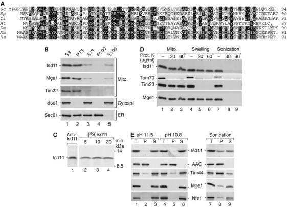
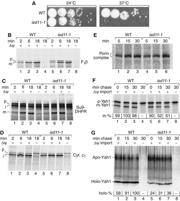
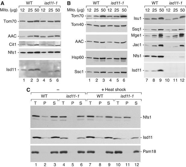
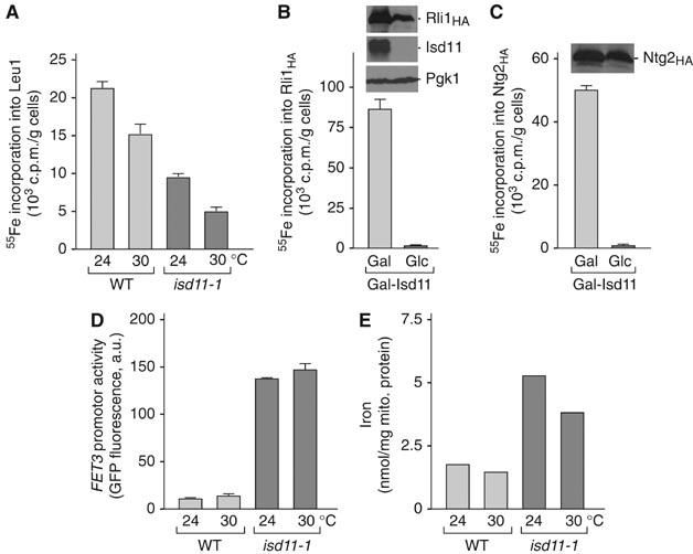
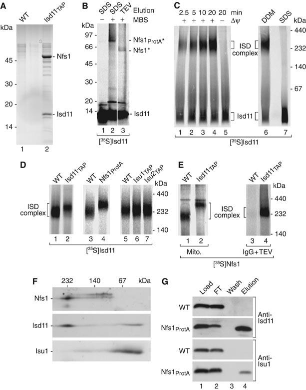
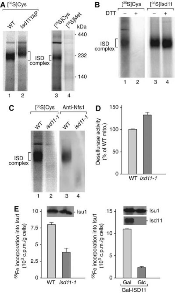
Mitochondria are indispensable for cell viability; however, major mitochondrial functions including citric acid cycle and oxidative phosphorylation are dispensable. Most known essential mitochondrial proteins are involved in preprotein import and assembly, while the only known essential biosynthetic process performed by mitochondria is the biogenesis of iron-sulfur clusters (ISC). The components of the mitochondrial ISC-assembly machinery are derived from the prokaryotic ISC-assembly machinery. We have identified an essential mitochondrial matrix protein, Isd11 (YER048w-a), that is found in eukaryotes only. Isd11 is required for biogenesis of cellular Fe/S proteins and thus is a novel subunit of the mitochondrial ISC-assembly machinery. It forms a complex with the cysteine desulfurase Nfs1 and is required for formation of an Fe/S cluster on the Isu scaffold proteins. We conclude that Isd11 is an indispensable eukaryotic component of the mitochondrial machinery for biogenesis of Fe/S proteins.


Figures

Figure 1
Figure 1
Isd11 is located in the mitochondrial matrix. (A) Deduced primary structures of Isd11 and homologs from S. cerevisiae (Sc), Schizosaccharomyces pombe (Sp), Yarrowia lipolytica (Yl), Arabidopsis thaliana (At), Drosophila melanogaster (Dm), Mus musculus (Mm) and Homo sapiens (Hs). Black, identical amino-acid residues in at least four proteins; gray, similar residues. (B) Yeast cells were fractionated by differential centrifugation and analyzed by SDS–PAGE and Western blotting. S, supernatant; P, pellet (multiples of 1000 g). (C) 35S-labeled Isd11 precursor was imported into isolated mitochondria. The mitochondria were swollen, treated with proteinase K and separated by SDS–PAGE. Analysis was performed by immunodecoration (lane 1) or digital autoradiography (lanes 2–4). (D) Mitochondria were either left untreated or subjected to swelling or sonication. Then, a treatment with proteinase K was performed as indicated. (E) Mitochondria were incubated at alkaline pH or subjected to sonication, followed by centrifugation at 100 000 g. T, total; AAC, ADP/ATP carrier.
Figure 2
Figure 2
isd11-1 mutant mitochondria import preproteins but are impaired in maturation of Yah1. (A) A conditional yeast mutant of ISD11. Wild-type (WT) and isd11-1 yeast cells were spotted on YPG agar plates and grown for 3 days at 24 or 37°C. (B–D) Mitochondria were isolated from WT or isd11-1 cells grown at 19°C. 35S-labeled precursor proteins were imported and analyzed by SDS–PAGE and digital autoradiography. p, precursor; i, intermediate; m, mature; F1β, F1-ATPase subunit β. (E) [35S]porin was imported into isolated mitochondria and analyzed by BN-PAGE. (F) 35S-labeled precursor of Yah1 was imported into isolated mitochondria for 10 min, followed by dissipation of Δψ and a further incubation (chase). Imported mature-sized (m) Yah1 was analyzed by SDS–PAGE and digital autoradiography. The maximal amount formed was set to 100%. (G) The experiment was performed as described for panel F except that apo-Yah1 and the ISC-containing holo-Yah1 were analyzed by native PAGE (Lutz et al, 2001).
Figure 3
Figure 3
Reduced mitochondrial levels of Nfs1 in isd11-1 cells grown under non-permissive conditions. (A) Wild-type (WT) and isd11-1 cells were grown at 19°C. Isolated mitochondria (μg of protein) were analyzed by SDS–PAGE and Western blotting. Cit1, citrate synthase. (B) Cells were grown at 19°C and shifted to 37°C for 8 h on non-fermentable medium. (C) Mitochondria isolated from cells grown at 19°C were either kept on ice or heat shocked for 15 min at 37°C, lysed with digitonin and separated into pellet and supernatant by centrifugation.
Figure 4
Figure 4
isd11-1 cells show decreased activities of mitochondrial and cytosolic Fe/S proteins. (A) Mitochondria were isolated from WT and isd11-1 cells grown for 24 h in rich medium supplemented with glucose at 24 and 30°C. The enzymatic activities of aconitase, respiratory complexes II and III and malate dehydrogenase (MDH) were measured. The activity of aconitase in isd11-1 mitochondria isolated from cells grown at 19°C (as used in Figure 2B–G) was similarly diminished by 75% compared to WT mitochondria. (B) Levels of mitochondrial proteins detected by Western blotting. Aco1, aconitase; Rip1, Rieske Fe/S protein; Sdh2, subunit 2 of succinate dehydrogenase. (C, D) WT and isd11-1 cells were grown in minimal medium supplemented with glucose at 24 or 30°C for 24 h. In addition, Gal-ISD11 cells were cultured for 18 h in minimal medium containing galactose (Gal) or glucose (Glc) to induce or repress, respectively, synthesis of Isd11. Extracts were prepared by disruption of cells with glass beads, and aliquots were analyzed (C) for the enzymatic activities of Leu1 and alcohol dehydrogenase (ADH), or (D) for the protein levels of Isd11, Leu1 and the cytosolic marker protein Pgk1 by Western blotting.
Figure 5
Figure 5
isd11-1 cells are impaired in the de novo formation of Fe/S proteins and iron homeostasis. (A) Wild-type (WT) and isd11-1 cells were grown for 24 h in iron-poor minimal medium supplemented with glucose at 24 and 30°C. Cells were labeled with 55Fe, washed and a cell extract was prepared using glass beads. Leu1 was immunoprecipitated. The radioactivity associated with the immuno-beads was quantified by liquid scintillation counting. (B, C) A similar analysis as described in panel A was performed using Gal-ISD11 cells after growth for 40 h in iron-poor minimal medium containing galactose or glucose. After radiolabeling, cell extracts were analyzed for the de novo assembly of the Fe/S clusters on cytosolic Rli1HA (Kispal et al, 2005) and nuclear Ntg2HA. (D) Cellular iron homeostasis is perturbed in isd11-1 cells. WT and isd11-1 cells carrying plasmid p415-FET3-GFP were grown in minimal medium supplemented with 200 μM ferric ammonium citrate at 24 or 30°C. Cells were harvested at an optical density of 0.5 and the transcriptional activity of the FET3 promoter was determined by recording the fluorescence emission of the cell suspension at 513 nm (excitation at 480 nm). The signal of cells lacking plasmid p415-FET3-GFP was subtracted. (E) Mitochondrial iron accumulation in isd11-1 cells. Mitochondria were isolated from cells grown in rich medium containing glucose and the iron content was determined.
Figure 6
Figure 6
Nfs1 and Isd11 form a stable complex. (A) Mitochondria were lysed in 0.1% n-dodecyl-β-D-maltoside (DDM) and incubated with IgG-Sepharose. After washing of the beads and incubation with TEV protease, eluates were separated by SDS–PAGE, stained with colloidal Coomassie and analyzed by tandem mass spectrometry. (B) 35S-labeled Isd11 (non-tagged) was imported into Nfs1ProtA mitochondria. After incubation with MBS and quenching, mitochondria were lysed and subjected to IgG affinity chromatography. Elution was performed with SDS sample buffer or TEV protease, followed by SDS–PAGE and digital autoradiography. Asterisks, crosslinking products. (C) [35S]Isd11 was imported into WT mitochondria, followed by lysis with 0.1% DDM (samples 1–6) or 0.1% SDS (sample 7) and BN-PAGE analysis. (D) [35S]Isd11 was imported into energized mitochondria. Upon lysis with DDM, the samples were analyzed by BN-PAGE. (E) [35S]Nfs1 (non-tagged) was imported into energized mitochondria. After lysis with DDM, samples 1 and 2 were directly analyzed by BN-PAGE, whereas samples 3 and 4 were first subjected to IgG affinity chromatography, followed by elution with TEV protease and BN-PAGE. (F) WT mitochondria were lysed and proteins were separated by BN-PAGE, followed by a second-dimension SDS–PAGE and Western blotting. (G) WT and Nfs1ProtA mitochondria were subjected to IgG affinity chromatography. Isd11 and Isu1 were analyzed by SDS–PAGE and Western blotting. FT, flow-through.
Figure 7
Figure 7
Isd11 is part of the active desulfurase complex and required for ISC formation on Isu1. (A) Wild-type (WT) and Isd11TAP mitochondria were treated with chloramphenicol. Mitochondria were lysed, incubated with [35S]cysteine or [35S]methionine and subjected to BN-PAGE and digital autoradiography. (B) Samples 1 and 2, WT mitochondrial lysate was incubated with [35S]cysteine as described for panel A. Samples 3 and 4, [35S]Isd11 was imported into WT mitochondria. Before BN-PAGE analysis, half of the samples were treated with 10 mM DTT. (C) WT and isd11-1 mitochondrial lysates were either directly subjected to BN-PAGE and Western blotting (samples 3 and 4) or incubated with [35S]cysteine and analyzed by BN-PAGE and digital autoradiography (samples 1 and 2). (D) isd11-1 cells are not impaired in cysteine desulfurase activities. Mitochondria isolated from cells grown at 30°C were lysed and incubated in the presence of 4 mM cysteine and 1 mM DTT for 20 min at 30°C. The amount of sulfide formed from cysteine by Nfs1 was determined (Mühlenhoff et al, 2004). The signal recorded in samples lacking cysteine was subtracted. (E) Isd11 is required for Fe/S cluster assembly on Isu1. WT and isd11-1 cells overexpressing ISU1 from vector p426-GPD were grown for 24 h at 30°C. Gal-ISD11 cells overexpressing ISU1 were grown in the presence of galactose or glucose. Fe/S cluster assembly on Isu1 was determined by 55Fe labeling and immunoprecipitation assay described in Figure 5A.

References

    1. Balk J, Pierik AJ, Netz DJ, Mühlenhoff U, Lill R (2004) The hydrogenase-like Nar1p is essential for maturation of cytosolic and nuclear iron–sulphur proteins. EMBO J 23: 2105–2115 - PMC - PubMed
    1. Chacinska A, Lind M, Frazier AE, Dudek J, Meisinger C, Geissler A, Sickmann A, Meyer HE, Truscott KN, Guiard B, Pfanner N, Rehling P (2005) Mitochondrial presequence translocase: switching between TOM tethering and motor recruitment involves Tim21 and Tim17. Cell 120: 817–829 - PubMed
    1. Chacinska A, Rehling P, Guiard B, Frazier AE, Schulze-Specking A, Pfanner N, Voos W, Meisinger C (2003) Mitochondrial translocation contact sites: separation of dynamic and stabilizing elements in formation of a TOM–TIM–preprotein supercomplex. EMBO J 22: 5370–5381 - PMC - PubMed
    1. Chen OS, Crisp RJ, Valachovic M, Bard M, Winge DR, Kaplan J (2004) Transcription of the yeast iron regulon does not respond directly to iron but rather to iron–sulfur cluster biosynthesis. J Biol Chem 279: 29513–29518 - PubMed
    1. Contamine V, Picard M (2000) Maintenance and integrity of the mitochondrial genome: a plethora of nuclear genes in the budding yeast. Microbiol Mol Biol Rev 64: 281–315 - PMC - PubMed

Publication types

MeSH terms