. 2006 May;235(5):1300-9.
doi: 10.1002/dvdy.20655.

Origin of the ultimobranchial body cyst: T/ebp/Nkx2.1 expression is required for development and fusion of the ultimobranchial body to the thyroid

Affiliations

Origin of the ultimobranchial body cyst: T/ebp/Nkx2.1 expression is required for development and fusion of the ultimobranchial body to the thyroid

Takashi Kusakabe et al. Dev Dyn. 2006 May.

Abstract

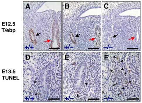
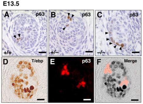
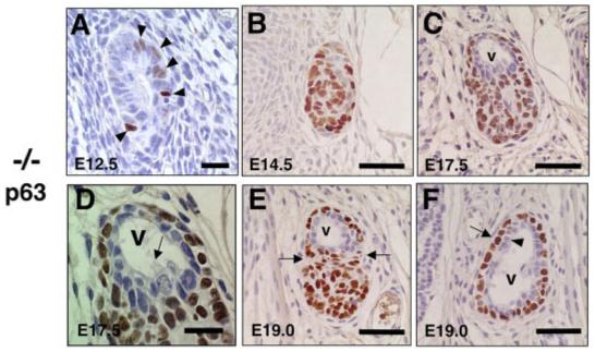
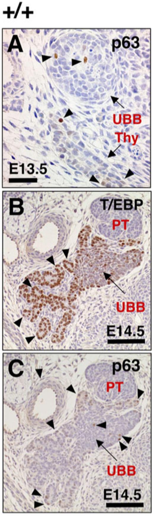
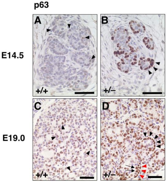
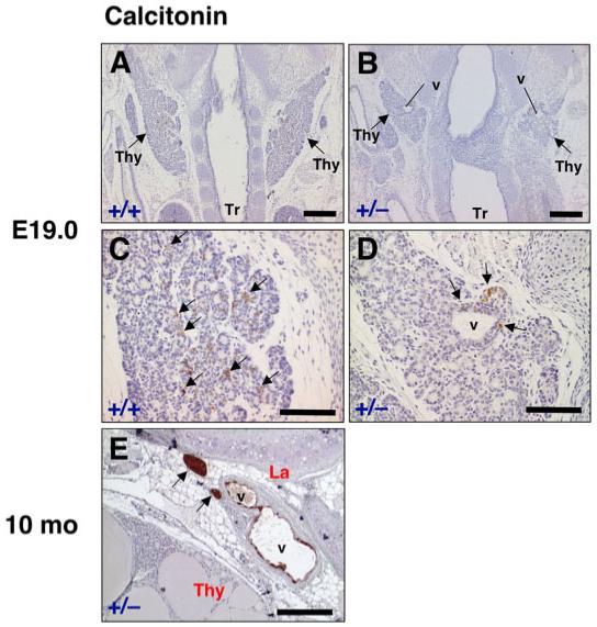
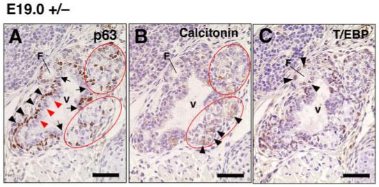
The ultimobranchial body (UBB) is an outpocketing of the fourth pharyngeal pouch that fuses with the thyroid diverticulum, giving rise to calcitonin-producing C-cells. In this study, we demonstrate that the UBB is composed of two types of cells: one expressing T/ebp/Nkx2.1 and the other expressing p63. The former cell type, accounting for a majority of the UBB, requires T/ebp/Nkx2.1 for their survival. In contrast, the p63-positive cells, even in the absence of T/ebp/Nkx2.1 expression, can proliferate and give rise to a vesicular structure that is lined by a monolayer of p63-negative cells, surrounded by a cluster and/or single layer of p63-positive cells, displaying the basal/stem cell phenotype. T/ebp/Nkx2.1 haploinsufficiency causes abnormal fusion of the UBB with the thyroid diverticulum, which stays as a cluster of C-cells around the vesicular structure, similar to the one observed in mice null for T/ebp/Nkx2.1 expression. These results demonstrate that T/ebp/Nkx2.1 plays a role in the survival of UBB cells, their dissemination into the thyroid diverticulum, and the formation of UBB-derived vesicular structure.


Figures

Fig. 1
Fig. 1
Ultimobranchial body (UBB) of wild-type, T/ebp-heterozygous, and T/ebp-null mutant mice. A-C: Immunostaining for T/ebp on transverse sections of embryonic day (E) 12.5 embryos. T/ebp-positive cells are shown in brown. Dorsal is up. Black arrow indicates UBB. Red arrow points to the laryngotracheal groove. Most cells in the UBB of wild-type (A) and T/ebp-heterozygous mutant mice (B) express T/ebp. Note that the UBB forms and migrates properly in both T/ebp-heterozygous and null mice (B,C) just like wild-type control, as seen in B,C. D-F: Terminal deoxynucleotidyl transferase-mediated deoxyuridinetriphosphate nick end-labeling (TUNEL) staining of UBBs using transverse sections of E13.5 embryos. Cells stained in brown represent apoptotic cells (shown by arrowheads). Dorsal is up. Note that many cells are positive for apoptosis in the UBB of T/ebp-null mutants (F), whereas a few apoptotic cells are observed in both wild-type (D) and T/ebp-heterozygous mice (E). Scale bar = 100 μm in A-C, 50 μm in D-F.
Fig. 2
Fig. 2
Expression of p63 and T/ebp in ultimobranchial body (UBB) cells. A-C: Transverse sections of UBBs from E13.5 wild-type, T/ebp-heterozygous and T/ebp-null mutant embryos. p63-positive cells are shown in brown. Dorsal is up. Only a few p63-positive cells can be detected per section in all genotypes, and a vast majority of UBB cells are negative for p63. D-F: Double staining for T/ebp (D, brown) and p63 (E, red) using wild-type UBB. Dorsal is up. F: The merged image demonstrates the complementary expression of T/ebp and p63 in the UBB. Scale bar = 20 μm.
Fig. 3
Fig. 3
Altered ultimobranchial body (UBB) development and characteristic pattern of p63 expression in T/ebp-null mutants. A-F: Transverse sections of UBBs obtained from various embryonic days of T/ebp-null mutants were subjected to p63 immunostaining. Dorsal is up. Cells stained in brown indicate p63 expression. A: No significant difference was observed in the number of p63-positive cells between E12.5 and E13.5 UBBs (Fig. 2C). B: The p63-positive cells make a solid, oval mass by E14.5 that have taken over most of the UBBs. C,D: By E17.5, a vesicular structure is generated at the dorsal part of the UBB (v in C), which is lined by a monolayer of cells without p63 expression, accompanied by occasional ciliated cells (D, arrow). E: By E19.0, the UBB becomes constricted between the vesicular structure and the mass of p63-positive cells (shown by arrows). F: Cranial to the section shown in E, the vesicular structure displays a well-defined double layer of cells composed of p63-negative inner layer (arrowhead) and p63-positive surrounding layer (arrow). Scale bar = 20 μm in A,D, 50 μm in B,C,E,F.
Fig. 4
Fig. 4
Images of p63 and T/ebp expression in the ultimobranchial body (UBB) and thyroid diverticulum of wild-type embryos. A: The p63 immunostaining can be detected in thyroid diverticulum as well as UBB (arrowheads, shown in brown) in transverse sections of embryonic day (E) 13.5 embryo. Dorsal is up. Thy, thyroid diverticulum. B,C: Immunostaining for T/ebp and p63 (both shown in brown) using serially prepared E14.5 embryo transverse sections. Dorsal is up. The UBB has already been fused but has not yet been completely incorporated into the thyroid diverticulum at this stage. The thyroid diverticulum begins to form many primitive follicles (B, arrowheads) with high levels of T/ebp expression, making a sharp contrast to solid appearance and faint T/ebp expression of partly fused UBB (B, C, arrows). C: P63-positive cells are sparsely distributed in both UBB and primitive thyroid follicles (arrowheads). PT, parathyroid. Scale bar = 50 μm in A,100 μm in B,C.
Fig. 5
Fig. 5
Immunostaining for p63 in primitive thyroid follicles of wild-type and T/ebp-heterozygous mutant embryos. A-D: The p63 expression is shown in brown. Dorsal is up. A,B: At embryonic day (E)14.5, the p63 expression level is mostly low or undetectable in primitive follicles of wild-type embryo thyroids (A, arrowheads), whereas almost all primitive follicles in T/ebp-heterozygous mutant thyroids are clearly rimmed with strongly p63-positive cells (B, arrowheads). C,D: At E19.0, many but not all follicular cells in primitive follicles of wild-type thyroid exhibit low p63 expression (C, arrowheads), whereas strong p63 expression is evident not only in follicular cells of primitive follicles (D, arrows) but also cells surrounding the primitive follicle (D, arrowheads) in T/ebp-heterozygous mutants, clearly showing a bilayered appearance in some part (D, arrows and red arrowheads). Scale bar = 50 μm.
Fig. 6
Fig. 6
Calcitonin expression in the thyroid of wild-type and T/ebp-heterozygous mutant mice. A-D: Frontal sections of embryonic day (E) 19.0 embryos from wild-type and T/ebp-heterozygous mice were subjected to calcitonin immunostaining. Arrows point to bilateral thyroid lobes (Thy). Tr, trachea. Calcitonin expression is shown in brown. A,B: Cranial is up. T/ebp-heterozygous mutants always form vesicular structures in bilateral thyroid lobes (v in B), whereas the vesicular structure never appears in wild-type (A). C,D: Higher magnification of the same section shown in A and B, respectively. In wild-type, calcitonin-positive cells are diffusely disseminated into the thyroid lobe (C, arrows), whereas in T/ebp-heterozygous mutant, calcitonin expression is found only around the vesicular structure (D, arrows). E: Immunostaining for calcitonin on a thyroid section from 10-month-old adult T/ebp-heterozygous mutant. Dorsal is right. Vesicular structures persist between the thyroid lobe (Thy) and the larynx (La). Calcitonin is expressed in some cells lining the vesicles and strong expression found in clumps of cells localized in the vicinity of the vesicles (arrows). Scale bar = 300 μm in A,B, 100 μm in C,D, 200 μm in E.
Fig. 7
Fig. 7
Defective ultimobranchial body (UBB) development and abnormal merging with the thyroid diverticulum. A-C: Immunostaining for p63, calcitonin, and T/ebp using serially prepared sections from embryonic day (E) 19.0 T/ebp-heterozygous mutant embryos. Dorsal is up. The expression is shown in brown for each staining. Vesicular structure (v) consistently appears at the dorsal part of the embryonic thyroid. Similar to the vesicular structures seen in T/ebp-null mutants (Fig. 3C-F), the vesicular wall consists of inner p63-negative (red arrowheads) and outer p63-positive cells (arrowheads). This wall is frequently intermittent (A, arrows) and interrupted by a cluster of C-cell precursors (A, B, circled) and small follicles (F in A-C), which express calcitonin (B, arrowhead) and T/ebp (C, arrowhead), respectively. Scale bar = 50 μm.

References

    1. Barbareschi M, Pecciarini L, Cangi MG, Macri E, Rizzo A, Viale G, Doglioni C. p63, a p53 homologue, is a selective nuclear marker of myoepithelial cells of the human breast. Am J Surg Pathol. 2001;25:1054–1060. - PubMed
    1. Biddinger PW, Ray M. Distribution of C cells in the normal and diseased thyroid gland. Pathol Annu. 1993;28(Pt 1):205–229. - PubMed
    1. Burstein DE, Nagi C, Wang BY, Unger P. Immunohistochemical detection of p53 homolog p63 in solid cell nests, papillary thyroid carcinoma, and hashimoto’s thyroiditis: a stem cell hypothesis of papillary carcinoma oncogenesis. Hum Pathol. 2004;35:465–473. - PubMed
    1. Cameselle-Teijeiro J, Varela-Duran J, Sambade C, Villanueva JP, Varela-Nunez R, Sobrinho-Simoes M. Solid cell nests of the thyroid: light microscopy and immunohistochemical profile. Hum Pathol. 1994;25:684–693. - PubMed
    1. Damante G, Tell G, Di Lauro R. A unique combination of transcription factors controls differentiation of thyroid cells. Prog Nucleic Acid Res Mol Biol. 2001;66:307–356. - PubMed

Publication types

MeSH terms

LinkOut - more resources