. 2005 Dec 12:2:29.
doi: 10.1186/1742-2094-2-29.

Stimulation of cannabinoid receptor 2 (CB2) suppresses microglial activation

Affiliations

Stimulation of cannabinoid receptor 2 (CB2) suppresses microglial activation

Jared Ehrhart et al. J Neuroinflammation. .

Abstract

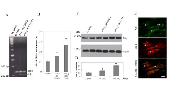
Background: Activated microglial cells have been implicated in a number of neurodegenerative disorders, including Alzheimer's disease (AD), multiple sclerosis (MS), and HIV dementia. It is well known that inflammatory mediators such as nitric oxide (NO), cytokines, and chemokines play an important role in microglial cell-associated neuron cell damage. Our previous studies have shown that CD40 signaling is involved in pathological activation of microglial cells. Many data reveal that cannabinoids mediate suppression of inflammation in vitro and in vivo through stimulation of cannabinoid receptor 2 (CB2).

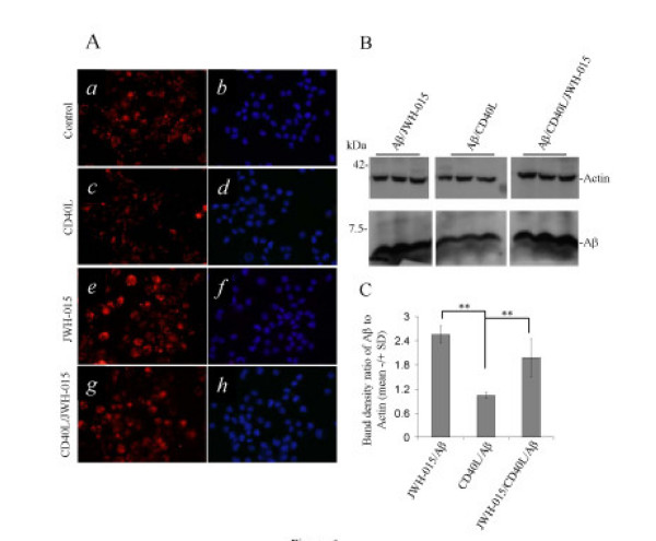
Methods: In this study, we investigated the effects of a cannabinoid agonist on CD40 expression and function by cultured microglial cells activated by IFN-gamma using RT-PCR, Western immunoblotting, flow cytometry, and anti-CB2 small interfering RNA (siRNA) analyses. Furthermore, we examined if the stimulation of CB2 could modulate the capacity of microglial cells to phagocytise Abeta1-42 peptide using a phagocytosis assay.

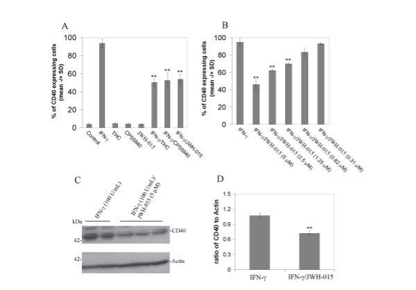
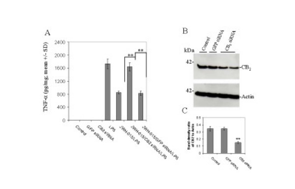
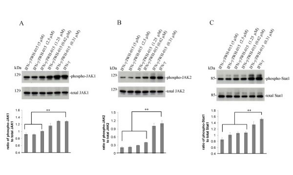
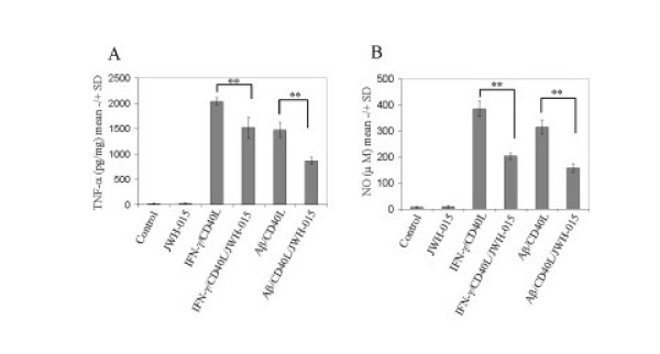
Results: We found that the selective stimulation of cannabinoid receptor CB2 by JWH-015 suppressed IFN-gamma-induced CD40 expression. In addition, this CB2 agonist markedly inhibited IFN-gamma-induced phosphorylation of JAK/STAT1. Further, this stimulation was also able to suppress microglial TNF-alpha and nitric oxide production induced either by IFN-gamma or Abeta peptide challenge in the presence of CD40 ligation. Finally, we showed that CB2 activation by JWH-015 markedly attenuated CD40-mediated inhibition of microglial phagocytosis of Abeta1-42 peptide. Taken together, these results provide mechanistic insight into beneficial effects provided by cannabinoid receptor CB2 modulation in neurodegenerative diseases, particularly AD.


Figures

Figure 1
Figure 1
Cannabinoids inhibit microglial CD40 expression induced by IFN-γ. A, Mouse primary microglial cells were cultured in 6-well tissue-culture plates (5 × 10⁵/well) and treated with THC (0.6 μM), CP55940 (5 μM) or selective cannabinoid CB2 agonist (JWH015; 5 μM) in the presence or absence of IFN-γ (100 U/mL), or treated with vehicle (1% DMSO Control) or IFN-γ alone (100 U/mL); B, In parallel 6-well tissue-culture plates, microglial cells were incubated with IFN-γ (100 U/mL) in the presence or absence of JWH-015 at the indicated doses. After 12 hr-treatments, these cells were prepared for FACS analysis of CD40 expression as described in Materials and methods. For A, ANOVA and post hoc testing showed significant differences of mean fluorescence (+/- SD with n = 3 for each condition) between IFN-γ treatment and IFN-γ treatment in the presence of THC, CP55940 or JWH-015 (p < 0.001). However, there was not a significant difference between IFN-γ/THC and either IFN-γ/CP55940 or IFN-γ/JWH-015 (p > 0.05). For B, ANOVA and post hoc testing showed significant differences of mean fluorescence (+/- SD with n = 3 for each condition) between IFN-γ treatment and IFN-γ treatment in the presence of JWH-015 at 5 μM, 2.5 μM and 1.25 μM (** p < 0.001). C, Western blot analysis by anti-mouse CD40 antibody shows CD40 protein expression and, by anti-β-actin antibody, shows β-actin protein (internal reference). D, Densitometric quantification of Western immunoblotting analysis from independent experiments (n = 2 for IFN-γ; n = 3 for IFN-γ/JWH-015 treatment) indicated that doses of JWH-015 of 1.25 μM or greater significantly (** p < 0.05) reduced IFN-γ-induced CD40 expression. CD40 expression is shown normalized to β-actin.
Figure 2
Figure 2
Cannabinoid receptor CB2 is expressed by cultured microglial cells. A, RT-PCR analysis of murine primary cultured microglial cells. A 239-bp band corresponding to CB2 was specifically generated with primers described in the Materials and methods section. B, Graphical representation of RT-PCR band density ratio of CB2 expression normalized to β-actin (mean +/- SD) is shown (n = 3 for each condition). ANOVA revealed significant between-group differences (control versus IFN-γ (50 U/mL) and IFN-γ (50 U/mL) versus IFN-γ (100 U/mL); p < 0.005). C, Western immunoblot analysis of murine primary cultured microglial cells using specific antibodies targeting CB2 and β-actin proteins. D, Western blot band density is represented as ratio of CB2 to β-actin (mean +/- SD; n = 4 for each condition). ANOVA revealed significant between-group differences [Control versus IFN-γ (50 U/mL) and IFN-γ (50 U/mL) versus IFN-γ (100 U/mL); ** p < 0.005]. E, Cannabinoid receptor CB2 is expressed in microglial cells in situ. In white matter, microglial cells are positive in their somata and processes for CB2. White arrowheads show positive cells as indicated. The expression of CB2 (FITC; green) was co-localized with Iba-1, microglial cell marker (TRITC; red) as indicated. Bottom panel denotes merge signals. Bar denotes 10 μm.
Figure 3
Figure 3
Cultured microglial cells (N9) treated with LPS and 100 nM anti-murine CB2 siRNA lose their ability to respond to CB2 agonist, JWH-015. A, Microglial cells treated with LPS (100 ng/mL) secreted large quantities of TNF-α (n = 3, **p < 0.005). Co-treatment with JWH-015 (5 μM) attenuated LPS-induced TNF-α release. Pre-treatment with anti-CB2 siRNA abolished JWH-015's ability to reduce LPS-induced TNF-α release (n = 3, ** p < 0.05). Non-targeting anti-GFP siRNA control had no effect. B and C, Western blot using an anti-murine CB2 antibody demonstrates that 100 nM anti-CB2 siRNA significantly reduced expression of CB2 protein by N9 microglial cells after 48 hr (n = 2, ** p < 0.05).
Figure 4
Figure 4
Cannabinoid CB2 agonist treatment opposes IFN-γ-induced phosphorylation of JAK/STAT1 in microglial cells. A, B, Primary microglial cells were seeded in 6-well tissue-culture plates (5 × 10⁵/well) and treated with IFN-γ (100 U/mL) in the presence or absence of CB2 agonist (JWH-015) at the indicated doses for 30 min. Cell lysates were prepared from these cells and subjected to Western immunoblotting using antibodies against phospho-JAK1 (Tyr1022/1023) and JAK2 (Tyr1007/1008), or total JAK1 and JAK2, as indicated. Densitometric quantification of all Western immunoblots results are summarized by the histograms below, representative of Western immunoblots from two independent experiments. Dose-dependent reductions in phospho-JAK1/total JAK1 and phosphor-JAK2/total JAK2 correlated with JWH-015 treatments, becoming significant (** p < 0.05) at doses greater than or equal to 1.25 μM and 0.62 μM for JAK1 and JAK2, respectively. C, In parallel experiments, cell lysates were subjected to Western immunoblotting using anti-phospho-STAT1 (Ser727) or anti-total STAT1 antibody as indicated. Dose-dependent reductions in phospho-Stat1/total Stat1 correlated with JWH-015 treatments, becoming significant (** p < 0.05) at doses greater than or equal to 0.62 μM.
Figure 5
Figure 5
CB2 stimulation attenuates microglial proinflammatory cytokine release. Mouse primary microglial cells were seeded in 24-well tissue-culture plates (1 × 10⁵/well) and co-treated with either IFN-γ (100 U/mL)/CD40L protein (2 μg/mL) or Aβ1–42 (1 μM)/CD40L protein (2 μg/mL) in the presence or absence of cannabinoid receptor CB2 agonist (JWH015, 5 μM) for 24 hr. Cell cultured supernatants were collected and subjected to TNF-α cytokine ELISA (A) and NO release assay (B) as indicated. TNF-α production was represented as mean pg of TNF-α per mg of total cellular protein (+/- SD). Similar results were obtained in three independent experiments. ANOVA and post hoc testing revealed significant differences between IFN-γ/CD40L and IFN-γ/CD40L and JWH-015 (** p < 0.005); Aβ1–42/CD40L and Aβ1–42/CD40L plus JWH-015 treatment (** p < 0.001).
Figure 6
Figure 6
CB2 stimulation modulates microglial phagocytic function. A, Mouse primary microglial cells were seeded in 6-well tissue culture plates with glass inserts (5 × 10⁵cells/well) and treated with 3 μM Cy3™-Aβ1–42 in the absence (a and b; Control) or presence of either CD40L protein (c and d 2.5 μg/mL) or JWH-015 (e and f; 5 μM), or both JWH-015 and CD40L protein (g and h). After 3 hr these cells were washed and fixed (see Materials and Methods). Subsequently, immunofluorescence microscopy examination was performed using a 40 X objective with appropriate filter selection. The darkfield images a, c, e, and g show the fluorescence of Cy3™ labeled Aβ1–42 whereas, b, d, f, and h show only the DAPI nuclear stain of the same fields. B, In parallel experiments, under the same treatment conditions, microglial cell lysates were prepared for Western immunoblotting analysis (see Materials and methods) of cell-associated Aβ1–42 using anti-Aβ antibody (BAM-10, Sigma). C, Aβ mean band densities are graphically represented as ratios to β-actin +/- SD (n = 3 for each condition). ANOVA revealed significant between-group differences (JWH-015/Aβ versus CD40L/Aβ and Aβ/CD40L versus JWH-015/CD40L/Aβ; ** p < 0.005), and post hoc testing showed significant differences between CD40L/Aβ and JWH-015/CD40L/Aβ (** p < 0.005).

References

    1. Polazzi E, Contestabile A. Reciprocal interactions between microglia and neurons: from survival to neuropathology. Rev Neurosci. 2002;13:221–242. - PubMed
    1. Schonrock LM, Kuhlmann T, Adler S, Bitsch A, Bruck W. Identification of glial cell proliferation in early multiple sclerosis lesions. Neuropathol Appl Neurobiol. 1998;24:320–330. doi: 10.1046/j.1365-2990.1998.00131.x. - DOI - PubMed
    1. Mackenzie IR, Hao C, Munoz DG. Role of microglia in senile plaque formation. Neurobiol Aging. 1995;16:797–804. doi: 10.1016/0197-4580(95)00092-S. - DOI - PubMed
    1. Gendelman HE, Tardieu M. Macrophages/microglia and the pathophysiology of CNS injuries in AIDS. J Leukoc Biol. 1994;56:387–388. - PubMed
    1. Nelson PT, Soma LA, Lavi E. Microglia in diseases of the central nervous system. Ann Med. 2002;34:491–500. doi: 10.1080/078538902321117698. - DOI - PubMed