. 2005 Dec 12:5:17.
doi: 10.1186/1471-2490-5-17.

Stromal mesenchyme cell genes of the human prostate and bladder

Affiliations

Stromal mesenchyme cell genes of the human prostate and bladder

Young Ah Goo et al. BMC Urol. .

Abstract

Background: Stromal mesenchyme cells play an important role in epithelial differentiation and likely in cancer as well. Induction of epithelial differentiation is organ-specific, and the genes responsible could be identified through a comparative genomic analysis of the stromal cells from two different organs. These genes might be aberrantly expressed in cancer since cancer could be viewed as due to a defect in stromal signaling. We propose to identify the prostate stromal genes by analysis of differentially expressed genes between prostate and bladder stromal cells, and to examine their expression in prostate cancer.

Methods: Immunohistochemistry using antibodies to cluster designation (CD) cell surface antigens was first used to characterize the stromas of the prostate and bladder. Stromal cells were prepared from either prostate or bladder tissue for cell culture. RNA was isolated from the cultured cells and analyzed by DNA microarrays. Expression of candidate genes in normal prostate and prostate cancer was examined by RT-PCR.

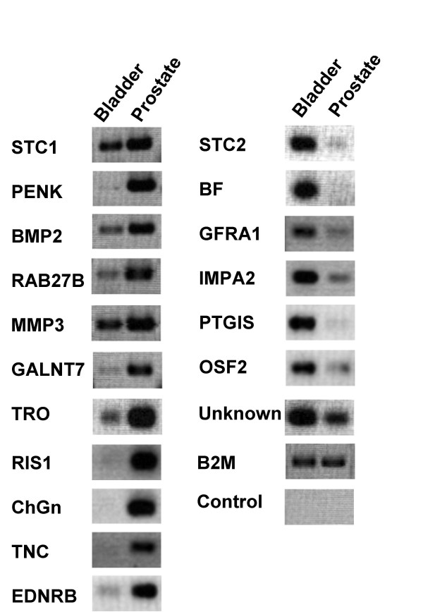
Results: The bladder stroma was phenotypically different from that of the prostate. Most notable was the presence of a layer of CD13+ cells adjacent to the urothelium. This structural feature was also seen in the mouse bladder. The prostate stroma was uniformly CD13-. A number of differentially expressed genes between prostate and bladder stromal cells were identified. One prostate gene, proenkephalin (PENK), was of interest because it encodes a hormone. Secreted proteins such as hormones and bioactive peptides are known to mediate cell-cell signaling. Prostate stromal expression of PENK was verified by an antibody raised against a PENK peptide, by RT-PCR analysis of laser-capture microdissected stromal cells, and by database analysis. Gene expression analysis showed that PENK expression was down-regulated in prostate cancer.

Conclusion: Our findings show that the histologically similar stromas of the prostate and bladder are phenotypically different, and express organ-specific genes. The importance of these genes in epithelial development is suggested by their abnormal expression in cancer. Among the candidates is the hormone PENK and the down-regulation of PENK expression in cancer suggests a possible association with cancer development.


Figures

Figure 1
Figure 1
CD13 immunohistochemistry of the prostate and bladder. (a) Human bladder: CD13 stains a subpopulation of stromal cells (black arrow) in the lamina propria. The partially denuded urothelium is indicated by the red arrow. (b) Mouse bladder: CD13 also stains a similar region (black arrow) in the mouse bladder as in the human bladder. (c) Human prostate: CD13 stains only luminal epithelial cells (black arrow) of prostatic glands.
Figure 2
Figure 2
Organ-specific stromal genes. Shown are the results of the genes tested for their specificity, (cultured prostate vs. bladder stromal cells). Differential expression is gauged by the band intensity of the PCR products. B2M is β2-microglobulin, which was used as a positive control, and H2O was used as a negative control for the reaction.
Figure 3
Figure 3
Expression of PENK in prostate stromal cell. (a) RT-PCR of laser-captured stromal cells. Cells were taken from non-cancer (NP), and PENK was detected in these cells. H2O was used as a negative control for the reaction. (b) PENK expression in sorted prostate cells. The various prostate cell types were sorted from tissue: CD26+ luminal cells, CD104+ basal cells, CD49a+ stromal cells, and CD31+ endothelial cells. Their transcriptomes were determined by microarray analysis using the Affymetrix Human Genome U133 Plus 2.0 GeneChips. PENK expression is localized to the CD49a+ stromal cells.
Figure 4
Figure 4
PENK immunohistochemistry of prostate and bladder. (a) In prostate, the PENK antibody stains the stroma in a pattern that is similar to that by CD56 [13]. The smooth muscle wall of a large blood vessel is also stained (black arrow, left panel). Benign glands appear to be stained at the luminal surface, but this staining is likely non-specific because it was present in the control without the primary antibody (in which the stromal staining was not seen). (b) In bladder, both the urothelium (blue arrow) and stroma (red arrow) of the lamina propria are not stained. Stained are the muscle bundles of the muscularis propria.
Figure 5
Figure 5
Expression of PENK and STC1 in prostate cancer. NP and CP are matched non-cancer and cancer specimens processed into cDNA. CP1 is a Gleason 4+5 (G9) tumor, CP2 a Gleason 3+3 (G6) tumor, and CP3 a Gleason 3+4 (G7) tumor. Bone and liver metastasis were obtained from end-stage diseases. PC3 and C4-2 are prostate cancer cell lines. PENK is detectable in all NP samples, it is lowered in G6, and barely detectable or absent in G7 and G9; as well as the metastases and cancer cell lines. PENK is found in placenta but not kidney. No significant differential expression was found for STC1 in these same samples, though the bone metastasis had lower expression than the liver metastasis, and lower expression in CP2 compared to NP2. STC1 is known to be expressed in the kidney. cDNA quantity of each sample used was monitored by B2M and αSMA (not shown).

References

    1. Cunha GR, Alarid ET, Turner T, Donjacour AA, Boutin EL, Foster BA. Normal and abnormal development of the male urogenital tract. Role of androgen, mesenchymal-epithelial interactions, and growth factors. J Androl. 1992;13:465–475. - PubMed
    1. Aboseif S, El-Sakka A, Young P, Cunha GR. Mesenchymal reprogramming of adult human epithelial differentiation. Differentiation. 1999;65:113–118. doi: 10.1046/j.1432-0436.1999.6520113.x. - DOI - PubMed
    1. Bissell MJ, Ram TG. Regulation of functional cytodifferentiation and histogenesis in mammary epithelial cells: role of the extracellular matrix. Environ Health Persp. 1989;80:61–70. - PMC - PubMed
    1. Lee C. Role of androgen in prostate growth and regression: stromal-epithelial interaction. Prostate. 1996:52–56. doi: 10.1002/(SICI)1097-0045(1996)6+<52::AID-PROS10>3.0.CO;2-Q. - DOI - PubMed
    1. Kedinger M, Lefebvre O, Duluc I, Freund JN, Simon-Assmann P. Cellular and molecular partners involved in gut morphogenesis and differentiation. Phil Trans R Soc Lond B Biol Sci. 1998;353:847–856. doi: 10.1098/rstb.1998.0249. - DOI - PMC - PubMed

Publication types