. 2005 Dec 19;171(6):919-24.
doi: 10.1083/jcb.200509095. Epub 2005 Dec 12.

Regulation of Sar1 NH2 terminus by GTP binding and hydrolysis promotes membrane deformation to control COPII vesicle fission

Affiliations

Regulation of Sar1 NH2 terminus by GTP binding and hydrolysis promotes membrane deformation to control COPII vesicle fission

Anna Bielli et al. J Cell Biol. .

Abstract

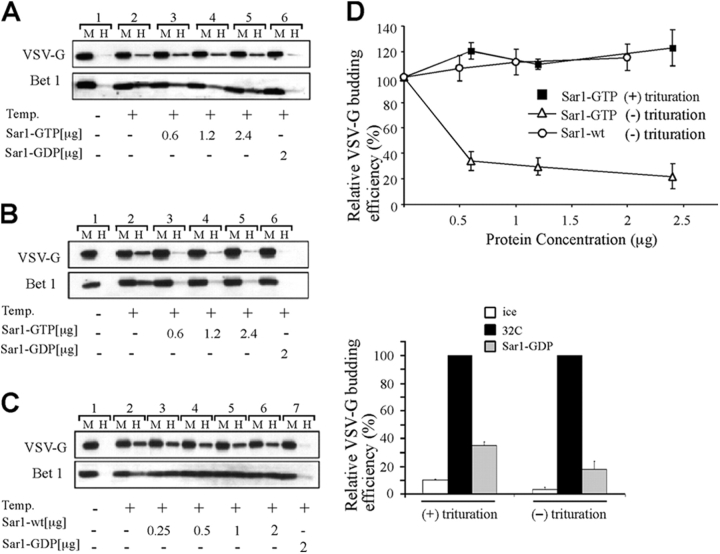
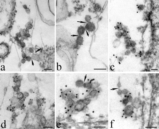
The mechanisms by which the coat complex II (COPII) coat mediates membrane deformation and vesicle fission are unknown. Sar1 is a structural component of the membrane-binding inner layer of COPII (Bi, X., R.A. Corpina, and J. Goldberg. 2002. Nature. 419:271-277). Using model liposomes we found that Sar1 uses GTP-regulated exposure of its NH2-terminal tail, an amphipathic peptide domain, to bind, deform, constrict, and destabilize membranes. Although Sar1 activation leads to constriction of endoplasmic reticulum (ER) membranes, progression to effective vesicle fission requires a functional Sar1 NH2 terminus and guanosine triphosphate (GTP) hydrolysis. Inhibition of Sar1 GTP hydrolysis, which stabilizes Sar1 membrane binding, resulted in the formation of coated COPII vesicles that fail to detach from the ER. Thus Sar1-mediated GTP binding and hydrolysis regulates the NH2-terminal tail to perturb membrane packing, promote membrane deformation, and control vesicle fission.


Figures

Figure 1.
Figure 1.
Sar1 is capable of constricting lipid bilayers. (A) Helical wheel representation of amino acids 1–18 of the NH2 terminus of hamster Sar1a. (B) The NH2-terminal tail of Sar1 destabilizes lipid membranes. Liposomes (100–120 nm, DOPC/DLPA, 80/20 mol percent) were incubated with 10 μM GST (a) or with 10 μM GST-Sar1-N-Tail (b) at 37°C for 1 h. At the end of incubations, liposomes were absorbed on charged grids and negatively stained with 1% phosphotungstic acid for EM analysis. (C) Sar1-GTP is capable of constricting liposome membranes. Liposomes (80–100 nm, DOPC and DLPA, 80/20 mol percent) were incubated in buffer (control; a), 10 μM Sar1-GDP (Sar1-T39N, b), 15 μM Sar1-GTP (Sar1-H79G, c), or 10 μM Δ9-Sar1 (d) mutants with GTP or GDP for 1 h at 37°C. Liposomes were stained and analyzed by EM. (D) A gallery of Sar1-GTP–induced tubulating (a–d) and fused (e and f) DOPC/DLPA liposomes. (E) Cholesterol/DOPC/DLPA (20/75/5 mol percent) liposomes (100–120 nm) were incubated as described above in the absence or presence of Sar1-GTP as indicated. (F) A gallery of tubulating cholesterol/DOPC/DLPA liposomes deformed during incubations with Sar1-GTP. Bars, 100 nm. (G) Sar1 uses its amphipathic NH2 terminus to bind liposomes in a GTP-dependent manner. Sar1 wt (1 μg) or Δ9-Sar1 (1 μg) was incubated with GTP or GDP in the absence or presence of liposomes as indicated. Liposomes were floated into a sucrose gradient and fractions collected from the top were numbered sequentially and analyzed by Western blot with Sar1 antibody. Fractions 1–4 contain liposome-associated Sar1, whereas fractions 5–12 contain unbound Sar1. (H) Liposome-bound Sar1 GTP can form lateral protein interactions. Liposome-bound Sar1 wt (1 μg) was cross-linked with DTSSP (100 μM) and loaded onto sucrose gradients as in G. Floated fractions were analyzed on nonreducing gels by Western blotting. When indicated, DTT (50 mM) was added to reverse cross-linking.
Figure 2.
Figure 2.
Sar1-GTP (H79G) inhibits vesicle release. (A) tsO45-VSV-G–containing membranes were incubated (40 μl final volume) in the presence of cytosol for 30 min on ice (lane 1) or at 32°C (lanes 2–6) in the absence (lanes 1 and 2) or presence of Sar1-GTP (lanes 3–5) or Sar1-GDP (lane 6). At the end of incubation, membranes were subjected to a physical trituration step. The vesicle fraction (H) was separated from the donor membranes (M) by differential centrifugation and analyzed by Western blotting with antibodies against VSV-G and the SNARE protein Bet1. (B and C) The budding assay was performed as above without physical trituration. The mobilization of VSV-G and Bet1 to the vesicular fraction was analyzed in the presence of increasing concentrations of either Sar1-GTP (B, lanes 3–5) or Sar1 wt (C, lanes 3–6) as indicated. (D) Quantitation of the budding efficiency for VSV-G in three independent experiments under the indicated conditions is shown (means ± SEM).
Figure 3.
Figure 3.
Vesicle release is regulated by endogenous Sar1 GTPase activity and requires functional NH2-terminal amphipathic domain. (A) Inhibition of endogenous Sar1 GTPase activity inhibits COPII vesicle release. VSV-G–containing membranes were incubated in the presence of cytosol for 30 min on ice (lane 1) or at 32°C (lanes 2–7) in the absence (lanes 1 and 2) or presence of increasing concentrations of GTP-γ-S as indicated. At the end of incubation, the vesicle fraction (H) was separated from the donor membranes (M) by differential centrifugation without trituration and analyzed by Western blotting as indicated. (B) COPII components are not limiting under conditions inhibitory to vesicle release. The vesicle formation reaction was carried in the absence or presence of GTP-γ-S (100 μM). At the end of incubation the vesicle fraction was separated from the membrane fraction. The supernatant of the vesicle fraction was collected and analyzed for available COPII component Sec23. Quantitation of the amount of VSV-G in the vesicle fraction (left) and COPII Sec23 subunit remaining in the supernatant of the vesicle fraction (right) averaged from three independent experiments ± SEM. (C) Cargo-free COPII vesicles are not produced when Sar1 GTPase activity is inhibited. Vesicles generated as described in A, in the presence or absence of 100 μM GTP-γ-S, were separated from donor membranes by centrifugation and floated into sucrose gradients. Fractions 1–4 (collected from the top of the gradient) contain floated vesicles (VSV-G– and Bet1-containing fractions). The presence of VSV-G, Sec23, and Bet1 in the vesicle fraction was determined as indicated. (D) Sar1FPF (Y9F, G11P, S14F) does not support efficient vesicle release. In the upper panel, VSV-G–containing membranes were incubated (40 μl final volume) with limiting cytosol in the presence of Sar1 wt or Sar1FPF (2.5 μg each) on ice or at 32°C for 30 min as indicated. At the end of incubation the vesicle fraction was prepared without physical trituration and analyzed by Western blotting. For the trituration assay (lower panel), membranes were incubated (40 μl final volume) with limiting cytosol in the presence of Sar1 wt or Sar1FPF (2 μg each) for 15 min on ice or at 32°C as indicated. The vesicle fraction was prepared with physical trituration and analyzed by Western blotting.
Figure 4.
Figure 4.
Inhibition of GTP hydrolysis by Sar1 leads to the formation of COPII-coated vesicles that fail to detach from the ER. NRK cells were permeabilized and incubated (200 μl final volume) in the presence of Sar1-GTP (5 μg, a–d) or GTP-γ-S (100 μM, e and f) and cytosol as described previously (Bannykh et al., 1996). At the end of incubation the cells were fixed and incubated with primary antibodies to Sar1, Sec23, Sec13, and Sec31 and protein A (5 nm, a, b, and d) or (10 nm, c, e, and f) conjugates. The cells were postfixed, stained, and embedded in Epon. 70 nm sections were analyzed by transmission EM. Clusters of 60 nm vesicles were formed in the presence of Sar1-GTP and cytosol. These vesicles contained both inner (Sar1 in a and b and Sec23 in c) and outer (Sec13 in d and Sec31 in e and f) COPII layers. Arrows indicated membrane continuity between the vesicles themselves and between vesicles and ER membranes. Bars, 100 nm.

References

    1. Aridor, M., S. Bannykh, T. Rowe, and W.E. Balch. 1995. Sequential coupling between COPII and COPI vesicle coats in endoplasmic reticulum to Golgi transport. J. Cell Biol. 131:875–893. - PMC - PubMed
    1. Aridor, M., J. Weissman, S. Bannykh, C. Nuoffer, and W.E. Balch. 1998. Cargo selection by the COPII budding machinery during export from the ER. J. Cell Biol. 141:61–70. - PMC - PubMed
    1. Aridor, M., K.N. Fish, S. Bannykh, J. Weissman, T.H. Roberts, J. Lippincott-Schwartz, and W.E. Balch. 2001. The Sar1 GTPase coordinates biosynthetic cargo selection with endoplasmic reticulum export site assembly. J. Cell Biol. 152:213–229. - PMC - PubMed
    1. Bannykh, S.I., T. Rowe, and W.E. Balch. 1996. The organization of endoplasmic reticulum export complexes. J. Cell Biol. 135:19–35. - PMC - PubMed
    1. Barlowe, C., L. Orci, T. Yeung, M. Hosobuchi, S. Hamamoto, N. Salama, M.F. Rexach, M. Ravazzola, M. Amherdt, and R. Schekman. 1994. COPII: a membrane coat formed by Sec proteins that drive vesicle budding from the endoplasmic reticulum. Cell. 77:895–907. - PubMed

Publication types

MeSH terms