. 2005 Dec 20;102(51):18391-6.
doi: 10.1073/pnas.0504954102. Epub 2005 Dec 12.

Structure of the streptococcal cell wall C5a peptidase

Affiliations

Structure of the streptococcal cell wall C5a peptidase

C Kent Brown et al. Proc Natl Acad Sci U S A. .

Abstract

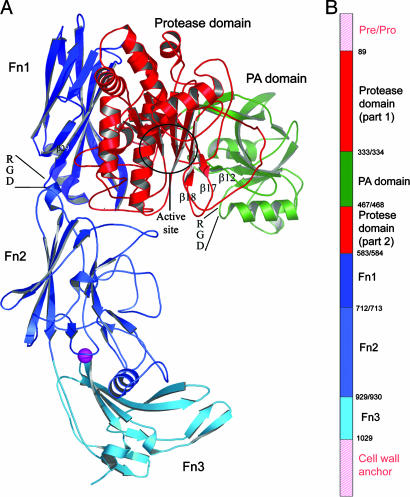
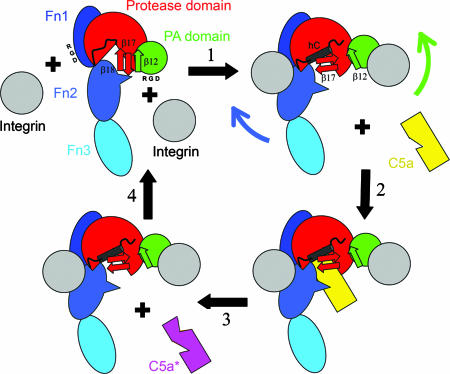
The structure of a cell surface enzyme from a gram-positive pathogen has been determined to 2-A resolution. Gram-positive pathogens have a thick cell wall to which proteins and carbohydrate are covalently attached. Streptococcal C5a peptidase (SCP), is a highly specific protease and adhesin/invasin. Structural analysis of a 949-residue fragment of the [D130A,S512A] mutant of SCP from group B Streptococcus (S. agalactiae, SCPB) revealed SCPB is composed of five distinct domains. The N-terminal subtilisin-like protease domain has a 134-residue protease-associated domain inserted into a loop between two beta-strands. This domain also contains one of two Arg-Gly-Asp (RGD) sequences found in SCPB. At the C terminus are three fibronectin type III (Fn) domains. The second RGD sequence is located between Fn1 and Fn2. Our analysis suggests that SCP binding to integrins by the RGD motifs may stabilize conformational changes required for substrate binding.


Figures

Fig. 1.
Fig. 1.
Domain structure of SCPBw. (A) Ribbon drawing of SCPBw monomer. Protease domain is red, PA domain is green, and three Fn domains are navy, sky blue, and cyan. Maroon sphere is calcium ion. The β12–β17–β18 sheet, the two RGD sequences, and the active-site region are indicated. (B) Schematic representation of SCPB domain organization. Domains are colored as in A. The domains not in SCPBw are indicated by pink labels and pink hatching. Numbers indicate domain boundaries in SCPB.
Fig. 2.
Fig. 2.
Ribbon drawings of individual SCPBw domains (colored as in Fig. 1) superimposed with nearest structural homolog (yellow, Protein Data Bank ID code and RMSD between Cαs are indicated). Secondary structural elements are labeled as in Fig. 3. Calcium binding sites are shown as spheres. Bound citrates and acetate are shown as sticks. (A) Protease domain with Bacillus M-protease. (B) PA domain with the apical domain from TfR (26). (C) Fn1 with human Rho guanine nucleotide dissociation inhibitor (37). (D) Fn2 with PapD from E. coli (38). (E) Fn3 with third Fn from human Kiaa0343 protein.
Fig. 3.
Fig. 3.
Modeling of the SCPB–C5a complex. (A) Stereo ribbon drawing of superposition of the protease domain and Fn2 of SCPBw (light blue with van der Waals surface) with B. mesentericus subtilisin (red)–eglin C (yellow) complex. (B) Superposition of C5a (green) onto eglin C (yellow) using residues around the cleavage site to determine orientation. (C) Model of the SCPB–C5a complex using results in A and B. Protease domain and Fn2 are shown as van der Waals surface in red and light blue, respectively; C5a is blue.
Fig. 4.
Fig. 4.
Cartoon showing hypothesized effect of integrins (gray) on SCPB structure. Domains of SCPW are indicated and colored as in Fig. 1. Positions of RGD sequences in PA domain and Fn1 are indicated as are the β12, β17, and β18 strands. In step 1, binding of integrins to RGD sequences disrupts the β12–β17 interactions, allowing the β17–β18 hairpin to rotate and promote formation of helix hC. Fn2 is pulled back, making space for C5a binding in step 2. In step 3, C5a is proteolyzed. In step 4, the integrins dissociate regenerating free SCPB.

References

    1. Walport, M. J. (2001) N. Engl. J. Med. 344, 1140–1144. - PubMed
    1. Walport, M. J. (2001) N. Engl. J. Med. 344, 1058–1066. - PubMed
    1. Hill, H. R., Bohnsack, J. F., Morris, E. Z., Augustine, N. H., Parker, C. J., Cleary, P. P. & Wu, J. T. (1988) J. Immunol. 141, 3551–3556. - PubMed
    1. Cheng, Q., Stafslien, D., Purushothaman, S. S. & Cleary, P. (2002) Infect. Immun. 70, 2408–2413. - PMC - PubMed
    1. Beckmann, C., Waggoner, J. D., Harris, T. O., Tamura, G. S. & Rubens, C. E. (2002) Infect. Immun. 70, 2869–2876. - PMC - PubMed

Publication types

Associated data

LinkOut - more resources