. 2006 Jan;16(1):97-105.
doi: 10.1101/gr.3690506. Epub 2005 Dec 12.

A missense mutation in the bovine SLC35A3 gene, encoding a UDP-N-acetylglucosamine transporter, causes complex vertebral malformation

Affiliations

A missense mutation in the bovine SLC35A3 gene, encoding a UDP-N-acetylglucosamine transporter, causes complex vertebral malformation

Bo Thomsen et al. Genome Res. 2006 Jan.

Abstract

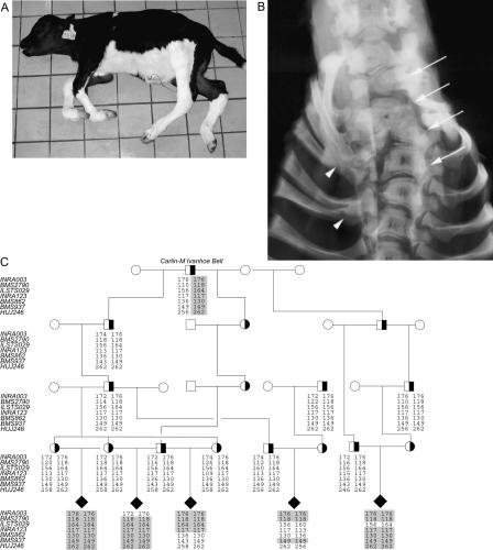
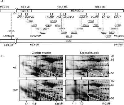
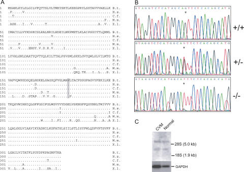
The extensive use of a limited number of elite bulls in cattle breeding can lead to rapid spread of recessively inherited disorders. A recent example is the globally distributed syndrome Complex Vertebral Malformation (CVM), which is characterized by misshapen and fused vertebrae around the cervico-thoracic junction. Here, we show that CVM is caused by a mutation in the Golgi-resident nucleotide-sugar transporter encoded by SLC35A3. Thus, the disease showed complete cosegregation with the mutation in a homozygous state, and proteome patterns indicated abnormal protein glycosylation in tissues of affected animals. In addition, a yeast mutant that is deficient in the transport of UDP-N-acetylglucosamine into its Golgi lumen can be rescued by the wild-type SLC35A3 gene, but not by the mutated gene. These results provide the first demonstration of a genetic disorder associated with a defective SLC35A3 gene, and reveal a new mechanism for malformation of the vertebral column caused by abnormal nucleotide-sugar transport into the Golgi apparatus.


Figures

Figure 1.
Figure 1.
Complex vertebral malformation. (A) Affected calf. Notice the short neck and contraction of the distal joints. (B) Radiograph of the cervico-thoracic part of the vertebral column (arch removed), showing multiple malformed vertebrae and scoliosis (arrows) and fusion of the proximal part of several ribs (arrowheads), ventro-dorsal projection. (C) Pedigree and haplotype analysis. All affected calves can be traced back to a common ancestor Carlin-M Ivanhoe Bell. Genotypes of seven polymorphic makers at the disease locus on BTA3 that helped to define the disease locus are shown. The combined genotyping results suggested that the disease gene is located near BMS2790 in an ∼5 cM region flanked by the markers INRA003 and ILSTS029. The shared disease-associated haplotype is shaded in gray. (Filled symbols) Affected; (open symbols) unaffected; (half-filled symbols) carrier.
Figure 2.
Figure 2.
Bovine-human comparative map. (A) Twenty-two BAC clones constitute the minimum tiling path between BMS2790 and INRA003. Genes and map positions in HSA1p21 are based on the human genome draft sequence. Dotted lines indicate the gene content of the BAC clones. The positions (in cM) of microsatellite markers ILSTS029, BMS2790, and INRA003 on the bovine linkage map are indicated. (B) Differential proteome patterns of cardiac and skeletal tissues from CVM affected and unaffected (wt) calves. The four images display segments from silver-stained high-resolution 2DE maps. The encircled areas show the characteristic electrophoretic pattern of α1-antitrypsin that was observed only in tissues from affected CVM calves. The mobility changes with a shift in pI as well as in MW indicate that aberrant protein glycosylation is associated with CVM. Moreover, the increased spot intensity of α1-antitrypsin in disease-affected tissue relative to normal tissue suggests that the abnormally glycosylated variants accumulate in the tissues of affected calves. The pH spans are indicated along the x-axis and the approximate molecular weight is on the y-axis.
Figure 3.
Figure 3.
Mutation detection in SLC35A3. (A) The deduced amino acid sequence of bovine SLC35A3 and comparison with its homologs in human (Homo sapiens; BAA77841), dog (Canis familiaris; AAC39260), mouse (Mus musculus; AAH24110), and frog (Xenopus laevis; CAD47803). Dots indicate residues that match the Bos taurus sequence. Dashes indicate gaps that have been introduced to optimize the alignment. The valine at position 180 substituted by phenylalanine in CVM is indicated in bold type and shaded in gray. (B) Electropherograms showing the nucleotide sequences across the G→T mutation (indicated by an asterix) in normal (+/+), carrier (+/-), and CVM-affected (-/-) animals. (C) Northern blot analysis of SLC35A3 mRNA. Poly(A)+ RNA was isolated from kidney tissue of CVM calves and unaffected calves and analyzed by hybridizing Northern blots with a 32P-labeled SLC35A3 probe. Subsequently, the membranes were stripped and reprobed with a 32P-labeled GAPDH (glyceraldehyde-3-phosphate dehydrogenase) fragment.
Figure 4.
Figure 4.
Analysis of K. lactis cell-surface labeling by flow cytometry. (A) The K. lactis mutant mnn2-2 lacks terminal N-acetylglucosamine in its cell surface glycoproteins. Wild-type (MG1/2) and mnn2-2 (KL3) cells can therefore be differentially labeled by incubation with FITC-conjugated wheat-germ agglutinin (WGA), which has an affinity for N-acetylglucosaminyl residues. Phenotypic correction of the cell-surface defect was observed only in mnn2-2 transformants expressing wild-type SLC35A3. (Panel 1) Wild-type MG1/2 transformed with empty pE4 vector; (Panel 2) mutant mnn2-2 transformed with empty pE4 vector; (Panel 3) mutant mnn2-2 expressing the wild-type SLC35A3 gene; (Panel 4) mutant mnn2-2 expressing the mutated SLC35A3 gene with the V180F substitution. (B) Northern blot analysis. Total RNA extracted from the transformed yeast cells was separated by gel electrophoresis, blotted onto nylon membranes, and hybridized with a radioactively labeled full-length SLC35A3 cDNA probe. Positions of the 26S and 18S ribosomal RNA are indicated. (Lane 1) Wild-type MG1/2 transformed with empty pE4 vector; (lane 2) mutant mnn2-2 transformed with empty pE4 vector; (lane 3) mutant mnn2-2 expressing the wild-type SLC35A3 gene; (lane 4) mutant mnn2-2 expressing the mutated SLC35A3 gene with the V180F substitution. Ethidium bromide stained 26S and 18S rRNA to control for equal loading is shown below the Northern blot.

Similar articles

Cited by

References

    1. Abeijon, C., Yanagisawa, K., Mandon, E.C., Hausler, A., Moremen, K., Hirschberg, C.B., and Robbins, P.W. 1993. Guanosine diphosphatase is required for protein and sphingolipid glycosylation in the Golgi lumen of Saccharomyces cerevisiae. J. Cell. Biol. 122 307-323. - PMC - PubMed
    1. Abeijon, C., Robbins, P.W., and Hirschberg, C.B. 1996. Molecular cloning of the Golgi apparatus uridine diphosphate-N-acetylglucosamine transporter from Kluyveromyces lactis. Proc. Natl. Acad. Sci. 93 5963-5968. - PMC - PubMed
    1. Abeijon, C., Mandon, E.C., and Hirschberg, C.B. 1997. Transporters of nucleotide sugars, nucleotide sulfate and ATP in the Golgi apparatus. Trends Biochem. Sci. 22 203-207. - PubMed
    1. Agerholm, J.S., Bendixen, C., Andersen, O., and Arnbjerg, J. 2001. Complex vertebral malformation in holstein calves. J. Vet. Diagn. Invest. 13 283-289. - PubMed
    1. Agerholm, J.S., Bendixen, C., Arnbjerg, J., and Andersen, O. 2004. Morphological variation of “complex vertebral malformation” in Holstein calves. J. Vet. Diagn. Invest. 16 548-553. - PubMed

Web site references

    1. http://prowl.rockefeller.edu/profound_bin/WebProFound.exe; ProFound tool for searching protein sequence databases.
    1. http://www.phrap.org; PHRAP assembly engine.

Publication types

Substances

Associated data