. 2005 Dec;1(4):e42.
doi: 10.1371/journal.ppat.0010042. Epub 2005 Dec 9.

The beta-glucan receptor dectin-1 recognizes specific morphologies of Aspergillus fumigatus

Affiliations

The beta-glucan receptor dectin-1 recognizes specific morphologies of Aspergillus fumigatus

Chad Steele et al. PLoS Pathog. 2005 Dec.

Abstract

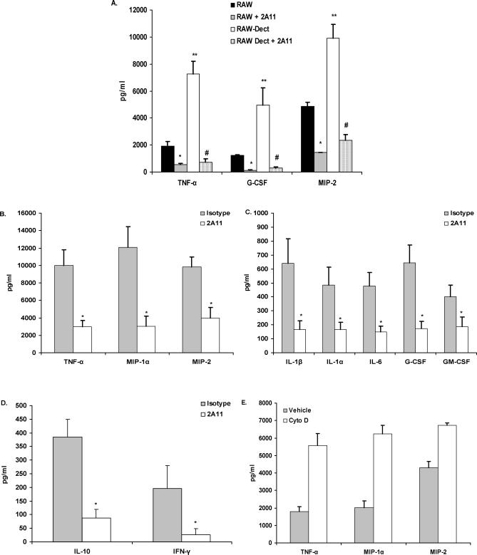
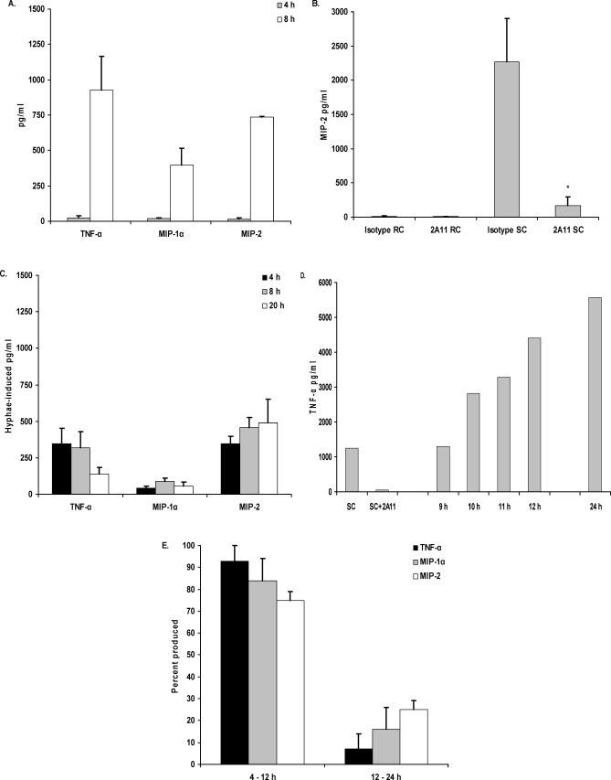
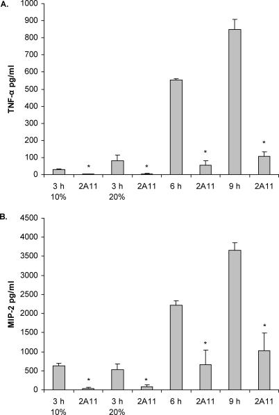
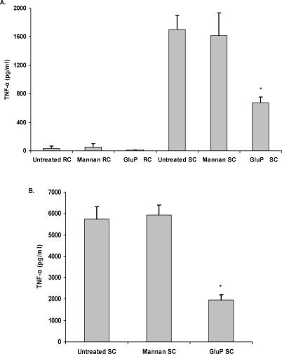
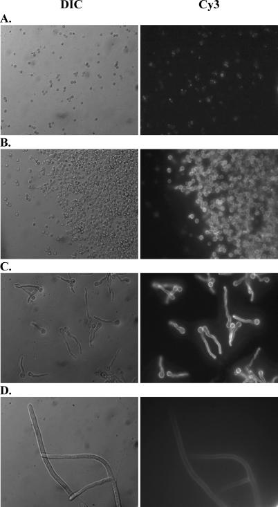
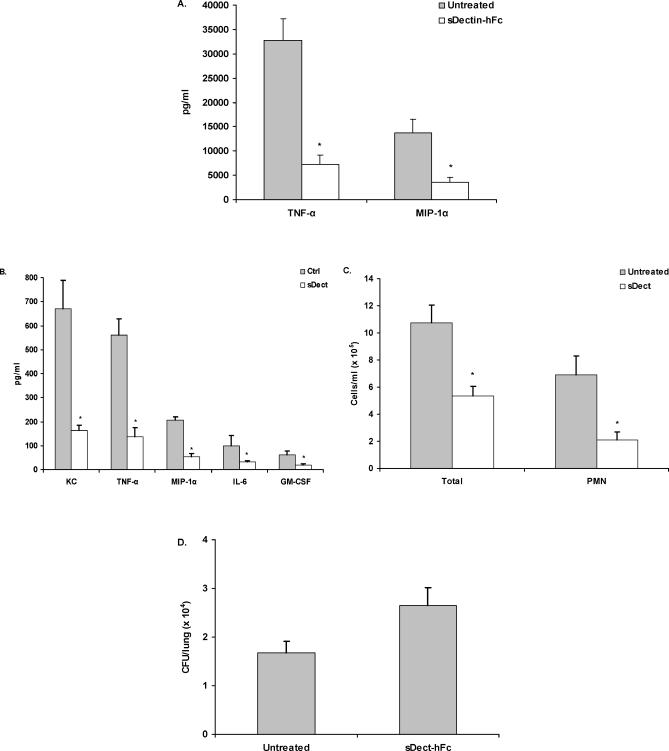
Alveolar macrophages represent a first-line innate host defense mechanism for clearing inhaled Aspergillus fumigatus from the lungs, yet contradictory data exist as to which alveolar macrophage recognition receptor is critical for innate immunity to A. fumigatus. Acknowledging that the A. fumigatus cell wall contains a high beta-1,3-glucan content, we questioned whether the beta-glucan receptor dectin-1 played a role in this recognition process. Monoclonal antibody, soluble receptor, and competitive carbohydrate blockage indicated that the alveolar macrophage inflammatory response, specifically the production of tumor necrosis factor-alpha (TNF-alpha), interleukin-1alpha (IL-1alpha), IL-1beta, IL-6, CXCL2/macrophage inflammatory protein-2 (MIP-2), CCL3/macrophage inflammatory protein-1alpha (MIP-1alpha), granulocyte-colony stimulating factor (G-CSF), and granulocyte monocyte-CSF (GM-CSF), to live A. fumigatus was dependent on recognition via the beta-glucan receptor dectin-1. The inflammatory response was triggered at the highest level by A. fumigatus swollen conidia and early germlings and correlated to the levels of surface-exposed beta glucans, indicating that dectin-1 preferentially recognizes specific morphological forms of A. fumigatus. Intratracheal administration of A. fumigatus conidia to mice in the presence of a soluble dectin-Fc fusion protein reduced both lung proinflammatory cytokine/chemokine levels and cellular recruitment while modestly increasing the A. fumigatus fungal burden, illustrating the importance of beta-glucan-initiated dectin-1 signaling in defense against this pathogen. Collectively, these data show that dectin-1 is centrally required for the generation of alveolar macrophage proinflammatory responses to A. fumigatus and to our knowledge provides the first in vivo evidence for the role of dectin-1 in fungal innate defense.


Conflict of interest statement

Competing interests. The authors have declared that no competing interests exist.

Figures

Figure 1
Figure 1. Dectin-1 Dependent Cytokine and Chemokine Production in Response to A. fumigatus
(A) Native RAW 264.7 macrophages or RAW 264.7 over-expressing murine dectin-1 were pretreated with isotype or 2A11 and co-cultured for 24 h with live A. fumigatus. Supernatant cytokine and chemokine levels were determined by Bio-Plex or ELISA. (A) illustrates cumulative results from five separate experiments. Asterisks, double asterisks, and the hash sign represent significant differences between RAW and RAW + 2A11, RAW and RAW-Dect, and RAW-Dect and RAW-Dect + 2A11, respectively (p < 0.05). Data are expressed as mean pg/ml + SEM. (B–D) Identical experimental design with alveolar macrophages. (B–D) illustrate cumulative results from five separate experiments. Asterisks represent significant differences between isotype and 2A11 (p < 0.05). Data are expressed as mean pg/ml + SEM. (E) Alveolar macrophages were pre-treated with 10-μM cytochalasin D and co-cultured with live A. fumigatus for 16 h. Supernatant cytokine and chemokine levels were determined by Bio-Plex or ELISA. (E) illustrates cumulative results from three separate experiments. Data are expressed as mean pg/ml + SEM.
Figure 2
Figure 2. Cytokine and Chemokine Production Is Dependent on Beta-Glucan Recognition in A. fumigatus SC and Pre-Competent Hyphae
Alveolar macrophages were added to (A) live A. fumigatus RC and incubated for 4 h and 8 h, (B) live RC or SC and incubated for 4 h in the presence of isotype or 2A11, or (C) live A. fumigatus hyphae and incubated for 4 h, 8 h, and 24 h. (D and E) Alveolar macrophages were added to A. fumigatus RC and incubated for 24 h. Samples for cytokine analysis were taken between 9 h and 12 h, and at 24 h, as indicated. Also shown in (D) is a representative 4-h response to pre-generated SC, and inhibition of this response by 2A11. Thereafter, cytokine and chemokine levels were determined in supernatants by Bio-Plex or ELISA. Percentage data expressed in (E) were calculated by dividing the pg/ml cytokine/chemokine level in 12-h samples by that in 24-h samples. Asterisks represent significant differences between isotype (rat IgG) and 2A11 (p < 0.05). (A–C,E, and F) are cumulative results from four experiments.
Figure 3
Figure 3. Cytokine and Chemokine Induction in Response to Different Heat-Killed Morphologies of A. fumigatus
A. fumigatus was grown for 3 h (in 10% or 20% serum), 6 h, or 9 h and thereafter subjected to heat-killing. Alveolar macrophages were added and incubated in the presence or absence of isotype or 2A11 for 6 h. Thereafter, cytokine and chemokine levels were determined in supernatants by Bio-Plex or ELISA. Asterisks represent significant differences between isotype (rat IgG) and 2A11 (p < 0.05). Figure 3 shows cumulative results from three experiments. Data are expressed as mean pg/ml + SEM.
Figure 4
Figure 4. Competitive Carbohydrate Blockage Indicates No Role of Mannan Receptors in the Inflammatory Response
Alveolar macrophages (A) or RAW 264.7 macrophages over-expressing dectin-1 (B) were pretreated with 250 μg/ml S. cerevisiae-derived mannan or 100 μg/ml glucan phosphate and then added to RC or SC. Thereafter, cytokine and chemokine levels were determined in supernatants by Bio-Plex or ELISA. Each graph illustrates cumulative results from four separate experiments. Asterisks represent significant differences between untreated and glucan-phosphate treated (p < 0.05).
Figure 5
Figure 5. Heightened Binding of Dectin-1 to A. fumigatus SC
A soluble fusion protein consisting of the extracellular carbohydrate recognition domain of dectin-1 fused with the Fc portion of murine IgG1 (s-dectin-mFc) was constructed and incubated with live A. fumigatus RC and SC. Binding of s-dectin-mFc was detected by Cy3-conjugated, goat anti-mouse IgG antibody followed by imaging with a Zeiss Axioplan 2 upright fluorescent deconvolution microscope (Zeiss), and images were captured using 3i Slidebook Version 4.0 software (Optical Analysis). Representative micrographs show s-dectin-mFc binding to A. fumigatus grown for 2 h (A), 6 h (B), 10 h (C), and 24 h (D). Left lane images are differential interference contrast (DIC) images, and right lane images are Cy3 staining. Magnification is 630 × oil emersion for all frames.
Figure 6
Figure 6. Blockage of Dectin-1-Mediated Inflammation In Vivo after A. fumigatus Lung Challenge
C57BL/6 mice were intratracheally administered live A. fumigatus conidia in the presence or absence of s-dectin-hFc. Mice were sacrificed 24 h later, and a BAL was performed. (A) Alveolar macrophages were co-cultured for 24 h with live A. fumigatus in the presence or absence of s-dectin-hFc (10 μg/ml). Supernatant cytokine and chemokine levels were determined by Bio-Plex or ELISA. (A) illustrates cumulative results from four separate experiments. Asterisks represent significant differences between untreated and s-dectin-1–containing wells (p < 0.05). Data are expressed as mean pg/ml + SEM. (B) Cytokine and chemokine levels in clarified BAL fluid from untreated and soluble (s-dect)–treated mice was measured by Bio-Plex. (B) illustrates representative results from three independent experiments (n = 5–7 mice per group). Asterisks represent significant differences between untreated and s-dectin-1–treated mice (p < 0.05). Data are expressed as mean pg/ml + SEM. (C) Total cell counts (Total) in BALF fluid as enumerated on a hemacytometer. Neutrophil (PMN) concentrations were determined by calculating the percentages of neutrophils in three to five sets of 100 cells and multiplying the percentage with the total BALF cell number. (D) Lungs were excised from non-lavaged–untreated and s-dectin-hFc–treated mice, homogenized, followed by serial 1:10 dilutions, and plated onto potato dextrose agar. CFU/lung were determined after incubating the plates for 24 h at 37 °C. (D) illustrates representative results from three independent experiments (n = 5–7 mice per group).

References

    1. Kontoyiannis DP, Bodey GP. Invasive aspergillosis in 2002: An update. Eur J Clin Microbiol Infect Dis. 2002;21:161–172. - PubMed
    1. Baddley JW, Stroud TP, Salzman D, Pappas PG. Invasive mold infections in allogeneic bone marrow transplant recipients. Clin Infect Dis. 2001;32:1319–1324. - PubMed
    1. Denning DW. Invasive aspergillosis. Clin Infect Dis. 1998;26:781–803. - PubMed
    1. Patterson TF, Kirkpatrick WR, White M, Hiemenz JW, Wingard JR, et al. Invasive aspergillosis. Disease spectrum, treatment practices, and outcomes. I3 Aspergillus Study Group. Medicine. 2000;79:250–260. - PubMed
    1. Marr KA, Patterson T, Denning D. Aspergillosis pathogenesis, clinical manifestations and therapy. Infect Dis Clin North Am. 2002;16:875–894. - PubMed

Publication types