. 1998 Mar;64(3):890-5.
doi: 10.1128/AEM.64.3.890-895.1998.

Purification and Characterization of Exo-beta-d-Glucosaminidase from a Cellulolytic Fungus, Trichoderma reesei PC-3-7

Affiliations

Purification and Characterization of Exo-beta-d-Glucosaminidase from a Cellulolytic Fungus, Trichoderma reesei PC-3-7

M Nogawa et al. Appl Environ Microbiol. 1998 Mar.

Abstract

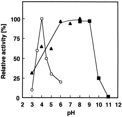
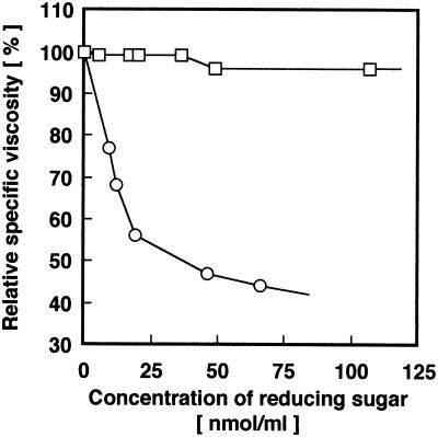
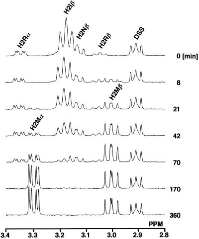
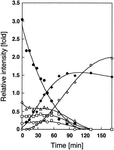
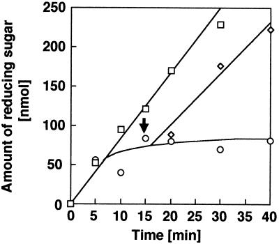
Chitosan-degrading activities induced by glucosamine (GlcN) or N-acetylglucosamine (GlcNAc) were found in a culture filtrate of Trichoderma reesei PC-3-7. One of the chitosan-degrading enzymes was purified to homogeneity by precipitation with ammonium sulfate followed by anion-exchange and hydrophobic-interaction chromatographies. The enzyme was monomeric, and its molecular mass was 93 kDa. The optimum pH and temperature of the enzyme were 4.0 and 50 degrees C, respectively. The activity was stable in the pH range 6.0 to 9.0 and at a temperature below 50 degrees C. Reaction product analysis from the viscosimetric assay and thin-layer chromatography and H nuclear magnetic resonance spectroscopy clearly indicated that the enzyme was an exo-type chitosanase, exo-beta-d-glucosaminidase, that releases GlcN from the nonreducing end of the chitosan chain. H nuclear magnetic resonance spectroscopy also showed that the exo-beta-d-glucosaminidase produced a beta-form of GlcN, demonstrating that the enzyme is a retaining glycanase. Time-dependent liberation of the reducing sugar from partially acetylated chitosan with exo-beta-d-glucosaminidase and the partially purified exo-beta-d-N-acetylglucosaminidase from T. reesei PC-3-7 suggested that the exo-beta-d-glucosaminidase cleaves the glycosidic link of either GlcN-beta(1-->4)-GlcN or GlcN-beta(1-->4)-GlcNAc.


Figures

FIG. 1
FIG. 1
Homogeneity of purified chitosanase. Proteins containing chitosanase fractions from each purification step were detected by SDS-PAGE. Lane 1, marker proteins containing soybean trypsin inhibitor (20 kDa), carbonic anhydrase (30 kDa), ovalbumin (43 kDa), bovine serum albumin (67 kDa), and phosphorylase (94 kDa); lane 2, culture broth (5.9 μg of protein); lane 3, (NH4)2SO3 precipitate (7.7 μg of protein); lane 4, Bio-Gel P-6 column mixture (4.9 μg of protein); lane 5, Q-Sepharose column mixture (1.5 μg of protein); lane 6, butyl-Sepharose column mixture (0.5 μg of protein).
FIG. 2
FIG. 2
Effects of pH on 93-kDa chitosanase. The chitosanase activity (○) was assayed at 37°C for 20 min in 50 mM acetate buffers with various pHs (3.0 to 6.0) and with chitosan 10B (0.1%) as the substrate. The residual activities of the enzyme after incubation at 37°C for 1 h at various pHs between 3.0 and 11.0 were measured. Buffers used were 50 mM citrate buffer (▴; pH 3.0 to 8.0) and 50 mM borate buffer (▪; pH 7.0 to 11.0).
FIG. 3
FIG. 3
Relationship between reduction in viscosity of and liberation of reducing sugars from a chitosan solution with the 93-kDa chitosanase and crude enzyme. Reductions in viscosity were determined with an Ostwald viscosimeter, and amounts of reducing sugar were assayed by the method of Imoto and Yagishita (16). Relative specific viscosity is described in Materials and Methods. Twenty milliunits of the purified exo-β-d-glucosaminidase (□) or crude enzyme (○) was used.
FIG. 4
FIG. 4
Analysis of enzymatic hydrolysates by TLC. Enzymatic hydrolysis of GlcN6 was performed in 50 mM acetate buffer (pH 4.0) at 37°C for various times. Lane S, standards containing GlcN and chitooligosaccharides from GlcN2 to GlcN6; lane 1, unhydrolyzed substrate; lanes 2, 3, 4, 5, 6, and 7, hydrolysates obtained after 2 min, 5 min, 30 min, 1 h, 10 h, and 15 h of reaction, respectively.
FIG. 5
FIG. 5
Time-dependent 1H-NMR spectra in hydrolysis of GlcN6. The enzyme (320 mU) was mixed with 630 μl of 10 mM acetate buffer (pH 4.0) containing 5.2 μmol of GlcN6 and 2.9 μmol of DSS as the standard. The reaction was performed directly in an NMR tube at 30°C. The signals derived from 2-H protons were assigned by two-dimensional COSY. H2Rα, α-form reducing-end residue of the oligomer; H2Rβ, β-form reducing-end residue of the oligomer; H2Iβ, β-form internal residue of the oligomer; H2Nβ, β-form nonreducing-end residue of the oligomer; H2Mα, α-form of GlcN; H2Mβ, β-form of GlcN.
FIG. 6
FIG. 6
Time course of 2-H signals during GlcN6 degradation. The relative peak areas of the 2-H signals to the standard DSS peak were determined from the NMR spectra and plotted against reaction times. □, the α-form reducing-end residue of the oligomer; ○, the β-form reducing-end residue of the oligomer; •, the β-form internal residue of the oligomer; ▵, the β-form nonreducing-end residue of the oligomer; ◊, the α-form of GlcN; and ⧫, the β-form of GlcN.
FIG. 7
FIG. 7
Time course of chitosan hydrolysis with exo-β-d-glucosaminidase. The hydrolysis pattern of completely deacetylated chitosan (chitosan 10B) or chitosan with a D.A. of 30% (chitosan 7B) with exo-β-d-glucosaminidase was observed. Exo-β-d-N-acetylglucosaminidase was added to the reaction mixture of chitosan 7B after 15 min of reaction. The arrow indicates the addition of exo-β-d-N-acetylglucosaminidase. □, chitosan 10B with exo-β-d-glucosaminidase; ○, chitosan 7B with exo-β-d-glucosaminidase; ◊, chitosan 7B with exo-β-d-glucosaminidase after addition of exo-β-d-N-acetylglucosaminidase.

References

    1. Alfonso C, Martinez M J, Reyes F. Purification and properties of two endochitosanases from Mucor rouxii implicated in its cell wall degradation. FEMS Microbiol Lett. 1992;95:187–194.
    1. Bartnicki-Garcia S. Cell wall chemistry, morphogenesis, and taxonomy of fungi. Annu Rev Microbiol. 1968;22:87–108. - PubMed
    1. Bradford M M. A rapid and sensitive method for the quantitation of microgram quantities of protein utilizing the principle of protein-dye binding. Anal Biochem. 1976;72:248–254. - PubMed
    1. Carmen Limón M, Lora J M, García I, de la Cruz J, Llobell A, Benítez T, Pintor-Toro J A. Primary structure and expression pattern of the 33-kDa chitinase gene from the mycoparasitic fungus Trichoderma harzianum. Curr Genet. 1995;28:478–483. - PubMed
    1. El Ouakfaoui S, Asselin A. Multiple forms of chitosanase activities. Phytochemistry. 1992;31:1513–1518.

LinkOut - more resources