Comparative Study
. 2006 Mar 15;394(Pt 3):715-25.
doi: 10.1042/BJ20050905.

Comparing calpain- and caspase-3-mediated degradation patterns in traumatic brain injury by differential proteome analysis

Affiliations
Comparative Study

Comparing calpain- and caspase-3-mediated degradation patterns in traumatic brain injury by differential proteome analysis

Ming Cheng Liu et al. Biochem J. .

Abstract

A major theme of TBI (traumatic brain injury) pathology is the over-activation of multiple proteases. We have previously shown that calpain-1 and -2, and caspase-3 simultaneously produced alphaII-spectrin BDPs (breakdown products) following TBI. In the present study, we attempted to identify a comprehensive set of protease substrates (degradome) for calpains and caspase-3. We further hypothesized that the TBI differential proteome is likely to overlap significantly with the calpain- and caspase-3-degradomes. Using a novel HTPI (high throughput immunoblotting) approach and 1000 monoclonal antibodies (PowerBlottrade mark), we compared rat hippocampal lysates from 4 treatment groups: (i) naïve, (ii) TBI (48 h after controlled cortical impact), (iii) in vitro calpain-2 digestion and (iv) in vitro caspase-3 digestion. In total, we identified 54 and 38 proteins that were vulnerable to calpain-2 and caspase-3 proteolysis respectively. In addition, the expression of 48 proteins was down-regulated following TBI, whereas that of only 9 was up-regulated. Among the proteins down-regulated in TBI, 42 of them overlapped with the calpain-2 and/or caspase-3 degradomes, suggesting that they might be proteolytic targets after TBI. We further confirmed several novel TBI-linked proteolytic substrates, including betaII-spectrin, striatin, synaptotagmin-1, synaptojanin-1 and NSF (N-ethylmaleimide-sensitive fusion protein) by traditional immunoblotting. In summary, we demonstrated that HTPI is a novel and powerful method for studying proteolytic pathways in vivo and in vitro.


Figures

Figure 1
Figure 1. αII-Spectrin immunoblot, a positive control before HTPI
Pooled naïve rat hippocampal lysate, lysates digested with calpain-2 or caspase-3 in vitro, and rat TBI hippocampal lysate. Separate naïve and TBI hippocampi from 6 individual animals respectively were pooled. To confirm that the extent of proteolysis in the last 3 samples was comparable, we analysed 20 μg of protein from each of these samples by traditional SDS/6% PAGE, immunoblotting and probing with monoclonal anti-αII-spectrin antibody (Affiniti anti-fodrin). Intact protein (280 kDa) was observed under all conditions. Calpain-2 digestion produced major fragments SBDP150 (150 kDa) and SBDP145 (145 kDa) (solid arrows), whereas caspase-3 digestion produced SBP149 (SBDP150i, 149 kDa) and SBDP120 (120 kDa) (open arrow heads) [5]. In TBI samples, a mixture of SBDP150, SBDP149, SBDP145 and SBDP120 was observed. M, molecular mass marker.
Figure 2
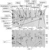
Figure 2. Example of the calpain-2 degradome (Template A)
A-templates from naïve hippocampus (upper panel) and calpain-2-digested hippocampal lysate (lower panel) were compared in triplicate (9 comparisons in total). One set of representative blots is shown. MM markers (lane 40) are indicated on the right. Protein bands with sufficient intensity were subsequently decoded and quantified using computer software as described in the Experimental section. We noted that for a number of proteins (solid box; upper panel), their average intensity decreased more than 2-fold after calpain digestion and several BDPs were also observed (BDP, dotted box; lower panel). ZO, zona occludin; for further definitions see Table 1, legend.
Figure 3
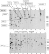
Figure 3. Example of the caspase-3 degradome (Template A)
A-templates from naïve hippocampus (upper panel) and caspase-3-digested hippocampal lysate (lower panel) were compared in triplicate. Only 1 set of representative blots is shown. MM markers (lane 40) are indicated on the right. Similarly to Figure 3, the expression of 9 parent proteins in template A (solid box, upper panel) was significantly decreased after caspase-3 digestion as a result of proteolysis, and several BDPs were observed (dotted box; lower panel). For definitions see Table 1, legend.
Figure 4
Figure 4. Example of the TBI differential proteome (Template A)
Template A for naïve hippocampus (upper panel) was compared with that for the TBI (1.6 mm deformation distance, 48 h) counterpart (lower panel). Comparisons were made in triplicate. A set of representative blots is shown. MM markers (lane 40) are indicated on the right. A total of 13 proteins in Template A were decreased in average intensity (down-regulated) after TBI (solid box; upper panel). In addition, several BDPs were readily observed (dotted box; lower panel). The only 2 proteins found to be up-regulated after TBI were CASK (A3) and Psme3 (A29) (dotted boxes). For definitions please see Table 1, legend.
Figure 5
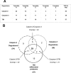
Figure 5. Summary of the calpain-2 and caspase-3 degradomes and differential TBI proteome results from HTPI
(A) The number of putative degradomic hits for each template based on calpain-2 versus naïve, caspase-3 versus naïve, and TBI versus naïve comparisons were tabulated. The total number of degradome hits is listed on the far right. (B) Venn diagram showing overlap of protein targets in the 3 degradomes (calpain, dashed line; caspase-3, dotted line; TBI, solid line). The total number of protein targets is in brackets. Overlaps and triple overlap numbers are indicated.
Figure 6
Figure 6. An HTPI approach allows rapid target confirmation
(A) Extracted lanes (E7) from template E of the HTPI gel: intact βII-spectrin (240 kDa) expression level was shown to be significantly decreased by calpain-2 digestion, caspase-3 digestion and after TBI. A calpain-mediated BDP of 110 kDa (black label) and 2 caspase-mediated BDPs of 108 and 85 kDa respectively (grey labels) were tentatively identified. These 3 BDPs were also tentatively identified in the TBI samples. (B) Traditional SDS/PAGE and Western blotting were also performed using identical monoclonal anti-βII-spectrin antibodies. Samples analysed were naïve (pooled) versus calpain-2 and capase-3 digestion (left 3 lanes), as well as 4 separate naïve and TBI samples. Again, BDPs of 110 kDa (solid arrow) and of 108 and 85 kDa (open arrow heads) were observed. * Indicates rat heavy-chain IgG and fragments from contaminating blood that cross-react with the secondary anti-(mouse IgG) antibody detection system used.
Figure 7
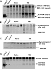
Figure 7. Examples of 4 proteins identified by HTPI as novel proteolytic targets
A total of 4 proteins were identified as proteolytic targets for either calpain-2, capase-3 and/or in TBI: striatin (A), synaptojanin-1 (B), synaptotagmin-1 (C) and NSF (D). Traditional SDS/PAGE and Western blotting were also performed using monoclonal antibodies against striatin, synaptojanin-1, synaptotagmin (isoform I) and NSF. Samples analysed were calpain-2 and capase-3 digestions (2 left-hand lanes), as well as 4 separate naïve and TBI samples. In (AD), intact proteins are shown with bold arrows (with MM in brackets). Calpain-2-mediated BDPs are shown with solid arrows. Caspase-3-mediated BDPs are shown with open arrow heads. MMs are as indicated. * In (A) indicates rat light-chain IgG from contaminating blood that cross-reacts with the secondary anti-(mouse IgG) antibody detection system used.
Figure 8
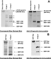
Figure 8. Examples of 2 purified proteins confirmed as proteolytic targets for calpain/caspase-3: βII-spectrin and synaptotagmin-1
Purified βII-spectrin (as a subunit of rat brain αII/βII-spectrin) (A) and recombinant GST–synaptotagmin-1 (B) were subjected to calpain-2/caspase-3 digestion, and calpain-2 only digestion respectively. Both Coomassie Blue stained blotting-membrane (left panels) and immunoblotting analysis with anti-βII-spectrin and anti-synaptotagmin-1 antibodies (right panels) were performed. Intact proteins and major BDPs are labelled with arrows. Several major αII-spectrin BDPs were also identified (open triangles).

Similar articles

Cited by

References

    1. Yamashima T. Implication of cysteine proteases calpain, cathepsin and caspase in ischemic neuronal death of primates. Prog. Neurobiol. 2000;62:273–295. - PubMed
    1. Asah M., Asahi K., Jung J. C., del Zoppo G. J., Fini M. E., Lo E. H. Role for matrix metalloproteinase 9 after focal cerebral ischemia, effects of gene knockout and enzyme inhibition with BB-94. J. Cereb. Blood Flow Metab. 2000;20:1681–1689. - PubMed
    1. Clark A. W., Krekoski C. A., Bou S. S., Chapman K. R., Edwards D. R. Increased gelatinase A (MMP-2) and gelatinase B (MMP-9) activities in human brain after focal ischemia. Neurosci. Lett. 1997;238:53–56. - PubMed
    1. Phillips J. B., Williams A. J., Adams J., Elliott P. J., Tortella F. C. Proteasome inhibitor PS519 reduces infarction and attenuates leukocyte infiltration in a rat model of focal cerebral ischemia. Stroke. 2000;31:1686–1693. - PubMed
    1. Wang K. K. W. Calpain and caspase, can you tell the difference. Trends Neurosci. 2000;23:20–26. - PubMed

Publication types