. 2004;107(2):153-68.
doi: 10.1016/j.zool.2004.02.003.

Evolution of ciliary patterns in the Oligotrichida (Ciliophora, Spirotricha) and its taxonomic implications

Affiliations

Evolution of ciliary patterns in the Oligotrichida (Ciliophora, Spirotricha) and its taxonomic implications

Sabine Agatha. Zoology (Jena). 2004.

Abstract

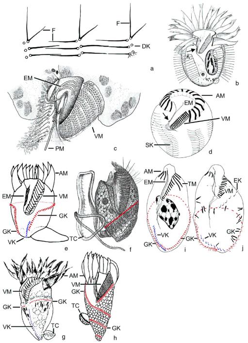
Although the somatic ciliature of the Oligotrichida typically comprises only a girdle and ventral kinety, a considerable diversity of ciliary patterns occurs. The four main girdle kinety patterns are identically found in tailed and tail-less species. The contractile tail has a complicated and unique ultrastructure and is potentially useful for the cell's movement and/or stabilization during feeding. Accordingly, I assume that this structure has evolved only once, namely, in the Tontoniidae nov. fam., and that the different girdle kinety patterns developed convergently in the tailed and tail-less taxa. Further distinct features suggest the establishment of the families Cyrtostrombidiidae nov. fam. (with cyrtos-like pharyngeal fibres and lack of ventral membranelles and endoral) and Pelagostrombidiidae nov. fam. (with neoformation organelle). An attempt is made to reconstruct the evolution of the kinety patterns based on morphologic, ontogenetic, and ultrastructural data. Some genera of tail-less Oligotrichida base on differences in the ciliary pattern; Omegastrombidium nov. gen. is erected for a further girdle kinety pattern. Likewise, the tailed genus Tontonia is split, resulting in two new genera, viz., Pseudotontonia nov. gen. and Spirotontonia nov. gen. Furthermore, the genus Spirostrombidium is split due to the different origin of the parallel course of girdle and ventral kinety, and Parallelostrombidium nov. gen. is established. However, the genus Thigmostrombidium is rejected because its enlarged thigmotactic membranelles are interpreted as an adaptation to the benthic lifestyle, which occurred several times within different girdle kinety patterns.


Figures

Fig. 1
Fig. 1
Ventral view of a generalized oligotrichid ciliate, illustrating some diagnostic features. Large anterior membranelles (AM) and small ventral membranelles (VM) along with their polykinetids form the membranellar zone. The endoral (EM) extends on the inner wall of the buccal lip. The somatic ciliature typically comprises a girdle kinety (GK) and a ventral kinety (VK). The distended cell surface (DC) occupies the cell portion underneath the girdle kinety in fixed and impregnated cells.
Fig. 2
Fig. 2
Evolution of oligotrichid ciliary patterns. (a) The ancestor had many longitudinal somatic kineties, whose dikinetids bore a distinct ciliumat each anterior basal body only (see detail). (b) Type I pattern. The kinety number was reduced to two. The rightwards torsion of the proximal end of the membranellar zone and the cell proper caused the dextrally spiralled pattern of the girdle kinety (GK; red in web version) and ventral kinety (VK; blue in web version). (c) Type II pattern. The ventral kinety orientated longitudinally. (d) Type III pattern. The right portion of the girdle kinety migrated posteriorly; both kinety ends are thus close to the cell’s posterior end. (e) Type IV pattern. The posterior portion of the girdle kinety curved anteriorly and is thus inversely orientated to the parallel ventral kinety. (f) Type V pattern. The left portion of the dextrally spiralled girdle kinety migrated anteriorly, causing a horizontal orientation. (g) Type VI pattern. The right portion of the horizontal girdle kinety spiralled sinistrally to the rear end. Arrows indicate the orientation of the kineties.
Fig. 3
Fig. 3
Ventral (a–h, j–l, p–r) and dorsal (i, m, n) views and oral primordium (o) of early (a, d, g, j, m, p), middle (b, e, h, k, n, q), and late (c, f, i, l, o, r) dividers after protargol impregnation. The hypotrichs (a–c; Diophrys scutum from Song and Packroff, 1993), stichotrichs (d–f; Lamtostyla halophila from Foissner et al., 2002), and halteriids (g–i; Halteria grandinella from Song, 1993) show a pronounced rightwards rotation of the anterior end of the oral primordium (arrows), whereas the oligotrichids (j–l; Strombidium arenicola from Agatha, 2003) and choreotrichids, including the strobilidiids (m–o; Strombidinopsis spiniferum from Dale and Lynn, 1998) and tintinnids (p–r; Tintinnopsis cylindrata from Petz and Foissner, 1993), show a pronounced rightwards rotation of the posterior end of the oral primordium (arrows).
Fig. 4
Fig. 4
Schematized illustrations of the main ontogenetic events in Strombidium (a–c), Novistrombidium (d–f), and Laboea strobila (g–i). During ontogenesis, the dextrally spiralled course of the girdle kinety found in Novistrombidium is apparently recapitulated in Strombidium and Laboea (for details, see Agatha et al., 2004). Thus, the latter taxa are assumed to be more derived. These observations support the proposed evolution of the ciliary patterns (cp. Fig. 2). The oral primordia are symbolized by dashed circles. GK/GK′, proter’s/opisthe’s girdle kinety (red and yellow in web version); VK/VK′, proter’s/opisthe’s ventral kinety (blue and light blue in web version).
Fig. 5
Fig. 5
Halteriid and oligotrichid genera from live (b, f, g) and after protargol impregnation (a, c–e, h–j). (a, c) Halteria grandinella. Schema of bristles (from Grim, 1974) and oral cavity (from Grain, 1972). (b, d) Meseres corlissi. Arrows denote paroral (from Petz and Foissner, 1992). (e, i, j) Paratontonia gracillima (e) and Omegastrombidium elegans (i, j). Both ends of the girdle kinety are near the rear end (e from Lynn et al., 1988; i, j from Song et al., 2000). (f) Tontonia appendiculariformis has a dextrally spiralled girdle kinety (from Fauré-Fremiet, 1924). (g) Pseudotontonia cornuta has a horizontal girdle kinety (from Suzuki and Song, 2001). (h) Spirotontonia grandis has a sinistrally spiralled girdle kinety (from Suzuki and Han, 2000). AM, anterior membranelles; DK, dikinetids; EK, extra kinety; EM, endoral; F, fibres; GK, girdle kinety; PM, paroral; SK, somatic kineties; TC, tail cilia; TM, thigmotactic membranelles; VK, ventral kinety; VM, ventral membranelles.
Fig. 6
Fig. 6
Halteriid genera in the scanning electron microscope. The minute paroral is marked by arrowheads. (a, b) Halteria grandinella, ventral view of a morphostatic specimen and posterior polar view of a middle divider (from Petz and Foissner, 1992). (c) Ventral view of Meseres corlissi (from Petz and Foissner, 1992). AM, anterior membranelles; B, bristles; OB, opisthe’s bristle; OP, oral primordium; SK, somatic kineties; VM, ventral membranelles.
Fig. 7
Fig. 7
Strombidiid genera after protargol impregnation. (a) Ventral view of Novistrombidium apsheronicum (from Agatha, 2003). The girdle kinety is dextrally spiralled, the ventral kinety longitudinal. (b, c) Ventral and dorsal view of Parallelostrombidium rhyticollare. The posterior portion of the girdle kinety and the ventral kinety have the same orientation (from Petz et al., 1995). (d, e) Ventral and dorsal view of Spirostrombidium urceolare (from Lei et al., 1999a). The posterior portion of the girdle kinety is parallel to the ventral kinety, but inversely orientated. (f–h, j) Ventral views of genera with a horizontal girdle kinety: Strombidium (S. arenicola from Agatha, 2003), Pelagostrombidium (P. mirabile from Krainer, 1991), Limnostrombidium (L. pelagicum from Krainer, 1991), and Cyrtostrombidium (C. longisomum from Lynn and Gilron, 1993). Instead of ventral membranelles and an endoral, Cyrtostrombidium species have cyrtos-like pharyngeal fibres (arrow). (i) Ventral view of Laboea strobila, showing sinistrally spiralled girdle kinety (from Montagnes et al., 1988). AM, anterior membranelles/polykinetids; E, extrusomes/extrusome attachment sites; GK, girdle kinety; NF, neoformation organelle; TM, thigmotactic membranelles; VK, ventral kinety; VM, ventral membranelles/ polykinetids.
Fig. 8
Fig. 8
Strombidiid genera in the scanning electron microscope. (a) Ventral view of Novistrombidium, showing dextrally spiralled course of the girdle kinety (N. apsheronicum from Agatha, 2003). (b, e) Ventral view of Strombidium and detail of its horizontal girdle kinety (S. arenicola from Agatha, 2003). (c, f) Ventral view of Limnostrombidium and detail of its horizontally orientated girdle kinety (L. viride; c from Foissner et al., 1999; f kindly supplied by W. Foissner). Arrowheads denote opening of neoformation organelle (tube in which the oral primordium develops). (d) Ventral view of Pelagostrombidium (P. mirabile from Foissner et al., 1999). Note that no somatic ciliature is recognizable. (g) Ventral view of L. strobila, showing the sinistrally spiralled girdle kinety (from Agatha et al., 2004). AM, anterior membranelles; E, just ejected extrusomes; GK, girdle kinety; VK, ventral kinety; VM, ventral membranelles.

References

    1. Aescht E. Catalogue of the generic names of ciliates (Protozoa, Ciliophora) Denisia. 2001;1
    1. Agatha S. Morphology and ontogenesis of Novistrombidium apsheronicum nov. comb. and Strombidium arenicola (Protozoa: Ciliophora): a comparative light microscopical and SEM study. Eur. J. Protistol. 2003;39:245–266.
    1. Agatha S. A cladistic approach for the classification of oligotrichid ciliates (Ciliophora, Spirotricha) Acta Protozool. 2004 in press. - PMC - PubMed
    1. Agatha S, Riedel-Lorjé JC. Morphology, infraciliature, and ecology of halteriids and strombidiids (Ciliophora, Oligotrichea) from coastal brackish water basins. Arch. Protistenkd. 1997;148:445–459.
    1. Agatha S, Strüder-Kypke MC, Beran A. Morphologic and genetic variability in the marine planktonic ciliate Laboea strobila Lohmann, 1908 (Ciliophora, Oligotrichia), with notes on its ontogenesis. J. Eukaryot. Microbiol. 2004;51:267–282. - PMC - PubMed

LinkOut - more resources