. 2006 Jan;80(1):38-50.
doi: 10.1128/JVI.80.1.38-50.2006.

Herpes simplex virus DNA synthesis is not a decisive regulatory event in the initiation of lytic viral protein expression in neurons in vivo during primary infection or reactivation from latency

Affiliations

Herpes simplex virus DNA synthesis is not a decisive regulatory event in the initiation of lytic viral protein expression in neurons in vivo during primary infection or reactivation from latency

N M Sawtell et al. J Virol. 2006 Jan.

Abstract

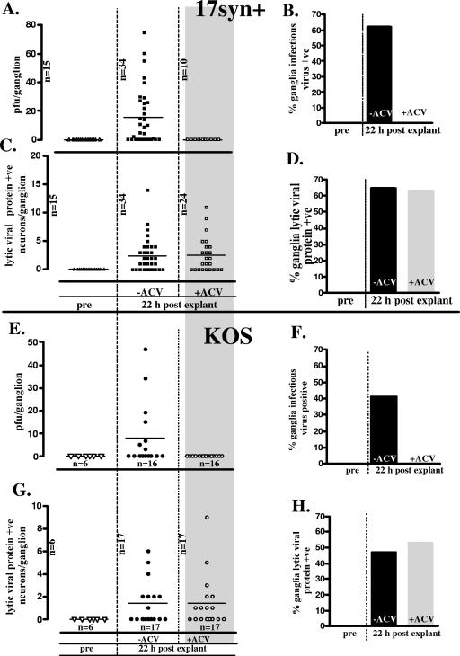
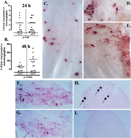
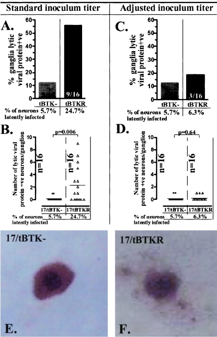
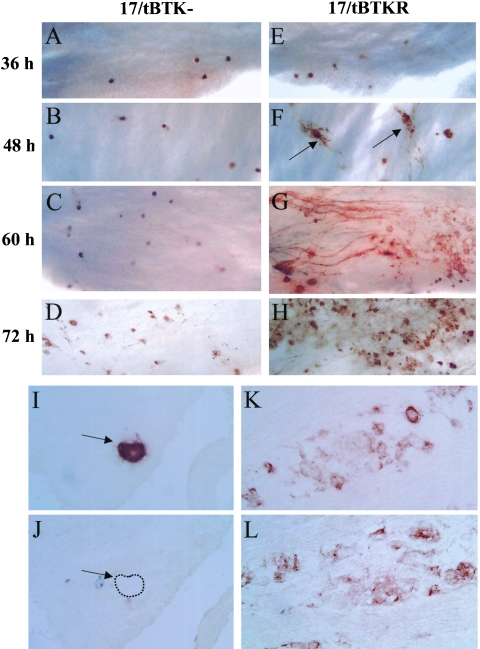
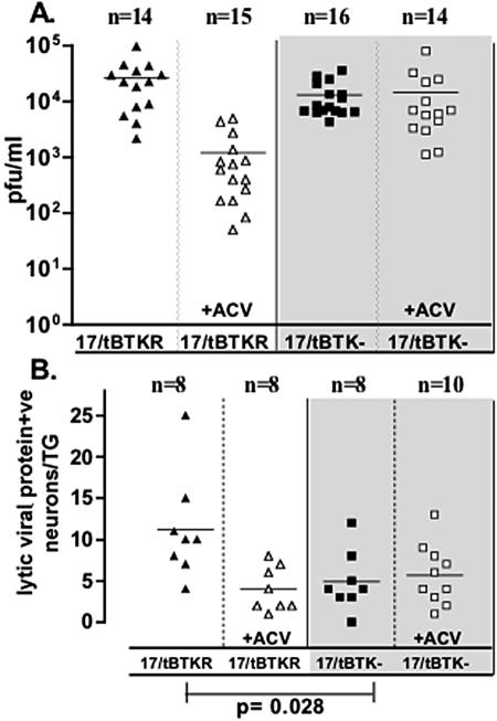
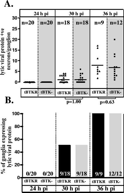
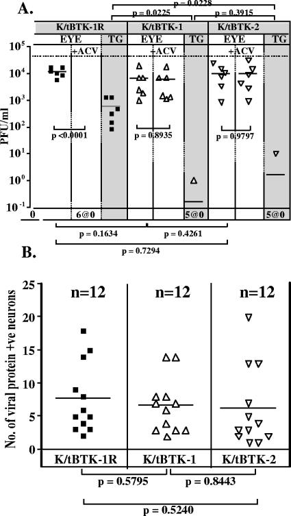
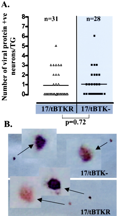
The herpes simplex virus genome can enter a repressed transcriptional state (latency) in sensory neurons of the host nervous system. Although reduced permissiveness of the neuronal environment is widely accepted as a causal factor, the molecular pathway(s) directing and maintaining the viral genome in the latent state remains undefined. Over the past decade, the field has been strongly influenced by the observations of Kosz-Vnenchak et al., which have been interpreted to indicate that, in sensory neurons in vivo, a critical level of viral DNA synthesis within the neuron is required for sufficient viral immediate-early (IE) and early (E) gene expression (M. Kosz-Vnenchak, J. Jacobson, D. M. Coen, and D. M. Knipe, J. Virol. 67:5383-5393, 1993). The levels of IE and E genes are, in turn, thought to regulate the decision to enter the lytic cycle or latency. We have reexamined this issue using new strategies for in situ detection and quantification of viral gene expression in whole tissues. Our results using thymidine kinase-null and rescued mutants as well as wild-type strains in conjunction with viral DNA synthesis blockers demonstrate that (i) despite inhibition of viral DNA replication, many neurons express lytic viral proteins, including IE proteins, during acute infection in the ganglion; (ii) at early times postinoculation, the number of neurons expressing viral proteins in the ganglion is not reduced by inhibition of viral DNA replication; and (iii) following a reactivation stimulus, the numbers of neurons and apparent levels of lytic viral proteins, including IE proteins, are not reduced by inhibition of viral DNA replication. We conclude that viral DNA replication in the neuron per se does not regulate IE gene expression or entry into the lytic cycle.


Figures

FIG. 1.
FIG. 1.
Quantification of infectious virus and lytic viral protein-expressing neurons in ganglia latently infected with either HSV-1 strain 17syn+ (squares) or KOS (circles) 22 h postexplant in the presence (open symbols) or absence (closed symbols) of the viral DNA replication inhibitor ACV. (A and E) Infectious virus titers in individual latently infected TGs before and 22 h postexplant. (B and F) Percentage of ganglia positive for infectious virus. (C and G) Number of lytic viral protein-expressing neurons in individual latently infected TGs before and 22 h postexplant. (D and H) Percentage of ganglia containing lytic viral protein-containing neurons.
FIG. 2.
FIG. 2.
Comparison of the effect of ACV treatment on the number of lytic viral protein-expressing neurons in TGs latently infected with 17syn+ at 24 h (A) and 48 h (B) postexplant. At 24 h postexplant there is no difference between ACV-treated (□) and untreated (▪) ganglia. However, at 48 h postexplant, the number of lytic viral protein-expressing neurons in untreated ganglia (▪) is significantly greater than that observed for ganglia treated with ACV (□) (P = 0.004, Student's t test). Panels C to E are photomicrographs of whole ganglia probed with a polyclonal antibody directed against HSV lytic proteins. This method allows for a comprehensive assessment of HSV lytic gene expression in a single view of the ganglia. As shown in panel C, at 48 h postexplant, ACV-treated ganglia contain discrete viral protein-expressing neurons with no evidence of lateral spread within the TG. In contrast, in the absence of ACV, clusters of viral protein-expressing neurons (D) and spread of lytic viral protein expression within the TG (E) are readily apparent. Panels F to I show serial sectioned frozen ganglia at 5 days postexplant in the absence (F and G) and presence (H and I) of ACV, immunostained for ICP4 (F and H) and gC (G and I). There is abundant ICP4 and gC detected in the TG after 5 days in the absence of ACV (F and G). In contrast, in the presence of ACV, only discrete neurons express ICP4 (H, arrows) and gC expression is not detected in these neurons (I).
FIG. 3.
FIG. 3.
Quantification of lytic viral protein-expressing neurons 22 h post-hyperthermic stress in ganglia latently infected with 17/tBTK (gray bars and squares) or 17/tBTKR (black bars and triangles). Mice were inoculated as detailed in Materials and Methods, and the percentage of neurons latently infected in the TG was determined using CXA. Using standard inoculum titers (A and B), the percentage of neurons latently infected in 17/tBTK-infected ganglia was more than fourfold lower than the percentage in 17/tBTKR-infected ganglia (A), and the number of neurons expressing lytic viral proteins post-hyperthermic stress was different (P = 0.006, Student's t test) in these two groups (B). When the inoculum titer was adjusted such that levels of latency were similar in the two groups, both the percentage of ganglia and the number of neurons per TG expressing lytic viral protein at 22 h post-HS were similar (C and D). Representative neurons expressing lytic viral proteins in 17/tBTK- and 17/tBTKR-infected ganglia at 22 h post-HS are shown in panels E and G, demonstrating that the intensity of staining is not different between the groups.
FIG. 4.
FIG. 4.
Lytic viral protein expression in 17/tBTK-infected (A to D) and 17/TBTKR-infected (E to H) ganglia 36 h (A and E), 48 h (B and F), 60 h (C and G), and 72 h (D and H) p.i. using WGIHC as described in Materials and Methods. In 17/tBTK-infected ganglia, lytic viral protein-expressing neurons remain discrete with no evidence of lateral spread. In contrast, spread of virus in TGs of 17/tBTKR-infected mice is apparent by 48 h p.i. (arrows). Panels I to L show serial sectioned frozen ganglia from 17/tBTK (I and J)- and 17/tBTKR (K and L)-infected mice harvested on day 4 day p.i. and immunostained for ICP27 (I and K) and gC (J and L). Arrows in panels I and J indicate neurons containing detectable ICP27 but no gC (dashed line indicates cell boundary).
FIG. 5.
FIG. 5.
Comparison of viral titers in the eyes (A) and the number of lytic viral protein-expressing neurons in the TGs (B) of mice infected with 17/tBTKR (triangles) and 17/tBTK (squares) at 48 h p.i. Open symbols indicate groups of mice treated with ACV as detailed in Materials and Methods (+ACV). The difference in the number of lytic protein-positive neurons in TGs of 17/tBTKR- and 17/tBTK-infected ganglia was significant (P = 0.028, Student's t test).
FIG. 6.
FIG. 6.
Comparison of the number of lytic viral protein-expressing neurons (A) and the percentage of TGs expressing viral proteins (B) in mice infected with 17/tBTK (squares) and 17/tBTKR (triangles) at 24 h, 30 h, and 36 h p.i. There is no significant difference between the groups at any of these times p.i. (Student's t test).
FIG. 7.
FIG. 7.
Comparison of viral titers (A) (n = 6 mice) and the number of lytic viral protein-expressing neurons in the TGs (B) (n = 12 TGs) at 42 h p.i. in mice infected with the TK-null and TK-restored mutants, K/tBTK-1 (triangles), K/tBTK-2 (inverted triangles), and K/tBTK-1R (squares). Groups of mice were inoculated with 106 PFU of the indicated virus, and at 42 h p.i. tissues were harvested and processed as detailed in Materials and Methods. Some mice (open symbols) were treated with ACV as detailed in Materials and Methods.
FIG. 8.
FIG. 8.
(A) Comparison of the number of neurons expressing IE proteins in TGs infected with 17/tBTK (squares) and 17/tBTKR (triangles) 30 h p.i. TGs were probed with antibodies directed against ICP27 and ICP0 as described in Materials and Methods. The difference between the groups was not significant (Student's t test). (B) Positive neurons representative of the intensity of staining in 17/tBTK- and 17/tBTKR-infected ganglia.
FIG. 9.
FIG. 9.
Viral genome copies in the ganglia of 17/tBTK (squares)- and 17/tBTKR (triangles)-infected mice from 16 to 60 h p.i. (black solid line). The viral replication contributing to this total number of viral genomes in the ganglia is also shown (eyes, open symbols and black dashed line; ganglia, gray solid symbols and gray solid line). A minimum of three mice per time point was examined. The means ± standard errors of the means of values from three to four mice per time point per group are plotted. The central shaded region indicates the time p.i. during which ganglia from mice infected with 17/tBTK and 17/tBTKR contain equivalent numbers of the viral protein-expressing neurons.

References

    1. Coen, D. M., M. Kosz-Vnenchak, J. G. Jacobson, D. A. Leib, C. L. Bogard, P. A. Schaffer, K. L. Tyler, and D. M. Knipe. 1989. Thymidine kinase-negative herpes simplex virus mutants establish latency in mouse trigeminal ganglia but do not reactivate. Proc. Natl. Acad. Sci. USA 86:4736-4740. - PMC - PubMed
    1. Davido, D. J., and D. A. Leib. 1996. Role of cis-acting sequences of the ICPO promoter of herpes simplex virus type 1 in viral pathogenesis, latency and reactivation. J. Gen. Virol. 77:1853-1863. - PubMed
    1. Field, H. J., and P. Wildy. 1978. The pathogenicity of thymidine kinase-deficient mutants of herpes simplex virus in mice. J. Hyg. (London) 81:267-277. - PMC - PubMed
    1. Gage, P. J., B. Sauer, M. Levine, and J. C. Glorioso. 1992. A cell-free recombination system for site-specific integration of multigenic shuttle plasmids into the herpes simplex virus type 1 genome. J. Virol. 66:5509-5515. - PMC - PubMed
    1. Honess, R. W., and B. Roizman. 1974. Regulation of herpesvirus macromolecular synthesis. I. Cascade regulation of the synthesis of three groups of viral proteins. J. Virol. 14:8-19. - PMC - PubMed

Publication types

MeSH terms

LinkOut - more resources