. 2006 Jan;80(1):252-61.
doi: 10.1128/JVI.80.1.252-261.2006.

Defective assembly of influenza A virus due to a mutation in the polymerase subunit PA

Affiliations

Defective assembly of influenza A virus due to a mutation in the polymerase subunit PA

John F Regan et al. J Virol. 2006 Jan.

Abstract

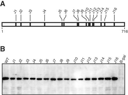
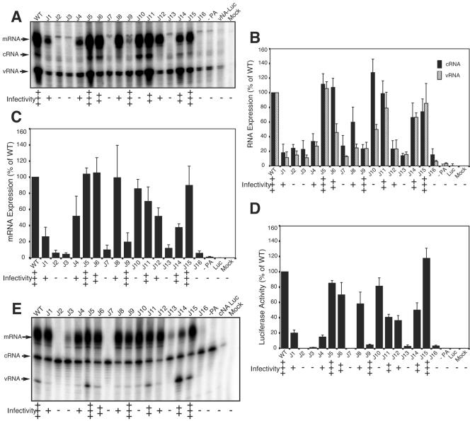
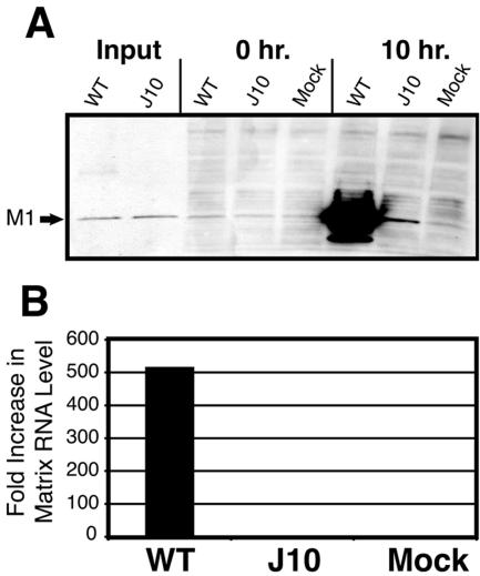
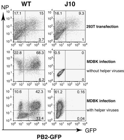
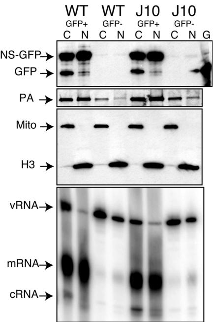
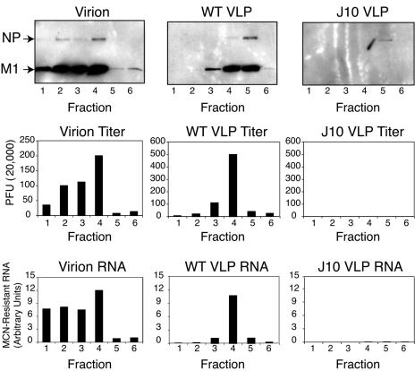
The RNA-dependent RNA polymerase of influenza A virus is composed of three subunits that together synthesize all viral mRNAs and also replicate the viral genomic RNA segments (vRNAs) through intermediates known as cRNAs. Here we describe functional characterization of 16 site-directed mutants of one polymerase subunit, termed PA. In accord with earlier studies, these mutants exhibited diverse, mainly quantitative impairments in expressing one or more classes of viral RNA, with associated infectivity defects of varying severity. One PA mutant, however, targeting residues 507 and 508, caused only modest perturbations of RNA expression yet completely eliminated the formation of plaque-forming virus. Polymerases incorporating this mutant, designated J10, proved capable of synthesizing translationally active mRNAs and of replicating diverse cRNA or vRNA templates at levels compatible with viral infectivity. Both the mutant protein and its RNA products were appropriately localized in the cytoplasm, where influenza virus assembly occurs. Nevertheless, J10 failed to generate infectious particles from cells in a plasmid-based influenza virus assembly assay, and hemagglutinating material from the supernatants of such cells contained little or no nuclease-resistant genomic RNA. These findings suggest that PA has a previously unrecognized role in assembly or release of influenza virus virions, perhaps influencing core structure or the packaging of vRNAs or other essential components into nascent influenza virus particles.


Figures

FIG. 1.
FIG. 1.
Structure and expression of PA mutants. (A) Schematic depiction of mutations studied here, which were introduced into the PA protein of strain A/WSN/33. (B) Western blot detection of WT and mutant PA proteins in lysates of 293T cells transfected with the PA expression vector and a β-Gal control vector. Samples were normalized by β-Gal activity and analyzed using a polyclonal anti-PA antibody. β-Gal, cells transfected with the control vector alone.
FIG. 2.
FIG. 2.
Reporter RNA expression by mutant influenza virus polymerase in cells. 293T cells underwent five-plasmid transfections that included either the WT PA protein vector or the indicated mutants, along with a luciferase reporter vector representing either the vRNA (A to D) or cRNA (E) product of the influenza virus NA gene. Total RNA was harvested 36 h (for the vRNA reporter) or 44 h (for the cRNA reporter) after transfection and was probed for NA-specific vRNA, cRNA, and mRNA by primer extension assay. Representative products from the vRNA reporter are shown in panel A. Expression of each RNA type was quantified by phosphorimaging from three independent transfections; the relative amounts of cRNA (black bars) and vRNA (striped bars) are presented in panel B, and those of mRNA are shown in panel C, each relative to the corresponding WT. Luciferase expression from triplicate transfections is depicted in panel D. Representative products from the cRNA reporter are shown in panel E. Mock, sham-transfected cells. Luc, reporter plasmid (vRNA or cRNA) only. −PA, PA vector omitted. Relative infectivities of the PA mutants (from Table 1) are indicated at the bottom of each panel.
FIG. 3.
FIG. 3.
Protein and RNA expression in infected MDCK cells. Aliquots of 293T supernatants from 17-plasmid transfections with WT or J10 PA vectors were normalized for influenza virus matrix (M1) protein expression and used to infect MDCK cells. (A) Western blot detection of M1 protein in 293T supernatants (Input) and in the MDCK cells at 0 h and 10 h postinfection. (B) Expression of M1-specific RNA as determined using quantitative RT-PCR, normalized to 5.8S rRNA, indicated as the fold increase at 5.5 h compared to 0 h postinfection. Mock, sham-infected MDCK cells.
FIG. 4.
FIG. 4.
Expression and transduction of influenza virus NP and of a vRNA-derived reporter in 293T producer and MDBK target cells. 293T cells underwent 17-plasmid transfections that included WT or J10 mutant PA vectors and a PB2-derived vRNA reporter vector that encoded GFP. Supernatants harvested after 48 h were used to infect MDCK cells. Two-color flow cytometry was used to score expression of immunoreactive NP protein and of GFP fluorescence in the 293T producer cells at 48 h posttransfection (top) and in the MDBK target cells at 15 h after inoculation with supernatant either alone (center) or together with authentic influenza virus helper virions at a multiplicity of infection of approximately 1.5 (bottom). Percentages of cells expressing NP only, GFP only, and the two markers together are indicated in the upper left, lower right, and upper right quadrants, respectively, of each plot, based on counting 20,000 cells from each population. As expression of the GFP reporter is assumed to require NP, the small percentage of cells expressing GFP alone was disregarded in our analysis. NP expression in the bottom right panel presumably results from infection with the helper virus.
FIG. 5.
FIG. 5.
Subcellular localization of viral RNAs and PA protein in 293T transfectants. 293T cells underwent five-plasmid transfection that included either the WT or J10 form of the PA protein vector, along with an NS-derived vRNA reporter encoding an NS1-GFP fusion protein. At 31 h posttransfection, cells were sorted into GFP-positive (GFP+) and GFP-negative (GFP−) subpopulations, from which nuclear (N) and cytoplasmic (C) extracts were then prepared. G, cells transfected with GFP reporter alone. Expression of the NS1-GFP fusion (NS-GFP), unfused GFP, PA, and histone H3 proteins, and of a mitochondrial antigen (Mito), was determined by Western blotting (top three panels). The vRNA, mRNA, and cRNA products of the NS vRNA reporter were detected by primer extension (bottom panel).
FIG. 6.
FIG. 6.
Density gradient fractionation of virions and VLPs. Supernatants from 293T cells, collected 48 h after 17-plasmid transfection, were used as a source of WT or J10 VLPs. Supernatants from infected MDCK cells were used as a source of authentic influenza virus A/WSN/33 virions. These supernatants were fractionated by centrifugation through continuous glycerol density gradients, and corresponding fractions were analyzed for viral NP and matrix (M1) proteins by Western blotting (top panel), for plaque-forming activity (middle panel), and for MCN-resistant PB1-specific viral RNA by quantitative RT-PCR (bottom panel).
FIG. 7.
FIG. 7.
Enrichment of hemagglutinating particles by adsorption to chicken erythrocytes. Supernatants from infected MDCK cells were incubated with chicken red blood cells to adsorb authentic influenza virus virions (Vir) and other hemagglutinating material. Similar adsorption was performed using supernatants from 17-plasmid 293T transfections that included WT or mutant (J10) PA vectors or from which PA vectors had been omitted (−PA). Erythrocytes were pelleted, washed, and then lysed for Western blot analysis. Alternatively, the washed erythrocyte pellets were incubated for 1 h at 37°C either with (+) or without (−) MCN, and RNA was then extracted for analysis, along with RNA from lysates of the corresponding 293T transfectants (T). (A) Western blot detection of viral PA (top panel) and M1 (bottom panel) proteins in erythrocyte-adsorbed material. The polyclonal anti-PA antibody used here detects J10 protein when present (Fig. 1B and 5). (B) Detection of NA-specific RNA species by primer extension assay. (C) Detection of viral RNAs by quantitative RT-PCR. Aliquots of RNA from transfected 293T cells (top panel) were treated with DNase and normalized to expression of 5.8S rRNA prior to analysis. RNA from equal volumes of the corresponding supernatants (bottom panel) was analyzed following erythrocyte adsorption and MCN digestion. Segment-specific RT-PCR was performed using vRNA-specific primers for the RT phase. Data are expressed as the number of PCR cycles required to reach CT, which is inversely proportional to concentration of the target RNA; RNAs from virions and wild-type VLPs were diluted 50-fold for analysis, and their depicted CT values were adjusted accordingly. The mean difference in CT values shown here for all eight segments between J10 and WT supernatants corresponds to a 9,400-fold average difference in RNA concentration.

Similar articles

Cited by

References

    1. Bancroft, C. T., and T. G. Parslow. 2002. Evidence for segment-nonspecific packaging of the influenza A virus genome. J. Virol. 76:7133-7139. - PMC - PubMed
    1. Delaluna, S., C. Martinez, and J. Ortin. 1989. Molecular cloning and sequencing of influenza virus A/Victoria/3/75 polymerase genes—sequence evolution and prediction of possible functional domains. Virus Res. 13:143-156. - PubMed
    1. Fodor, E., M. Crow, L. J. Mingay, T. Deng, J. Sharps, P. Fechter, and G. G. Brownlee. 2002. A single amino acid mutation in the PA subunit of the influenza virus RNA polymerase inhibits endonucleolytic cleavage of capped RNAs. J. Virol. 76:8989-9001. - PMC - PubMed
    1. Fodor, E., L. J. Mingay, M. Crow, T. Deng, and G. G. Brownlee. 2003. A single amino acid mutation in the PA subunit of the influenza virus RNA polymerase promotes the generation of defective interfering RNAs. J. Virol. 77:5017-5020. - PMC - PubMed
    1. Gomez-Puertas, P., C. Albo, E. Perez-Pastrana, A. Vivo, and A. Portela. 2000. Influenza virus matrix protein is the major driving force in virus budding. J. Virol. 74:11538-11547. - PMC - PubMed

Publication types

MeSH terms

LinkOut - more resources