. 2006 Jan;80(1):353-9.
doi: 10.1128/JVI.80.1.353-359.2006.

Expanded tropism and altered activation of a retroviral glycoprotein resistant to an entry inhibitor peptide

Affiliations

Expanded tropism and altered activation of a retroviral glycoprotein resistant to an entry inhibitor peptide

Sean M Amberg et al. J Virol. 2006 Jan.

Abstract

The envelope of class I viruses can be a target for potent viral inhibitors, such as the human immunodeficiency virus type 1 (HIV-1) inhibitor enfuvirtide, which are derived from the C-terminal heptad repeat (HR2) of the transmembrane (TM) subunit. Resistance to an HR2-based peptide inhibitor of a model retrovirus, subgroup A of the Avian Sarcoma and Leukosis Virus genus (ASLV-A), was studied by examining mutants derived by viral passage in the presence of inhibitor. Variants with reduced sensitivity to inhibitor were readily selected in vitro. Sensitivity determinants were identified for 13 different isolates, all of which mapped to the TM subunit. These determinants were identified in two regions: (i) the N-terminal heptad repeat (HR1) and (ii) the N-terminal segment of TM, between the subunit cleavage site and the fusion peptide. The latter class of mutants identified a region outside of the predicted HR2-binding site that can significantly alter sensitivity to inhibitor. A subset of the HR1 mutants displayed the unanticipated ability to infect nonavian cells. This expanded tropism was associated with increased efficiency of envelope triggering by soluble receptor at low temperatures, as measured by protease sensitivity of the surface subunit (SU) of envelope. In addition, expanded tropism was linked for the most readily triggered mutants with increased sensitivity to neutralization by SU-specific antiserum. These observations depict a class of HR2 peptide-selected mutations with a reduced activation threshold, thereby allowing the utilization of alternative receptors for viral entry.


Figures

FIG. 1.
FIG. 1.
ASLV-A mutations with reduced sensitivity to an HR2 peptide. (A) Schematic diagram of the ASLV-A envelope, with an expanded view of the amino acid sequence between the subunit cleavage site (arrow) through the N-terminal heptad repeat (HR1). Shown below the sequence are the identified mutations; each was tested as a single amino acid substitution except for the double mutant V31I/D60N. The fusion peptide is marked by diagonal lines and the membrane-spanning domain by horizontal lines (upper segment); the location of the region from which R99 is derived is shown with a black bar. In the expanded view, the fusion peptide is denoted by the dashed line, and HR1 with a cylinder. (B) Sample R99 inhibition curves for wild-type ASLV-A envelope and three mutants in the N-terminal end of the TM subunit. Infectivity of pseudotyped virus is shown as % of control (no inhibitor) with each point representing the average from one experiment performed in triplicate. (C) Coiled-coil projection indicating location of HR1 mutants. Each of the three inner coils is provided by the HR1 domain from one TM subunit. The dots represent locations in which HR2 peptide resistance determinants have been identified, and correspond to the dots shown in Fig. 1A. Each small dot represents a mutation with low resistance (3- to 4-fold change in IC50), each medium-sized dot shows a mutation with moderate resistance (7- to 20-fold change in IC50), and the large dots indicate the V48D mutation (260-fold change in IC50). The dashed circles correspond to the HR2 helices, which pack antiparallel against the grooves of the central trimer. This projection is based on an alignment with the Ebola GP2 sequence (58) and includes a stutter in the helix at Thr-54 (ASLV numbering).
FIG. 2.
FIG. 2.
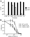
Expanded cell tropism of some of the R99-selected mutants. (A) Infectivity of pseudotyped virus particles on avian QT6 cells and mammalian 293T cells. Cells were infected in triplicate by centrifugal inoculation and data normalized to the amount of MLV Gag. Error bars show standard deviations. The pCB6 lane shows no detectable infectivity from a virus made with an empty vector plasmid instead of pCB6-EnvA. (B) Neutralization of MLV pseudotypes with either wild-type ASLV-A or V48D mutant envelopes by mc8C5-4, a monoclonal antibody that competes with the Tva receptor for envelope binding. Infectivity is shown as % of control (no antiserum) and error bars show standard deviations of experiment performed in triplicate.
FIG. 3.
FIG. 3.
Neutralization sensitivity of variant envelopes. Neutralization of MLV pseudotypes bearing the ASLV-A envelopes shown, using polyclonal antiserum directed against the SU subunit. Infectivity is shown as % of control (no antiserum) and error bars show standard deviations of experiment performed in quadruplicate on QT6 cells.
FIG. 4.
FIG. 4.
Altered thermodynamics of ASLV-A envelope triggering. (A) Increased protease sensitivity of SU subunit reveals receptor-triggered conformational change in envelope. Soluble receptor was bound to virus on ice where indicated, then the temperature was either shifted to 37°C or maintained on ice for 15 min as shown. Samples were returned to ice, digested with thermolysin, and resolved by SDS-PAGE and anti-SU Western blot. The positions of unprocessed (SU) and cleaved (SU*) subunit are shown. (B) Some of the mutant envelopes are efficiently triggered by receptor without elevating temperature. The bands from three separate gels were quantitated using [125I]protein A and the ratio of the amount of SU* detected at 2°C compared to 37°C was calculated. Shown are the average ratio and standard deviation for each envelope. The dashed line at a ratio of 1.0 indicates temperature independence.

References

    1. Balliet, J. W. 1998. Early events in subgroup A avian sarcoma and leukosis virus entry. Ph.D. thesis. University of Pennsylvania, Philadelphia, Pa.
    1. Balliet, J. W., J. Berson, C. M. D'Cruz, J. Huang, J. Crane, J. M. Gilbert, and P. Bates. 1999. Production and characterization of a soluble, active form of Tva, the subgroup A avian sarcoma and leukosis virus receptor. J. Virol. 73:3054-3061. - PMC - PubMed
    1. Bates, P., J. A. T. Young, and H. E. Varmus. 1993. A receptor for subgroup A Rous sarcoma virus is related to the low density lipoprotein receptor. Cell 74:1043-1051. - PubMed
    1. Bosch, B. J., B. E. E. Martina, R. van der Zee, J. Lepault, B. J. Haijema, C. Versluis, A. J. R. Heck, R. de Groot, A. D. M. E. Osterhaus, and P. J. M. Rottier. 2004. Severe acute respiratory syndrome coronavirus (SARS-CoV) infection inhibition using spike protein heptad repeat-derived peptides. Proc. Natl. Acad. Sci. USA 101:8455-8460. - PMC - PubMed
    1. Bosch, B. J., R. van der Zee, C. A. M. de Haan, and P. J. M. Rottier. 2003. The coronavirus spike protein is a class I virus fusion protein: structural and functional characterization of the fusion core complex. J. Virol. 77:8801-8811. - PMC - PubMed

Publication types

MeSH terms

LinkOut - more resources