. 2006 Jan;80(1):505-13.
doi: 10.1128/JVI.80.1.505-513.2006.

The herpes simplex virus type 1 vhs-UL41 gene secures viral replication by temporarily evading apoptotic cellular response to infection: Vhs-UL41 activity might require interactions with elements of cellular mRNA degradation machinery

Affiliations

The herpes simplex virus type 1 vhs-UL41 gene secures viral replication by temporarily evading apoptotic cellular response to infection: Vhs-UL41 activity might require interactions with elements of cellular mRNA degradation machinery

Ari Barzilai et al. J Virol. 2006 Jan.

Abstract

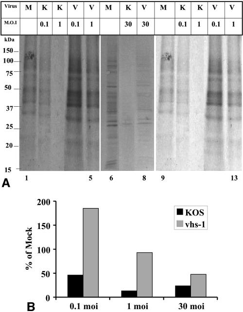
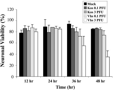
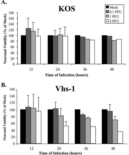
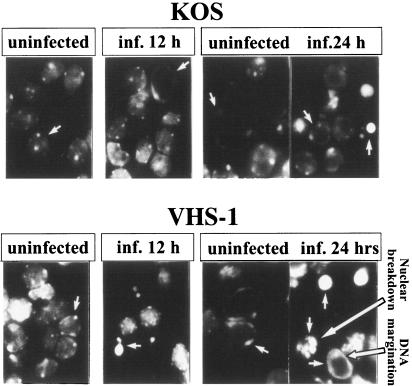
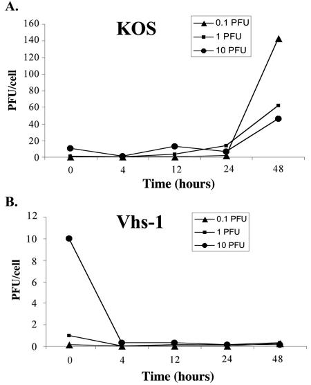
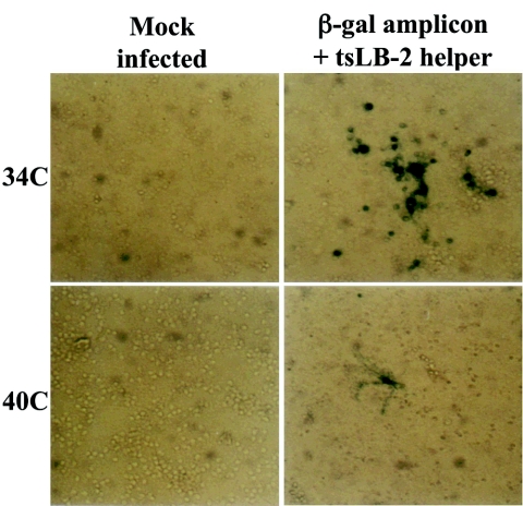
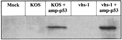
We have previously shown that herpes simplex virus type 1 (HSV-1) infection is associated with early destabilization/degradation of infected cell mRNAs and consequent shutoff of host protein synthesis by the activity of the virion-associated host shutoff (vhs) UL41 protein. Wild-type (wt) virus destabilized/degraded the housekeeping beta-actin and alpha-tubulin mRNAs as well host stress functions, like the heat shock 70 protein induced postinfection. vhs mutants did not degrade the mRNAs. Elaborate studies by others have been concerned with the mode of mRNA degradation and the mRNAs affected. We now describe vhs activity in primary cultures of mouse cerebellar granule neurons (CGNs). Specifically, (i) upon infection in the presence of actinomycin D to test activity of input viral particles, there was a generalized inhibition of protein synthesis, which depended on the input multiplicity of infection (MOI). (ii) Low-MOI infection with vhs-1 mutant virus was associated with increased synthesis of all apparent proteins. Higher MOIs caused some shutoff, albeit significantly lower than that of wt virus. This pattern could reflect an interaction(s) of vhs-1 protein with host machinery involved in cellular mRNA destabilization/degradation, sequestering this activity. (iii) wt virus infection was associated with cell survival, at least for a while, whereas mutant virus induced apoptotic cell death at earlier times. (iv) wt virus replicated well in the CGNs, whereas there was no apparent replication of the vhs-1 mutant virus. (v) The vhs-1 mutant could serve as helper virus for composite amplicon vectors carrying marker genes and the human p53 gene. Ongoing studies test the use of vhs-1-based composite oncolytic vectors towards cancer gene therapy.


Figures

FIG. 1.
FIG. 1.
A. Shutoff of host protein synthesis in cells infected with the wt HSV-1 (KOS) and vhs-1 mutant virus. CGNs plated at 1.5 × 10⁶ cells per well in 24-well plates were infected with the indicated virus at the indicated MOIs in the presence of 5 μg/ml actinomycin D. The cells were labeled from 4 to 5 h p.i. with 50 μCi/ml [35S]methionine. The cells were lysed, and 20-μg protein samples were loaded in each lane of 12.5% SDS-polyacrylamide gels. The gels were transferred to nitrocellulose membranes and exposed to film. Shown in this figure are longer (lanes 1 to 5) and shorter (lanes 9 to 13) exposures of the X-ray films for the gel containing the 0.1- and 1-PFU samples. Lanes 6 to 8 are from a gel with 30-PFU infections. B. Quantitative analysis of the shutoff of protein synthesis. The ImageJ program was used to quantitate the 35S-labeled proteins in the samples shown in panel A. The data are shown as percent relative to mock infected. M, mock; K, KOS; V, vhs1; moi, MOI measured in PFU/cell.
FIG. 2.
FIG. 2.
Cell death in infected cerebellar granule neurons. CGNs were infected with wt or vhs-1 mutant viruses at the indicated MOI. Neuronal viability was determined using trypan blue assay as described in Materials and Methods.
FIG. 3.
FIG. 3.
Viability of infected cerebellar granule neurons. CGNs were mock infected or infected with the wt HSV-1 (KOS) (A) or the vhs-1 mutant virus (B) at different MOIs. Neuronal viability was determined at the indicated times, using MTT assays as described in Materials and Methods.
FIG. 4.
FIG. 4.
Apoptosis of cerebellar granule neurons infected with wt HSV-1 (KOS) or the vhs-1 mutant virus. CGNs grown on glass coverslips were infected with 10 PFU/cell of the viruses. At the indicated time points, the cells were fixed and stained with DAPI, as described in Materials and Methods. Shown are fluorescent figures in the UV light microscope.
FIG. 5.
FIG. 5.
Infectious virus yields in the CGN infections. CGNs were infected with 0.1, 1, and 10 PFU/cell. (A) HSV-1 (KOS); (B) vhs-1 mutant virus. At the indicated times the infected cells were freeze-thawed three times, and resultant virus stock titers were determined in Vero cells, yielding infectious virus yields calculated in PFU/cell.
FIG. 6.
FIG. 6.
β-Gal expression in CGNs infected with composite amplicon β-Gal vector and the tsLB-2 helper virus. CGNs were infected or mock infected at 34 and 40°C with composite vector containing a mixture of the amplicon β-Gal and tsLB-2 helper virus. Shown is the β-Gal histochemical staining examined at 8 h p.i. in a Zeiss inverted microscope, photographed with an MC100 camera. Magnification, ×200.
FIG. 7.
FIG. 7.
Expression of the amplicon p53 vector in CGNs infected with composite amplicon vectors. CGN cells were plated at 1.5 × 10⁶ cells per well in 24-well plates. Infection was with 0.1 PFU/cell of the indicated viral stocks. Protein samples prepared at 5 h p.i. were electrophoresed in a 12.5% SDS-PAGE gel, blotted, and reacted with anti-p53 DO-1 antibody.

Similar articles

Cited by

References

    1. Aubert, M., and J. A. Blaho. 1999. The herpes simplex virus type 1 regulatory protein ICP27 is required for the prevention of apoptosis in infected human cells. J. Virol. 73:2803-2813. - PMC - PubMed
    1. Aubert, M., and J. A. Blaho. 2001. Modulation of apoptosis during herpes simplex virus infection in human cells. Microbes Infect. 3:859-866. - PubMed
    1. Aubert, M., J. O'Toole, and J. A. Blaho. 1999. Induction and prevention of apoptosis in human HEp-2 cells by herpes simplex virus type 1. J. Virol. 73:10359-10370. - PMC - PubMed
    1. Aubert, M., S. A. Rice, and J. A. Blaho. 2001. Accumulation of herpes simplex virus type 1 early and leaky-late proteins correlates with apoptosis prevention in infected human HEp-2 cells. J. Virol. 75:1013-1030. - PMC - PubMed
    1. Chou, J., and B. Roizman. 1994. Herpes simplex virus 1 gamma134.5 gene function, which blocks the host response to infection, maps in the homologous domain of the genes expressed during growth arrest and DNA damage. Proc. Natl. Acad. Sci. USA 91:5247-5251. - PMC - PubMed

Publication types