

Crystal structure of human T cell leukemia virus protease, a novel target for anticancer drug design
- PMID: 16352712
- PMCID: PMC1317974
- DOI: 10.1073/pnas.0509335102
Crystal structure of human T cell leukemia virus protease, a novel target for anticancer drug design
Abstract
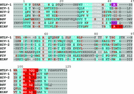
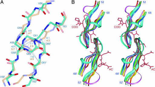
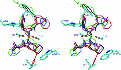
The successful development of a number of HIV-1 protease (PR) inhibitors for the treatment of AIDS has validated the utilization of retroviral PRs as drug targets and necessitated their detailed structural study. Here we report the structure of a complex of human T cell leukemia virus type 1 (HTLV-1) PR with a substrate-based inhibitor bound in subsites P5 through P5'. Although HTLV-1 PR exhibits an overall fold similar to other retroviral PRs, significant structural differences are present in several loop areas, which include the functionally important flaps, previously considered to be structurally highly conserved. Potential key residues responsible for the resistance of HTLV-1 PR to anti-HIV drugs are identified. We expect that the knowledge accumulated during the development of anti-HIV drugs, particularly in overcoming drug resistance, will help in designing a novel class of antileukemia drugs targeting HTLV-1 PR and in predicting their drug-resistance profile. The structure presented here can be used as a starting point for the development of such anticancer therapies.
Figures


Similar articles
-
Crystal structures of inhibitor complexes of human T-cell leukemia virus (HTLV-1) protease.J Mol Biol. 2010 Aug 27;401(4):626-41. doi: 10.1016/j.jmb.2010.06.052. Epub 2010 Jun 30. J Mol Biol. 2010. PMID: 20600105 Free PMC article.
-
Molecular insights on analogs of HIV PR inhibitors toward HTLV-1 PR through QM/MM interactions and molecular dynamics studies: comparative structure analysis of wild and mutant HTLV-1 PR.J Mol Recognit. 2014 Dec;27(12):696-706. doi: 10.1002/jmr.2395. J Mol Recognit. 2014. PMID: 25319617
-
Privileged Structures Meet Human T-Cell Leukemia Virus-1 (HTLV-1): C2-Symmetric 3,4-Disubstituted Pyrrolidines as Nonpeptidic HTLV-1 Protease Inhibitors.J Med Chem. 2015 Jun 11;58(11):4845-50. doi: 10.1021/acs.jmedchem.5b00346. Epub 2015 May 22. J Med Chem. 2015. PMID: 26000468
-
The protease of human T-cell leukemia virus type-1 is a potential therapeutic target.Curr Pharm Des. 2007;13(12):1285-94. doi: 10.2174/138161207780618849. Curr Pharm Des. 2007. PMID: 17504236 Review.
-
Understanding HTLV-I protease.Chem Biol. 2003 May;10(5):373-80. doi: 10.1016/s1074-5521(03)00104-2. Chem Biol. 2003. PMID: 12770819 Review. No abstract available.
Cited by
-
Viral proteases: Structure, mechanism and inhibition.Enzymes. 2021;50:301-333. doi: 10.1016/bs.enz.2021.09.004. Epub 2021 Nov 17. Enzymes. 2021. PMID: 34861941 Free PMC article.
-
Elucidation of the structure of retroviral proteases: a reminiscence.FEBS J. 2015 Nov;282(21):4059-66. doi: 10.1111/febs.13397. Epub 2015 Aug 28. FEBS J. 2015. PMID: 26258480 Free PMC article.
-
Riddelline from Tamarix articulate as a potential anti-bacterial lead compound for novel antibiotics discovery: A comprehensive computational and toxicological studies.PLoS One. 2024 Nov 14;19(11):e0310319. doi: 10.1371/journal.pone.0310319. eCollection 2024. PLoS One. 2024. PMID: 39541292 Free PMC article.
-
C-terminal residues of mature human T-lymphotropic virus type 1 protease are critical for dimerization and catalytic activity.Biochem J. 2008 Dec 15;416(3):357-64. doi: 10.1042/BJ20071132. Biochem J. 2008. PMID: 18636969 Free PMC article.
-
Kinetics of Bovine leukemia virus aspartic protease reveals its dimerization and conformational change.PLoS One. 2022 Jul 22;17(7):e0271671. doi: 10.1371/journal.pone.0271671. eCollection 2022. PLoS One. 2022. PMID: 35867649 Free PMC article.
References
-
- Franchini, G., Fukumoto, R. & Fullen, J. R. (2003) Int. J. Hematol. 78, 280–296. - PubMed
-
- Uchiyama, T. (1997) Annu. Rev. Immunol. 15, 15–37. - PubMed
-
- Ishikawa, T. (2003) Int. J. Hematol. 78, 304–311. - PubMed
-
- Kannagi, M., Harashima, N., Kurihara, K., Utsunomiya, A., Tanosaki, R. & Masuda, M. (2004) Exp. Rev. Anticancer Ther. 4, 369–376. - PubMed
-
- Nasr, R., El Sabban, M. E., Karam, J. A., Dbaibo, G., Kfoury, Y., Arnulf, B., Lepelletier, Y., Bex, F., de The, H., Hermine, O., et al. (2005) Oncogene 24, 419–430. - PubMed
Publication types
MeSH terms
Substances
Associated data
- Actions
Grants and funding
LinkOut - more resources
Full Text Sources
Research Materials
Miscellaneous
