. 2005 Dec 20;102(51):18455-60.
doi: 10.1073/pnas.0504343102. Epub 2005 Dec 13.

Epicardial retinoid X receptor alpha is required for myocardial growth and coronary artery formation

Affiliations

Epicardial retinoid X receptor alpha is required for myocardial growth and coronary artery formation

Esther Merki et al. Proc Natl Acad Sci U S A. .

Abstract

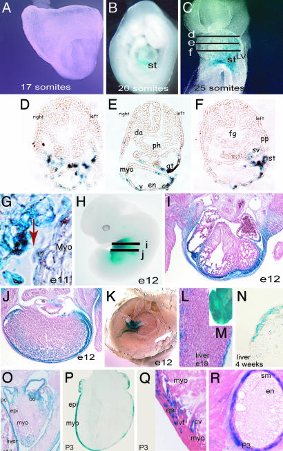
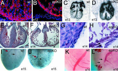
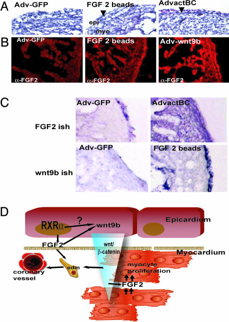
Vitamin A signals play critical roles during embryonic development. In particular, heart morphogenesis depends on vitamin A signals mediated by the retinoid X receptor alpha (RXRalpha), as the systemic mutation of this receptor results in thinning of the myocardium and embryonic lethality. However, the molecular and cellular mechanisms controlled by RXRalpha signaling in this process are unclear, because a myocardium-restricted RXRalpha mutation does not perturb heart morphogenesis. Here, we analyze a series of tissue-restricted mutations of the RXRalpha gene in the cardiac neural crest, endothelial, and epicardial lineages, and we show that RXRalpha signaling in the epicardium is required for proper cardiac morphogenesis. Moreover, we detect an additional phenotype of defective coronary arteriogenesis associated with RXRalpha deficiency and identify a retinoid-dependent Wnt signaling pathway that cooperates in epicardial epithelial-to-mesenchymal transformation.


Figures

Fig. 1.
Fig. 1.
Specificity of Cre expression in the epicardium of G5-Cre transgenic mice. (A and B) The onset of Cre activity from the G5-Cre line maps at the 20-somite stage (around E9.25) and is restricted to the distal portion of the septum transversum, as shown by intercross with the ROSA indicator mouse. (C) At the 25-somite stage, delamination of LacZ-positive cells from the septum transversum can be observed in whole-mount LacZ staining. These cells migrate rostrally and reach the left portion of the common atrium and left ventricle. Between day E9.5 (25 somites) and E10 (32 somites), LacZ-positive cells envelop the caudal surface of the common left ventricular myocardium. No expression is detected in hindgut, foregut, or the aorta. (DF) Transverse cryosections of a separate 25-somite embryo where performed at the approximate embryonic regions indicated in C and subsequently stained for β-galactosidase activity. LacZ-positive cells (Cre activity) in the septum transversum, sinus venosus, and over the myocardium in the caudal aspects of the heart are shown. (G) Delamination of epicardial cells to the outer surface of the myocardium and cytoplasmic expansions from the septum transversum to the myocardium (red arrow). (HJ) Whole-mount staining and histological sections of an E12 embryo showing Cre activity in the epicardium, pericardium, and body wall. (J) Staining also was detected in the hepatic capsule as well as some background staining within the liver. (K) Cre activity was not detected in the placenta. (L and M) Whole-mount and histological section of an E18 liver showing LacZ staining in the connective tissue covering the liver (not in the hepatocytes) (N) Frozen section of a 4-week-old liver, showing absence of staining in the hepatocytes. (O) Frozen section of an E17 embryonic heart illustrating LacZ staining in the epicardium, diaphragm, pericardium, and cardiac cushion. (PR) Frozen sections stained for β-galactosidase activity. (P) In postnatal hearts (postnatal day 3), LacZ activity is detected in the epicardium and subepicardial layers and in a subset of intermyocardial fibroblasts (Q) and in the smooth muscle of the coronary arteries (R). Note the absence of Cre activity in the coronary endothelium (R). st, septum transversum; LV, left ventricle; pa, pharyngeal arches; v, ventricle; epi, epicardium; myo, myocardium; ivf, interventricular fibroblasts; cv, coronary vessel; sm, smooth muscle; en, endocardium; cc, cardiac cushion; pc, pericardium. Note that, to avoid penetration artefacts, older samples were LacZ-stained on cryosectioned tissues in NR.
Fig. 2.
Fig. 2.
Histologic and morphologic analysis of epicardial mutant RXRα embryos. (A and B) A thin myocardium is detected as early as E12 and corresponds to decreased number of myocytes, as indicated by staining with the muscle marker sarcomeric α-actinin (red, α-actinin; blue, DAPI nuclear staining). (C and D) Epicardial RXRα mutation is sufficient to impair the normal down-regulation of mlc2a in E13 ventricles, and reflects deficient specification in the mutant myocardium. (E and F) Epi-RXRα mutant embryos at E14 display a lack of myocardial compaction, as indicated by hematoxylin–eosin staining. (G and H) E14 epi-RXRα mutant ventricles also exhibit epicardial detachment and thickened subepicardial space, suggesting deficient formation and/or activation of the epicardial epithelium (arrows). (IL) Epi-RXRα mutant ventricles at E15 present altered coronary arteriogenesis, with tortuous vessels (J, red arrow) and defective branching (J, black arrow) as shown by whole-mount immunohistochemistry using a Pecam-1 antibody. (K and L) Photographs of freshly dissected E16 hearts.
Fig. 3.
Fig. 3.
Altered EMT and defective maturation of myocardial progenitors in epicardial RXRα mutant embryos. (A) Epithelial monolayers cultured in collagen matrix were generated from E12–E12.5 mouse ventricles as described in Methods. After a period of 7 days in culture, the initial epicardial epithelium retracts and forms vessel-like structures under high serum conditions. Explants from wild-type embryos are largely negative for the transition marker vimentin (red signal; 15 vimentin-positive cells per 120 total cells) and positive for α-smooth muscle actin (green signal). Epicardial RXRα mutant explants fail to down-regulate vimentin (red signal, 117 of 122 total cells) and are deficient in α-smooth muscle expression (green). (B) Epicardial-mutant embryos display decreased β-catenin stability, concomitant with a specific decrease in wnt9b expression, as shown by RT-PCR on isolated hearts. No changes in the other Wnt genes, Wnt 8a, Wnt 8b, and Wnt 2a, were detected at the mRNA level. No changes in the epicardial marker Tbx18 were detected. GAPDH was used as control. FGF2 protein content is also decreased in mutant embryos as detected by Western blot in isolated hearts. (C) In serum-free conditions, simultaneous treatment of wild-type epicardial monolayers with all-trans retinoic acid (atRA) and the β-catenin activator lithium chloride (LiCl) induced differentiation of explanted monolayers to a vascular-like phenotype.
Fig. 4.
Fig. 4.
A retinoid–FGF–wnt pathway for epicardial activation and regulation of myocardial growth. (A) Histological analysis of chicken embryos overexpressing control GFP adenovirus (Adv-GFP), constitutively active β-catenin (AdvactBC), Wnt9b (Adv-wnt9b), or FGF2 beads. Observe the increased vascularization as demonstrated by the presence of blood cells in the subepicardial zone (arrowhead) upon β-catenin treatment and after grafting of FGF2-soaked beads. (B) Increased FGF2 expression is restricted to the epicardium upon FGF2 treatment. This finding is in contrast to induction of myocardial FGF2 expression after Wnt9b treatment. (C) Schematic showing the predicted relationship between wnt/β-catenin/FGF2 activation. Overexpression of active β-catenin induces FGF2 mRNA accumulation in the myocardium. Reciprocally, overexpression of epicardial FGF2 results in the accumulation of wnt9b mRNA in the epicardium. (D) Schematic of a proposed molecular mechanism for RXRα regulation of coronary formation and myocardial growth. RXRα activates the expression of epicardial FGF2. Epicardial FGF2 stimulates EMT of epithelial epicardial cells and induces epicardial wnt9b expression. Wnt9b then stabilizes myocardial β-catenin, inducing myocardial synthesis of FGF2, which in turn stimulates myocardial proliferation.

References

    1. Evans, R. M. (1988) Science 240, 889–895. - PMC - PubMed
    1. Green, S. & Chambon, P. (1988) Trends Genet. 4, 309–314. - PubMed
    1. Umesono, K., Murakami, K. K., Thompson, C. C. & Evans, R. M. (1991) Cell 65, 1255–1266. - PMC - PubMed
    1. Mark, M., Ghyselinck, N. B., Wendling, O., Dupe, V., Mascrez, B., Kastner, P. & Chambon, P. (1999) Proc. Nutr. Soc. 58, 609–613. - PubMed
    1. Dyson, E., Sucov, H. M., Kubalak, S. W., Schmid-Schonbein, G. W., DeLano, F. A., Evans, R. M., Ross, J., Jr., & Chien, K. R. (1995) Proc. Natl. Acad. Sci. USA 92, 7386–7390. - PMC - PubMed

Publication types

MeSH terms