Comparative Study
. 2005 Dec 19;202(12):1659-68.
doi: 10.1084/jem.20050768. Epub 2005 Dec 13.

Secretory leucoprotease inhibitor binds to NF-kappaB binding sites in monocytes and inhibits p65 binding

Affiliations
Comparative Study

Secretory leucoprotease inhibitor binds to NF-kappaB binding sites in monocytes and inhibits p65 binding

Clifford C Taggart et al. J Exp Med. .

Abstract

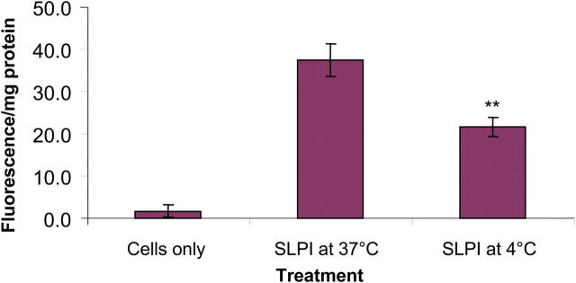
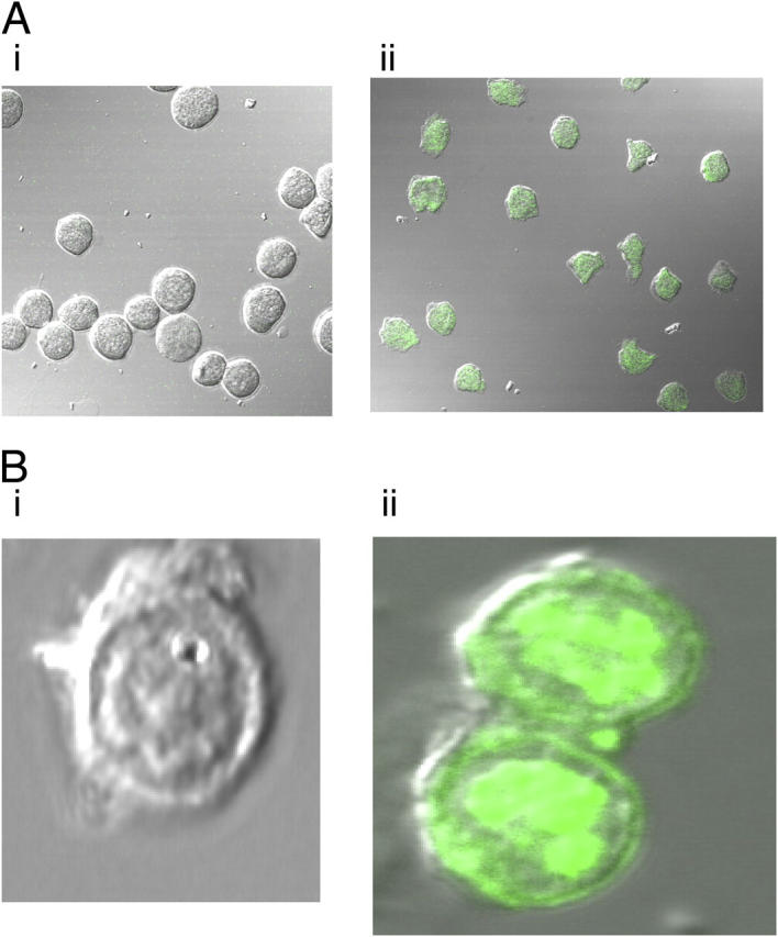
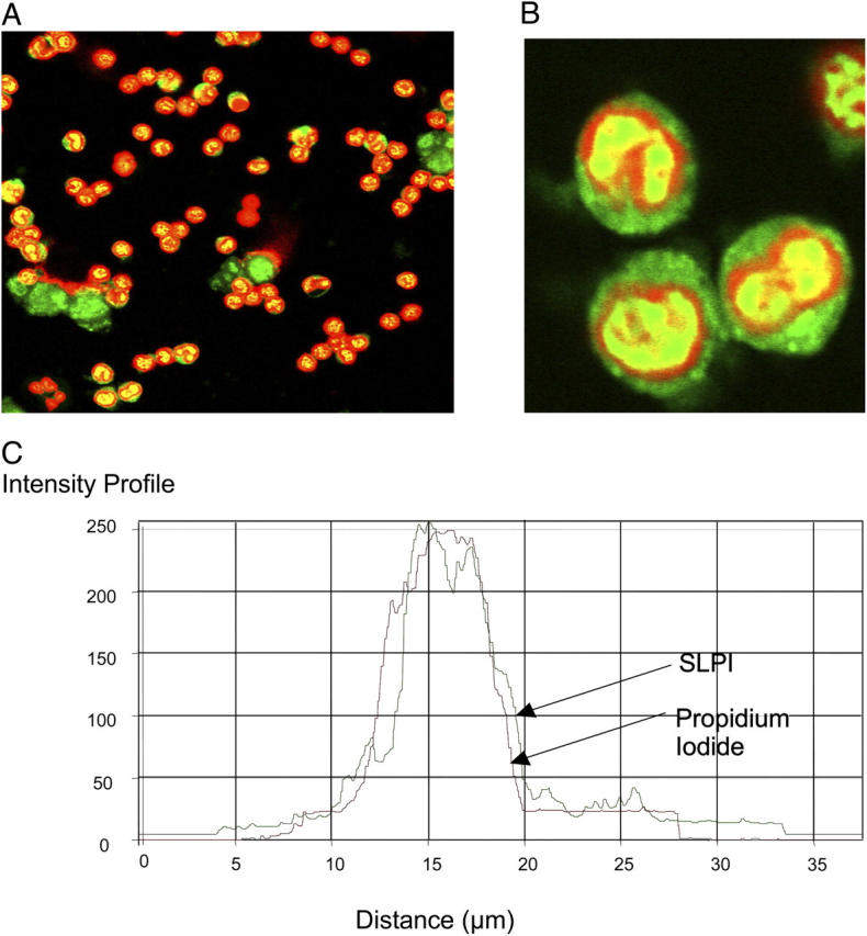
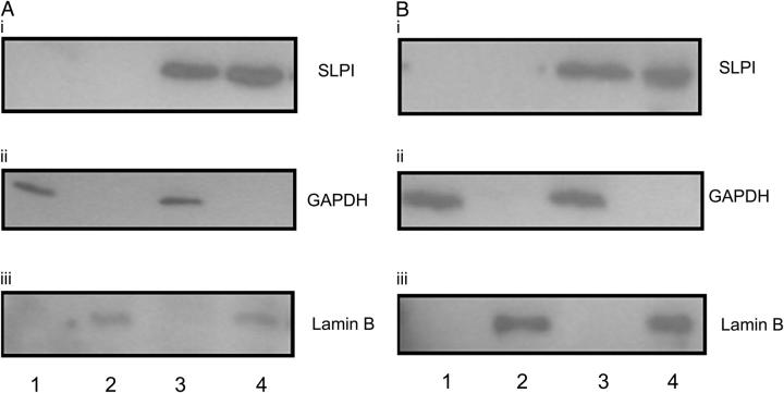
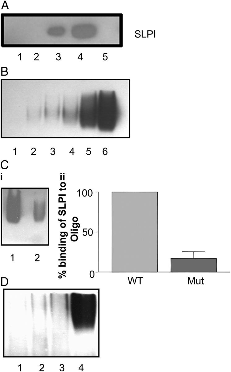
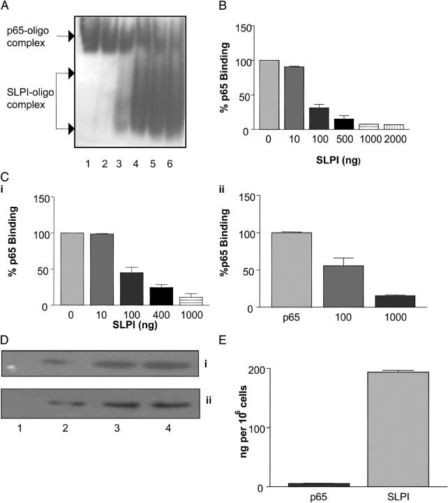
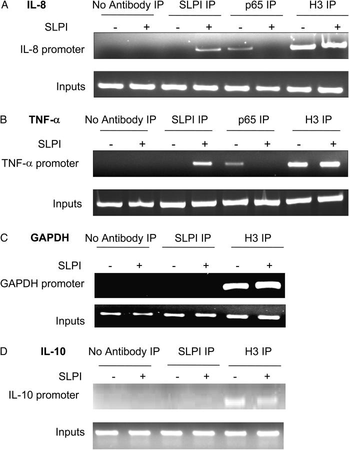
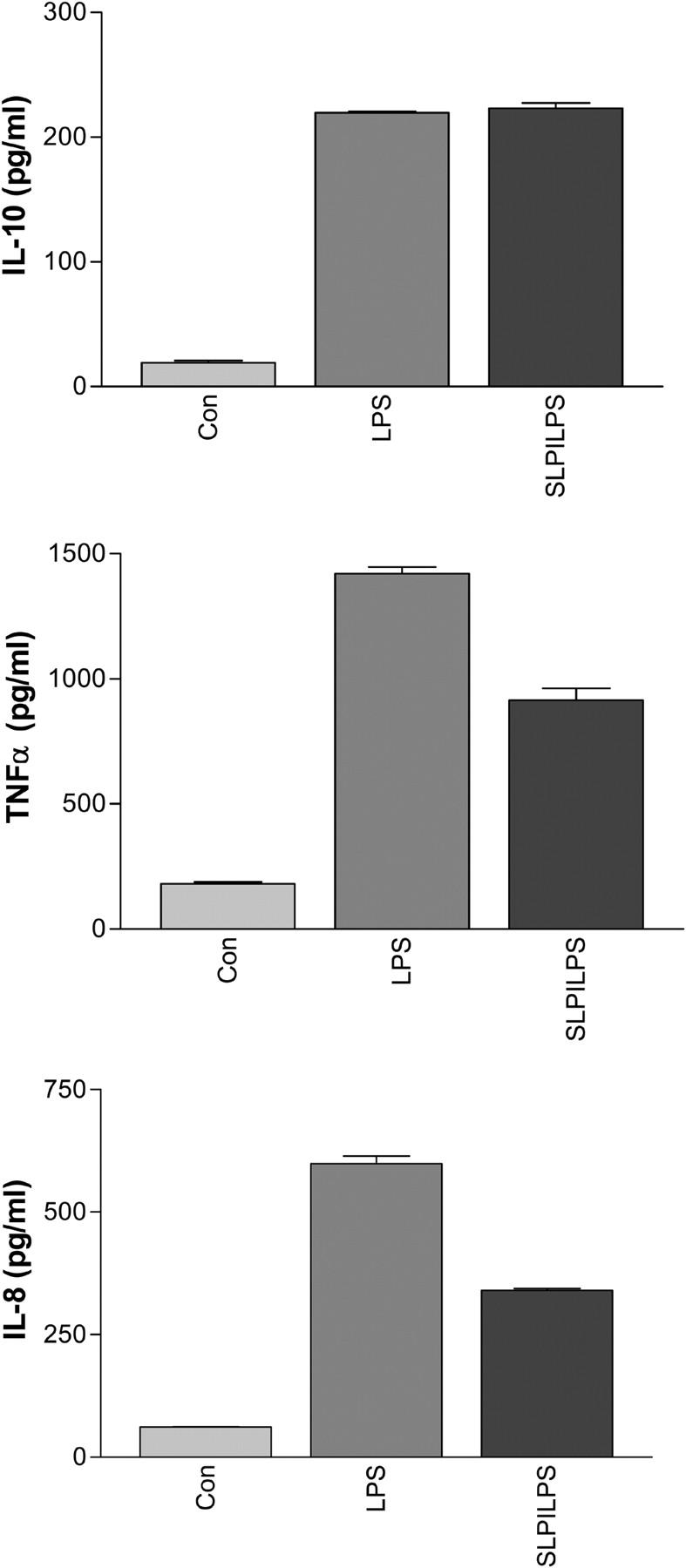
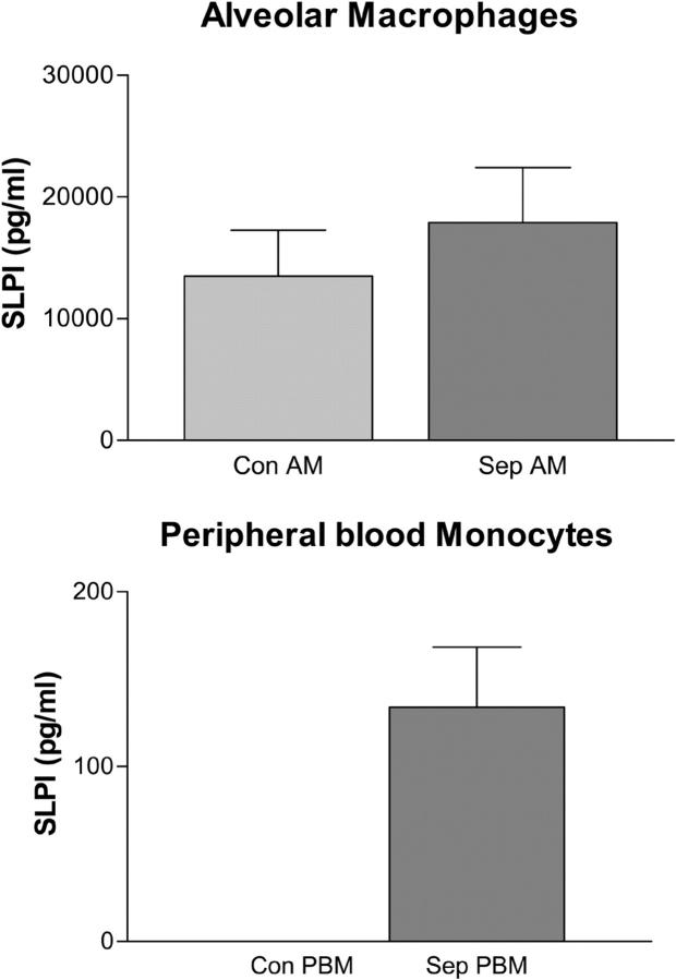
Secretory leucoprotease inhibitor (SLPI) is a nonglycosylated protein produced by epithelial cells. In addition to its antiprotease activity, SLPI has been shown to exhibit antiinflammatory properties, including down-regulation of tumor necrosis factor alpha expression by lipopolysaccharide (LPS) in macrophages and inhibition of nuclear factor (NF)-kappaB activation in a rat model of acute lung injury. We have previously shown that SLPI can inhibit LPS-induced NF-kappaB activation in monocytic cells by inhibiting degradation of IkappaBalpha without affecting the LPS-induced phosphorylation and ubiquitination of IkappaBalpha. Here, we present evidence to show that upon incubation with peripheral blood monocytes (PBMs) and the U937 monocytic cell line, SLPI enters the cells, becoming rapidly localized to the cytoplasm and nucleus, and affects NF-kappaB activation by binding directly to NF-kappaB binding sites in a site-specific manner. SLPI can also prevent p65 interaction with the NF-kappaB consensus region at concentrations commensurate with the physiological nuclear levels of SLPI and p65. We also demonstrate the presence of SLPI in nuclear fractions of PBMs and alveolar macrophages from individuals with cystic fibrosis and community-acquired pneumonia. Therefore, SLPI inhibition of NF-kappaB activation is mediated, in part, by competitive binding to the NF-kappaB consensus-binding site.


Figures

Figure 1.
Figure 1.
The level of cell association of SLPI-fluorescein with U937 cells. Cells were treated for 4 h with 10 μg/ml SLPI-fluorescein at 37 or 4°C. n = 3; mean ± SD; **, P < 0.01.
Figure 2.
Figure 2.
(A) Uptake of SLPI-fluorescein into U937 cells. 10 μg/ml SLPI-fluorescein was incubated with U937 cells for 1 h. (i) Untreated U937 cells. (ii) Treated U937 cells. (B) Uptake of SLPI-fluorescein into PBMs. 10 μg/ml SLPI-fluorescein was incubated with PBM for 1 h. (i) Untreated monocytes. (ii) Treated monocytes.
Figure 3.
Figure 3.
Nuclear import of SLPI-fluorescein in PBMs. 10 μg/ml SLPI-fluorescein (green) was incubated with monocytes for 1 h followed by counterstaining with propidium iodide. Red, nuclear stain. (A and B) Treated monocytes and (C) intensity profile. Red line, propidium iodide; green line, SLPI. (B) A magnification of the bottom left-hand quadrant of panel A.
Figure 4.
Figure 4.
SLPI cytoplasmic and nuclear distribution in (A) U937 cells and (B) PBMs. Cells were incubated with 10 μg/ml SLPI for 1 h followed by isolation of cytoplasmic and nuclear fractions. Lane 1, untreated cytoplasmic fraction; lane 2, untreated nuclear fraction; lane 3, SLPI-treated cytoplasmic fraction; lane 4, SLPI-treated nuclear fraction subjected to Western blot and probed using antibodies to (i) SLPI, (ii) GAPDH, and (iii) Lamin B.
Figure 5.
Figure 5.
(A) SLPI binds to DNA. 2 μg SLPI was incubated with DNA-cellulose matrix, eluted in increasing amounts of NaCl, and subjected to Western blotting. Lane 1, flow through; lane 2, 150 mM NaCl elution; lane 3, 300 mM NaCl elution; lane 4, 500 mM NaCl elution; lane 5, 750 mM NaCl elution. (B) SLPI binding to biotin-labeled NF-κB consensus oligonucleotide. Various amounts of SLPI (1–50 ng) were incubated with labeled NF-κB oligonucleotide in the presence of poly(dI-dC·dI-dC): poly(dI-dC·dI-dC) and then electrophoresed on a 5% polyacrylamide gel. Lane 1, labeled oligonucleotide alone; lane 2, labeled oligonucleotide plus 1 ng SLPI; lane 3, labeled, oligonucleotide plus 2 ng SLPI; lane 4, labeled oligonucleotide plus 5 ng SLPI; lane 5, labeled oligonucleotide plus 10 ng SLPI; lane 6, labeled oligonucleotide plus 50 ng SLPI. (C, i) SLPI interaction with wild-type (WT) and mutant (Mut) NF-κB oligonucleotides. 100 ng SLPI was incubated with equal amounts of biotin-labeled wild-type NF-κB consensus oligonucleotide (lane 1) or mutant NF-κB oligonucleotide (lane 2). (ii) Densitometry graph of SLPI binding to wild-type versus mutant NF-κB oligonucleotide, representative of five experiments. (D) SLPI interaction with biotin-labeled GATA-1 (lane 1), Sp-1 (lane 2), CREB (lane 3), and AP-1 (lane 4) consensus oligonucleotides.
Figure 6.
Figure 6.
(A) Increasing SLPI concentration prevents p65 binding to the NF-κB site. EMSA: 10–2,000 ng SLPI was incubated with NF-κB consensus binding site for 15 min followed by incubation with fixed amounts of p65 (100 ng) for an additional 20 min. Lane 1, p65 plus NF-κB DNA; lane 2, 10 ng SLPI plus p65 plus NF-κB DNA; lane 3: 100 ng SLPI plus p65 plus NF-κB DNA; lane 4, 500 ng SLPI plus p65 plus NF-κB DNA; lane 5, 1,000 ng SLPI plus p65 plus NF-κB DNA; lane 6, 2,000 ng SLPI plus p65 plus NF-κB DNA. P65–oligonucleotide and SLPI–oligonucleotide complexes are shown. (B) Densitometric evaluation of SLPI inhibition of p65 binding to NF-κB consensus oligonucleotide. (C, i) Increasing SLPI concentration prevents p65 binding to NF-κB site. p65 activity ELISA: 10–1,000 ng SLPI was incubated in wells containing the NF-κB consensus oligonucleotide for 15 min followed by incubation with fixed amounts of p65 (10 ng). p65 binding was detected by anti-p65 antibody/peroxidase-linked secondary antibody. (ii) SLPI displacement of p65 binding to NF-κB oligonucleotide. (D) p65 nuclear localization in U937 cells treated with (i) LPS or (ii) SLPI/LPS. U937 cells were incubated with 1 μg/ml LPS or 10 μg/ml SLPI for 1 h followed by LPS. Nuclear fractions were prepared and probed for NF-κB p65 over time. Lane 1, control; lane 2, 30 min; lane 3, 60 min; lane 4, 120 min. (E) SLPI and p65 concentrations in 106 cells. Nuclear extracts from SLPI-incubated U937 cells were subjected to Western blot, and SLPI and p65 levels were estimated by densitometry using SLPI and p65 standards. p65 = 5.61 ± 0.47 ng; SLPI = 194 ± 3.03 ng.
Figure 7.
Figure 7.
Interaction of SLPI with NF-κB sites in genomic DNA. U937 cells were incubated with or without 10 μg/ml SLPI for 1 h and then 1 μg/ml LPS for 1 h, and DNA was isolated by ChIP. U937 cells were incubated with anti-SLPI IgG, anti-p65 IgG, anti-acetyl histone H3 (positive control), or no antibody (negative control). DNA interacting with SLPI was isolated and subjected to PCR for detection of IL-8 promoter region (A), TNF-α promoter region (B), IL-10 promoter region (C), and GAPDH promoter (D). Lanes 1, 3, 5, and 7, cells incubated minus SLPI; lanes 2, 4, 6, and 8, cells incubated with SLPI; lanes 1 and 2, no antibody immunoprecipitation; lanes 3 and 4, SLPI antibody immunoprecipitation; lanes 5 and 6, p65 antibody immunoprecipitation (A and B only); lanes 7 and 8, acetyl histone H3 antibody immunoprecipitation.
Figure 8.
Figure 8.
Effect of SLPI on LPS-induced TNF-α (A), IL-8 (B), and IL-10 (C) protein production by U937 macrophages.
Figure 9.
Figure 9.
Measurement of SLPI in the nucleus of AMs and PBMs. Nuclear fractions were prepared from 106 AMs or PBMs from individuals with sepsis (cystic fibrosis and community-acquired pneumonia) and healthy controls. SLPI levels were determined by ELISA. Con, control; Sep, sepsis.

References

    1. Abe, T., N. Kobayashi, K. Yoshimura, B.C. Trapnell, H. Kim, R.C. Hubbard, M.T. Brewer, R.C. Thompson, and R.G. Crystal. 1991. Expression of the secretory leukoprotease inhibitor gene in epithelial cells. J. Clin. Invest. 87:2207–2215. - PMC - PubMed
    1. Gauthier, F., U. Fryksmark, K. Ohlsson, and J.G. Bieth. 1982. Kinetics of the inhibition of leukocyte elastase by the bronchial inhibitor. Biochim. Biophys. Acta. 700:178–183. - PubMed
    1. Seemuller, U., M. Arnhold, H. Fritz, K. Wiedenmann, W. Machleidt, R. Heinzel, H. Applehans, H.G. Gassen, and F. Lottspeich. 1986. The acid-stable proteinase inhibitor of human mucous secretions (HUSI-I, antileukoprotease). Complete amino acid sequence as revealed by protein and cDNA sequencing and structural homology to whey proteins and Red Sea turtle proteinase inhibitor. FEBS Lett. 199:43–48. - PubMed
    1. Hiemstra, P.S., B.A. Fernie-King, J. McMichael, P.J. Lachmann, and J.M. Sallenave. 2004. Antimicrobial peptides: mediators of innate immunity as templates for the development of novel anti-infective and immune therapeutics. Curr. Pharm. Des. 10:2891–2905. - PubMed
    1. Jin, F.Y., C. Nathan, D. Radzioch, and A. Ding. 1997. Secretory leukocyte protease inhibitor: a macrophage product induced by and antagonistic to bacterial lipopolysaccharide. Cell. 88:417–426. - PubMed

Publication types

MeSH terms