

Review
doi: 10.1128/JB.188.1.19-27.2006.
Septum enlightenment: assembly of bacterial division proteins
Affiliations
- PMID: 16352817
- PMCID: PMC1317574
- DOI: 10.1128/JB.188.1.19-27.2006
Item in Clipboard
Review
Septum enlightenment: assembly of bacterial division proteins
J Bacteriol.
2006 Jan.
No abstract available
Figures
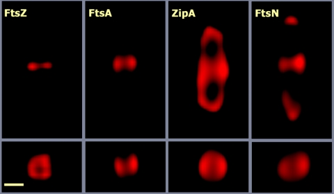
Imaging the division ring by three-dimensional deconvolution microscopy. The images correspond to E. coli MC1061 cells from an exponentially growing culture. The cells were stained with polyclonal antisera against FtsZ, FtsA, FtsN, or ZipA obtained in the authors' laboratory and an Alexa 594-conjugated anti-rabbit secondary antibody. Images from optical sections spaced 73 nm apart were taken with an Olympus BX-61 wide-field motorized microscope equipped with a DP70 charge-coupled-device camera. The image stacks were deconvoluted using the Huygens Professional software package. The top row shows images of the central, in-focus, section of each stack, whereas the bottom row shows the cross section of the same cells taken at the points of maximal fluorescence intensity. The elongation along the z axis introduced by the optical system was corrected in the deconvoluted stacks by multiplying the vertical scale by 0.42 (the correction factor calculated from the measurement of control spherical particles). The scale bar corresponds to 0.5 μm.
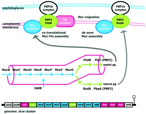
Schematic view of the genomic channeling hypothesis. Localized assembly of the enzymes that form the pathway of peptidoglycan (pg) precursor synthesis (indicated in the central part of the figure) into a multienzymatic complex (Mur complex) might drive the precursors directly to the sites of peptidoglycan synthesis by substrate channeling. This would be important in rod-shaped cells, where the highly localized machinery responsible for the synthesis of septal peptidoglycan should compete for precursors with the lateral peptidoglycan synthesis machinery, which is distributed over a much larger surface (both machineries are sketched at the top). The genomic channeling hypothesis (62, 63) states that the clustering of genes in rod-shaped cells (the E. coli dcw cluster is represented at the bottom) favors the cotranslational assembly and localization of the division protein complexes (Fts complex) and the precursor synthesis complex (Mur complex), driving the flux of precursors toward the septum synthesis machinery (PBP3-FtsW/PBP1b complexes) during cell division. After division the Mur complex might migrate to the sites of lateral peptidoglycan synthesis (PBP2-RodA/PBP1b complexes), or it might disassemble and assemble de novo at these sites. Note that in E. coli the murB gene is not found within the dcw cluster.
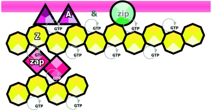
Proteins of the early assembly step of the division ring. Assembly of the FtsZ ring depends on either FtsA or ZipA or both, while the localization of these two depends on FtsZ. The localization of ZapA is dependent on FtsZ. FtsA and ZipA are bound to the inner cell membrane (magenta stripe), while FtsZ interacts with these two proteins. These three proteins assemble into the ring at the same time in a first step that might span a significant part of the cell cycle before the assembly of the late proteins shown in Fig. 4. Maintenance of the FtsZ ring is energy dependent (60), probably because FtsZ polymerization requires the presence of GTP at the nucleotide-binding site. Protein names are abbreviated as follows: A, FtsA; Z, FtsZ; Zip, ZipA; and Zap, ZapA. Icons are ordered from left to right following the linear assembly sequence of the proteins. An ampersand indicates that both proteins assemble simultaneously (see the text).
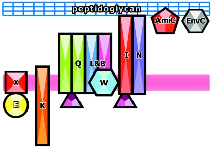
Proteins of the late assembly step of the division ring. Schematic view of the assembly of the late cell division proteins showing their relation to the cell membrane (magenta stripe) and peptidoglycan (blue grid). The protein icons are ordered from left to right according to the commonly accepted assembly sequence. An ampersand indicates that both proteins assemble simultaneously (see the text). Proteins that have been shown to interact physically are drawn in contact, while those whose assembly has been determined only by genetic methods are drawn separated. Protein names have been abbreviated by excluding “Fts” from them. FtsA is drawn as in Fig. 3.
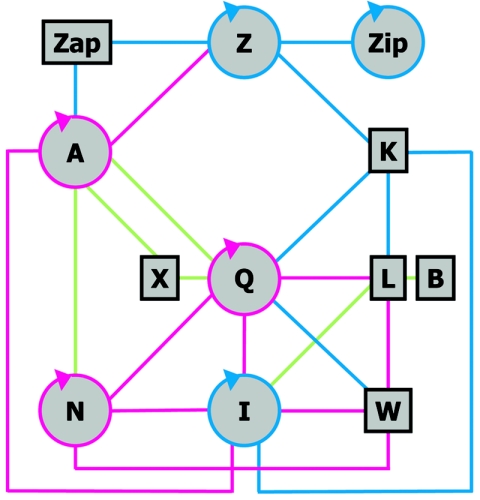
Network of interactions between division proteins. Schematic drawing of the protein-protein interactions among the E. coli cell division proteins found in systematic functional bacterial two-hybrid assays. The circular arrows indicate self-interactions. Blue lines indicate only those interactions described by Di Lallo et al. (25), green lines indicate only those described by Karimova et al. (46), while magenta lines are those detected in both studies. Note that the report of Karimova et al. (46) did not include FtsK. The FtsA self-interaction (96) and the ZipA-FtsZ interaction (38) are also found in functional yeast two-hybrid assays.
References
-
- Aarsman, M. E., A. Piette, C. Fraipont, T. M. Vinkenvleugel, M. Nguyen-Distéche, and T. den Blaauwen. 2005. Maturation of the Escherichia coli divisome occurs in two steps. Mol. Microbiol. 55:1631-1645. - PubMed
-
- Addinall, S. G., C. Cao, and J. Lutkenhaus. 1997. FtsN, a late recruit to the septum in Escherichia coli. Mol. Microbiol. 25:303-309. - PubMed
-
- Ayala, J. A., T. Garrido, M. A. de Pedro, and M. Vicente. 1994. Molecular biology of bacterial septation. .In J. M. Ghuysen and R. Habenneck (ed.), Bacterial cell wall. Elsevier Science, Amsterdam, The Netherlands.
-
- Barondess, J. J., M. Carson, L. M. Guzmán Verduzco, and J. Beckwith. 1991. Alkaline phosphatase fusions in the study of cell division genes. Res. Microbiol. 142:295-299. - PubMed
Publication types
MeSH terms
Substances
LinkOut - more resources
Full Text Sources
Other Literature Sources
