. 2006 Jan;188(1):211-22.
doi: 10.1128/JB.188.1.211-222.2006.

Transposon disruption of the complex I NADH oxidoreductase gene (snoD) in Staphylococcus aureus is associated with reduced susceptibility to the microbicidal activity of thrombin-induced platelet microbicidal protein 1

Affiliations

Transposon disruption of the complex I NADH oxidoreductase gene (snoD) in Staphylococcus aureus is associated with reduced susceptibility to the microbicidal activity of thrombin-induced platelet microbicidal protein 1

Arnold S Bayer et al. J Bacteriol. 2006 Jan.

Abstract

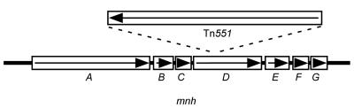
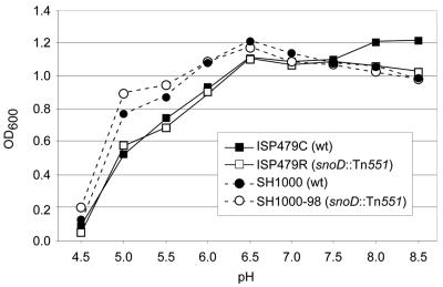
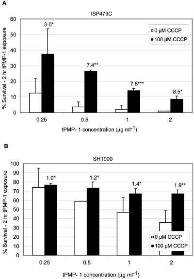
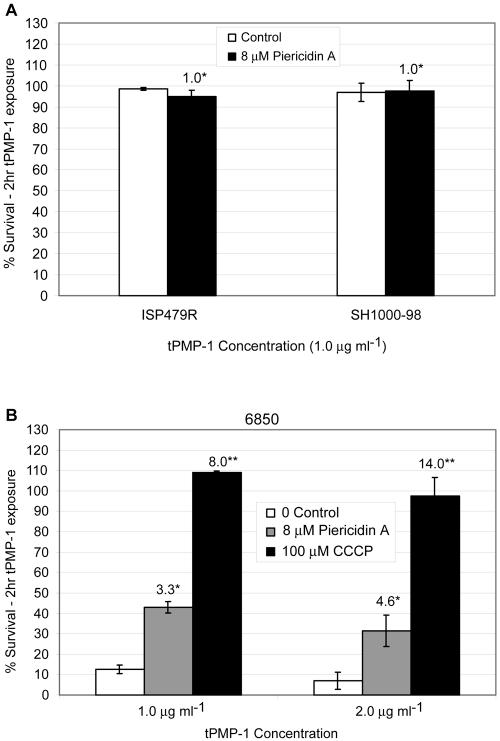
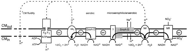
The cationic molecule thrombin-induced platelet microbicidal protein 1 (tPMP-1) exerts potent activity against Staphylococcus aureus. We previously reported that a Tn551 S. aureus transposon mutant, ISP479R, and two bacteriophage back-transductants, TxA and TxB, exhibit reduced in vitro susceptibility to tPMP-1 (tPMP-1(r)) compared to the parental strain, ISP479C (V. Dhawan, M. R. Yeaman, A. L. Cheung, E. Kim, P. M. Sullam, and A. S. Bayer, Infect. Immun. 65:3293-3299, 1997). In the current study, the genetic basis for tPMP-1(r) in these mutants was identified. GenBank homology searches using sequence corresponding to chromosomal DNA flanking Tn551 mutant strains showed that the fourth gene in the staphylococcal mnh operon (mnhABCDEFG) was insertionally inactivated. This operon was previously reported to encode a Na(+)/H(+) antiporter involved in pH tolerance and halotolerance. However, the capacity of ISP479R to grow at pH extremes and in high NaCl concentrations (1 to 3 M), coupled with its loss of transmembrane potential (DeltaPsi) during postexponential growth, suggested that the mnh gene products are not functioning as a secondary (i.e., passive) Na(+)/H(+) antiporter. Moreover, we identified protein homologies between mnhD and the nuo genes of Escherichia coli that encode components of a complex I NADH:ubiquinone oxidoreductase. Consistent with these data, exposures of tPMP-1-susceptible (tPMP-1(s)) parental strains (both clinical and laboratory derived) with either CCCP (a proton ionophore which collapses the proton motive force) or pieracidin A (a specific complex I enzyme inhibitor) significantly reduced tPMP-induced killing to levels seen in the tPMP-1(r) mutants. To reflect the energization of the gene products encoded by the mnh operon, we have renamed the locus sno (S. aureus nuo orthologue). These novel findings indicate that disruption of a complex I enzyme locus can confer reduced in vitro susceptibility to tPMP-1 in S. aureus.


Figures

FIG. 1.
FIG. 1.
Southern analysis of DNA isolated from the tPMP-1r transposon mutant ISP479R (lanes 1 and 3) and parental tPMP-1s, ISP479C (lanes 2 and 4), probed with a transposon-specific probe. Chromosomal DNA was digested with EcoRI (lanes 1 and 2) or PstI (lanes 3 and 4). A probe that hybridizes with the gene encoding the transposase of Tn551 detected a single 13-kb EcoRI fragment and a 10.2-kb PstI fragment in strain ISP479R that was not in parent strain ISP479C.
FIG. 2.
FIG. 2.
Southern analysis of S. aureus SH1000-98, a PMP-1r transductant derived from ISP479R. Lanes 1 and 2 contain chromosomal DNA from ISP479R, lanes 3 and 4 contain chromosomal DNA from strain SH1000, and lanes 5 and 6 contain DNA from SH1000-98. The even-numbered lanes contain PstI-digested DNA, while the odd-numbered lanes contain EcoRI-digested DNA. A Tn551-specific probe detected similarly sized DNA fragments in ISP479R and SH1000-98 that were not present in SH1000 or ISP479C (Fig. 1).
FIG. 3.
FIG. 3.
Amplification of the Tn551 insert site in strain ISP479R. A 0.75% agarose gel with the 1-kb ladder (lane 1) and five independent inverse PCRs that used a PstI-generated template and MgCl2 concentrations ranging from 0.0 to 9 mM in 1.5 mM increments (lanes 2 to 5). An arrow marks a 5-kb product. The size of this fragment is consistent with the Southern blot data (Fig. 1); the 10.2-kb PstI fragment corresponds to the size of the amplified product plus the 5.2 kb of DNA comprising Tn551. Similar DNA band patterns were observed for TxA, TxB, and SH1000-98 (data not shown).
FIG. 4.
FIG. 4.
Map of the Tn551 insertion site in strains ISP479R, TxA, TxB, and SH1000-98. The sno (mnh) operon is shown and consists of six genes (rectangles labeled A through G). Tn551 is depicted by the rectangle above the map of the sno (mnh) operon. The arrows within the depicted genes indicate the orientation of transcription of snoA-G (mnhA-G) and Tn551. The converging lines from the ends of the representation of Tn551 show the approximate location of the transposon insertion site.
FIG. 5.
FIG. 5.
Plasmid map of the integrative complementation plasmid, pJM798, showing relevant restriction sites and genes. The sno (mnh) operon, E. coli col E1 origin of replication (ori) (pSKII), bacteriophage L54a attachment site (attP), integrase gene (int), and the tetracycline resistance marker (tetM) are delineated by arrows or boxes.
FIG. 6.
FIG. 6.
Southern analysis of plasmid pJM798 second-site-complemented strains. EcoRV-digested chromosomal DNA from ISP479R (lane 1), ISP479R [chr::pJM798] (lane 2), TxB (lane 3), and TxB [chr::pJM798] (lane 4) was probed with the gene encoding staphylococcal glycerol ester hydrolase (geh). Upon integration into the chromosome, plasmid pJM798 introduces an EcoRV site that results in two fragments with a combined size equal to the sum of the geh EcoRV fragment in the parental strain plus the 13 kb of pJM798.
FIG. 7.
FIG. 7.
Halotolerance of parental versus respective PMP-1r mutants grown for 10 h at 37°C in NaCl-supplemented medium (3 M NaCl). Data represent the means (±SD) of three independent experiments.
FIG. 8.
FIG. 8.
pH tolerance of parental versus respective PMP-1r mutants grown for 24 h at 37°C in medium adjusted to a range of pHs from 4.5 to 8.5. Data represent the mean OD600 values obtained at 24 h growth for three independent experiments. SD bars are omitted for clarity of figure presentation.
FIG. 9.
FIG. 9.
Impact of CCCP treatment of tPMP-1s parental strains upon baseline tPMP-1 susceptibility profiles. Data represent the mean (±SD) of surviving CFU after exposure to various concentrations of tPMP-1 (0 to 2 μg/ml) in the presence or absence of CCCP (100 μM). (A) Data for parental strain ISP479C. *, not significantly different from untreated controls; **, P < 0.0001 versus untreated controls; ***, P < 0.001 versus untreated controls. (B) Data for parental strain SH1000. *, not significantly different from untreated controls; **, P < 0.05 versus untreated controls. Numbers above bars in panels A and B represent the mean fold increases in surviving CFU, comparing CCCP exposures to no-CCCP exposures. These data represent the means of at least two independent assays.
FIG. 10.
FIG. 10.
Impact of piericidin A treatment of tPMP-1s parental strains upon baseline tPMP-1 susceptibility profiles. Data represent the mean (±SD) of surviving CFU after tPMP-1 exposures (1 μg/ml) in the presence or absence of piericidin A (0, 4, or 8 μM). (A) Data for parental strain ISP479; (B) data for parental strain SH1000. Numbers above bars represent the mean fold increase in surviving CFU comparing piericidin A exposures to no-exposure controls. These data represent the means of at least two independent assays. *, not significantly different from untreated controls; **, P < 0.05 versus untreated controls.
FIG. 11.
FIG. 11.
(A) Demonstration of the impact of piericidin A treatment upon baseline tPMP-1 susceptibility profiles in two tPMP-1r strains. Numbers above bars represent the mean fold increases in surviving CFU comparing piericidin exposures to no-exposure controls. Data represent the mean (±SD) surviving CFU after tPMP-1 exposures (1 μg/ml) in the presence or absence of inhibitor. There were no significant changes in the presence of piericidin A. (B) Demonstration of the impact of two PMF inhibitors upon the tPMP-1 susceptibility profiles for clinical strain 6850. Numbers above bars represent the mean fold increases in surviving CFU comparing inhibitor exposures to no-exposure controls. Data represent the mean (±SD) of surviving CFU after tPMP-1 exposures (1 μg/ml) in the presence or absence of piericidin A (8 μM) or CCCP (100 μM). These data represent the means of at least two independent assays. *, P < 0.001; **, P < 0.0001.
FIG. 12.
FIG. 12.
Hypothetical model of the role of SnoA-G in electron transport in S. aureus. Depicted are the CM-associated components of a hypothetical electron transport chain: menaquinone (MK), the ATP synthase (F0 F1 ATPase), an aa3-type cytochrome (aa3), quinol oxidase (QoxA-D), the staphylococcal nuo-like orthologue complex (SnoA-G) coupled to an unidentified protein or proteins with NADH oxidase activity, a bd-type cytochrome, and nitrate reductase (NR). Reactions catalyzed by constituents of the hypothetical electron transport chain are shown in association with proteins or protein complexes. The thick arrows illustrate the flow of hydrogen ions (H+; dashed arrow). The aa3-type cytochromes are used in aerobic metabolism and are most closely linked to QoxA-D, an aerobic NADH oxidase. The bd-type cytochromes are used in microaerophilic and anaerobic metabolism and are linked to SnoA-G. Reduced susceptibility of S. aureus to tPMP-1 is predicted to be due to (i) the reduction in ΔΨ due to the loss of H+ translocation from the inside to the outside of the bacterium, from decreased electron flow to cytochrome bd, and (ii) an increase in diffusion of H+ from the outside to the inside of the bacterium caused by disordering (increased fluidity; dashed line) of the cytoplasmic membrane due to a shift from longer to shorter fatty acids and an increase in branched fatty acids.

References

    1. Altschul, S. F., W. Gish, W. Miller, E. W. Myers, and D. J. Lipman. 1990. Basic local alignment search tool. J. Mol. Biol. 215:403-410. - PubMed
    1. Ausubel, F. A., R. Brent, R. E. Kingston, R. E. Moore, D. D. Steidman, J. G. Smith, J. A. Smith, and K. Struhl (ed.). 1996. Current protocols in molecular biology. Greene Publishing and Wiley Interscience, New York, N.Y.
    1. Barquera, B., P. Hellwig, W. Zhou, J. E. Morgan, C. C. Hase, K. K. Gosink, M. Nilges, P. J. Bruesehoff, A. Roth, C. R. Lancaster, and R. B. Gennis. 2002. Purification and characterization of the recombinant Na+-translocating NADH:quinone oxidoreductase from Vibrio cholerae. Biochemistry 41:3781-3789. - PubMed
    1. Baumert, N., C. von Eiff, F. Schaaff, G. Peters, R. A. Proctor, and H. G. Sahl. 2002. Physiology and antibiotic susceptibility of Staphylococcus aureus small colony variants. Microb. Drug Resist. 8:253-260. - PubMed
    1. Bayer, A. S., R. P. Prasad, J. Chandra, A. Koul, M. Smriti, A. Varma, R. A. Skurray, N. Firth, M. H. Brown, S.-P. Koo, and M. R. Yeaman. 2000. In vitro resistance of Staphylococcus aureus to thrombin-induced platelet microbicidal protein in associated with alterations in cytoplasmic membrane fluidity. Infect. Immun. 68:3548-3553. - PMC - PubMed

Publication types

Substances