

Iron in skin of mice with three etiologies of systemic iron overload
- PMID: 16354190
- PMCID: PMC2243217
- DOI: 10.1111/j.0022-202X.2005.23949.x
Iron in skin of mice with three etiologies of systemic iron overload
Abstract
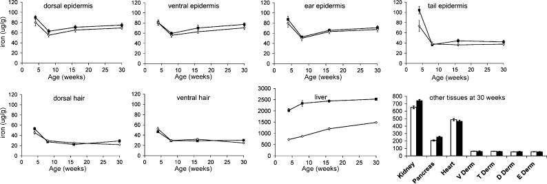
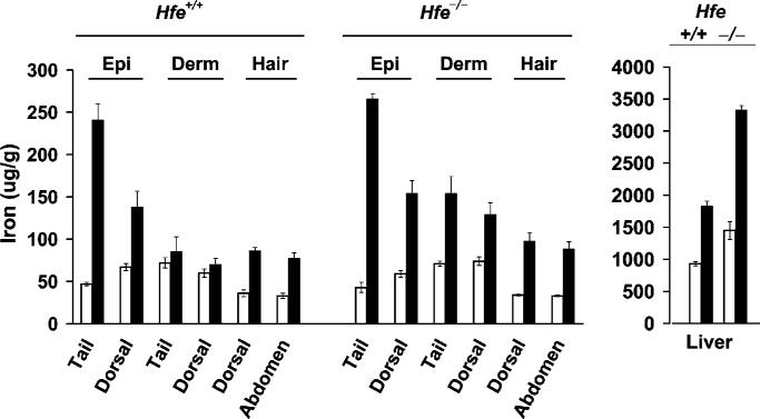
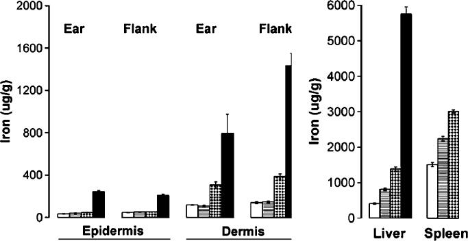
In human hemochromatosis, tissue toxicity is a function of tissue iron levels. Despite reports of skin toxicity in hemochromatosis, little is known about iron levels in skin of individuals with systemic iron overload. We measured skin iron and studied skin histology in three mouse models of systemic iron overload: mice with a deletion of the hemochromatosis (Hfe) gene, mice fed a high iron diet, and mice given parenteral injections of iron. In Hfe(-/-) mice, iron content in the epidermis and dermis was unexpectedly the same as in Hfe(+/+) mice, and there were no histological abnormalities detected after 30 wk. A high iron diet produced increased iron in the epidermis of both normal and Hfe(-/-) animals; a high diet increased iron in the dermis only in Hfe(-/-) mice. Increased skin iron was not associated with other histological changes, even after 19 wk. Parenteral administration of iron produced increased iron in the epidermis and dermis, and gave the skin a bronze hue. These results show that the amount and distribution of iron in the skin depends on the etiology of iron overload. It appears that neither Hfe deletion nor elevated skin iron alone can account for cutaneous manifestations reportedly seen in humans with hereditary hemochromatosis.
Figures
 ; 1 mg
  ; 5 mg (■).
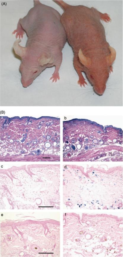
References
-
- Ajioka RS, Levy JE, Andrews NC, Kushner JP. Regulation of iron absorption in Hfe mutant mice. Blood. 2002;100:1465–1469. - PubMed
-
- Andrews NC. Disorders of iron metabolism. N Engl J Med. 1999;341:1986–1995. [published erratum appears in N Engl J Med 2000 Feb 3;342(5):364] [see comments]. - PubMed
-
- Bhasin G, Kausar H, Sarwar Alam M, Athar M. Progressive iron overload enhances chemically mediated tumor promotion in murine skin. Arch Biochem Biophys. 2003;409:262–273. - PubMed
-
- Bissett DL, Chatterjee R, Hannon DP. Chronic ultraviolet radiation-induced increase in skin iron and the photoprotective effect of topically applied iron chelators. Photochem Photobiol. 1991;54:215–223. - PubMed
-
- Cawley EP, Hsu YT, Wood BT, Weary PE. Hemochromatosis and the skin. Arch Dermatol. 1969;100:1–6. - PubMed
Publication types
MeSH terms
Substances
Grants and funding
LinkOut - more resources
Full Text Sources
Medical
