. 2006 Jan;26(1):39-49.
doi: 10.1128/MCB.26.1.39-49.2006.

Recruitment of DNA damage checkpoint proteins to damage in transcribed and nontranscribed sequences

Affiliations

Recruitment of DNA damage checkpoint proteins to damage in transcribed and nontranscribed sequences

Guochun Jiang et al. Mol Cell Biol. 2006 Jan.

Abstract

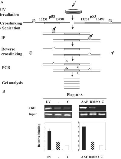
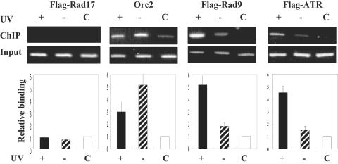
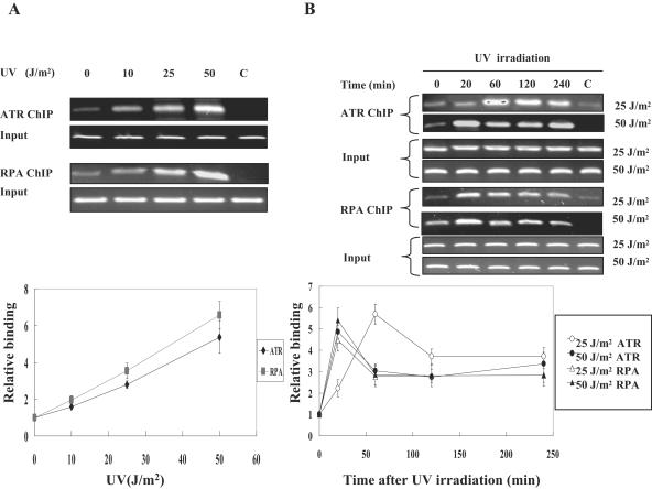
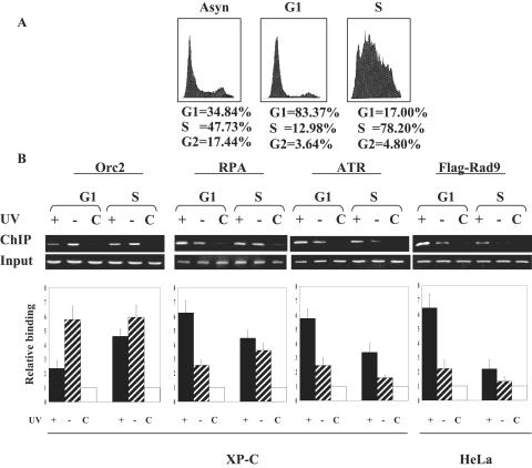
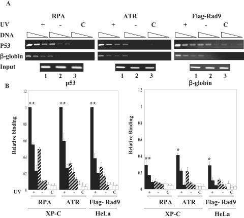
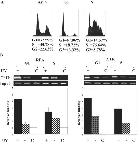
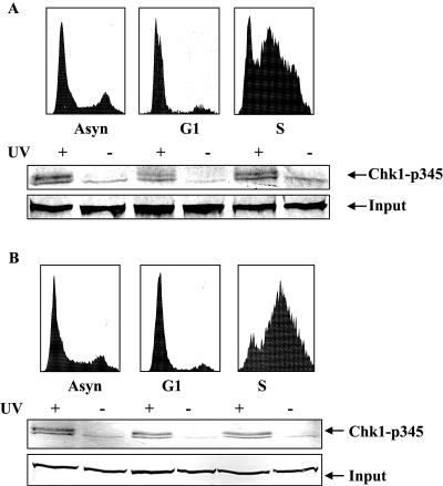
We developed a chromatin immunoprecipitation method for analyzing the binding of repair and checkpoint proteins to DNA base lesions in any region of the human genome. Using this method, we investigated the recruitment of DNA damage checkpoint proteins RPA, Rad9, and ATR to base damage induced by UV and acetoxyacetylaminofluorene in transcribed and nontranscribed regions in wild-type and excision repair-deficient human cells in G1 and S phases of the cell cycle. We find that all 3 damage sensors tested assemble at the site or in the vicinity of damage in the absence of DNA replication or repair and that transcription enhances recruitment of checkpoint proteins to the damage site. Furthermore, we find that UV irradiation of human cells defective in excision repair leads to phosphorylation of Chk1 kinase in both G1 and S phase of the cell cycle, suggesting that primary DNA lesions as well as stalled transcription complexes may act as signals to initiate the DNA damage checkpoint response.


Figures

FIG. 1.
FIG. 1.
(A) Method for detecting protein binding to UV photoproducts in a given genomic region. Cells are irradiated with UV (or treated with a UV-mimetic agent), incubated for an appropriate period of time, and then treated with formaldehyde, and ChIP is carried out by standard procedures. Binding of repair/checkpoint proteins to UV photoproducts (or chemical lesions) within ∼1 kbp on either side of the target sequence leads to enhancement of the target sequence in the immunoprecipitate. In most of our studies, a p53 fragment spanning the region of nt 13251 to 13498 (in intron 7) from the initiation codon was amplified, and thus, bindings of checkpoint proteins to the area extending from exon 4 to intron 9 are detected. (B) Binding of RPA to UV and N-AAAF lesions in the p53 of HEK293T cells. The cells were transfected with 10 μg pcDNA3-Flag-RPA32, then 72 h later, cells were either irradiated with 50 J/m2 or treated with 20 μg/ml N-AAAF for 30 min and then incubated with formaldehyde, ChIP was carried out by standard procedures. Top panel: ChIP data. UV and − indicate irradiated and unirradiated cells, respectively, and AAF and DMSO indicate cells treated with the carcinogen or with solvent, as indicated. C indicates the control reaction using cells that were transfected with pcDNA3 vector and used in ChIP with anti-Flag antibodies. Bottom panel: quantitative analysis of ChIP data. The averages of the results from three experiments, including the one shown in the top panel, are plotted. The values are relative to the vector-transfected cell control. Error bars indicate standard errors of the means.
FIG. 2.
FIG. 2.
Binding of checkpoint proteins to UV lesions in p53 in HEK293T cells. The cells were transfected with vectors expressing the indicated proteins containing Flag tag or with control (C) vector pcDNA3, irradiated where indicated with 50 J/m2, and 20 min after irradiation, treated with formaldehyde, and ChIP was carried out with anti-Flag antibodies. Top panel: representative ChIP data. +, irradiation; −, no irradiation. Bottom panel: average of the results from three experiments, including the one for which results are shown in the top panel. Error bars indicate standard errors of the means.
FIG. 3.
FIG. 3.
Binding of RPA and ATR to the p53 gene in XP-C cells as a function of UV dose and incubation time following UV irradiation. (A) Dose response. Cells were irradiated with the indicated doses of UV, and following a 20 min incubation, ATR- or RPA-bound DNA was immunoprecipitated with anti-ATR and anti-RPA antibodies using standard ChIP procedures. The C lane in the top panel contained DNA nonspecifically bound to agarose beads. The bottom panel shows quantitative analysis of the data from three experiments, including the one for which results are shown in the top panel. The values are relative to the irradiated control. (B) Time course. Cells were irradiated with either 25 J/m2 or 50 J/m2 and incubated for the indicated times before ChIP assays were performed with either anti-RPA or anti-ATR antibodies. The top panel shows representative ChIP assays, and the bottom panel shows quantitative analysis of data from three experiments. The values are relative to ChIP signals at 25 J/m2 and 50 J/m2 at time zero after irradiation. Note that, at both UV doses, RPA binding reaches a maximum at 20 min, whereas ATR binding reaches a maximum at 20 min at 50 J/m2 and gradually decreases thereafter, but the binding of ATR after 25 J/m2 proceeds at a slower rate and decays at a slower rate as well. Error bars indicate standard errors of the means.
FIG. 4.
FIG. 4.
Binding of checkpoint proteins to the p53 gene in G1 and S phases of the cell cycle. XP-C cells or HeLa cells expressing Flag-tagged Rad9 protein were synchronized by double thymidine block and then irradiated in either G1 or S phase, and 20 min after irradiation, ChIP was carried out with antibodies against Orc2, RPA, or ATR in XP-C cells and with anti-Flag antibodies in HeLa cells stably transfected with a Flag-Rad9 vector. The top panel shows the FACS analysis of XP-C cells before synchronization (Asyn) and of XP-C cells in G1 and S phases that were used for the ChIP experiments. The FACS profiles of G1- and S-phase HeLa cells were similar to that of XP-C cells and are not shown for clarity. The bottom panel shows the results of the ChIP experiments. The C columns contained ChIP material from control beads with no antibodies that were mixed with chromatin from irradiated cells. +, irradiation; −, no irradiation. Note the decreased binding of UV-irradiated DNA to all probed checkpoint proteins in S phase compared to the level of binding in G1 phase. Error bars indicate standard errors of the means.
FIG. 5.
FIG. 5.
Binding of checkpoint proteins to UV damage in transcribed and nontranscribed genes in G1 phase. For ChIP with RPA and ATR, XP-C cells were used. ChIP with Rad9 was carried out with HeLa cells expressing Flag-tagged Rad9. (A) ChIP assays performed with serial dilutions (4 μl of undiluted ChIP DNA and two- or fourfold dilutions, respectively) of the immunoprecipitated DNA to ensure the PCR amplification were within the linear range. The “input” gel in the RPA and ATR assays contains DNA from UV irradiated cells (lane 1), nonirradiated cells that were used for ChIP with anti-RPA and anti-ATR antibodies (lane 2), and DNA from irradiated cells that was used for the control no-antibody reaction (lane 3). In the Rad9 ChIP, the input DNAs were from cells that were transfected with Rad9 and UV irradiated (lane 1) or nonirradiated (lane 2) and cells that were transfected with control vector (lane 3). (B) Quantitative analysis of the ChIP data. Averages of the results from three experiments are plotted. The values were expressed relative to that of RPA, ATR, or Rad9 binding to UV-damaged p53 DNA at the highest DNA concentration used in the PCR. Significance is indicated by asterisks: **, P < 0.01; *, P < 0.05. +, irradiation; −, no irradiation.
FIG. 6.
FIG. 6.
Binding of checkpoint damage sensors, RPA and ATR, to DNA damage in transcribed DNA in the absence of transcription-coupled repair or replication. XP-A cells were synchronized by double thymidine block and were irradiated in G1 or S phase with 50 J/m2 of 254-nm light. Following irradiation, ChIP was performed at 20 min with either anti-RPA or anti-ATR antibodies. (A) FACS profile of asynchronous (Asyn) and synchronized XP-A cells. (B) ChIP of p53 intron 7. Top panels show ChIP data, and bottom panels show quantitative analysis of data from three experiments with standard errors of the means (error bars). +, irradiation; −, no irradiation.
FIG. 7.
FIG. 7.
Phosphorylation of Chk1-S345 residue in XP-A and XP-C cells that were irradiated with 50 J/m2 of 254-nm light while growing asynchronously (Asyn), in G1 phase, or in S phase. (A)The top panel shows the FACS analysis of XP-A cells used in the checkpoint activation experiment, and the bottom panel shows Western blot analysis of cell lysates that were used in ChIP assays probed with anti-S345P antibodies. The input gel represents a protein in the lysate that cross-reacts with anti-S345P antibody nonspecifically. (B) The top panel shows the FACS analysis of XP-C cells used in the checkpoint activation experiment, and the bottom panel shows Western blot analysis of cell lysates that were used in ChIP assays probed with anti-S345P antibodies. +, irradiation; −, no irradiation.

Similar articles

Cited by

References

    1. Abraham, R. T. 2001. Cell cycle checkpoint signaling through the ATM and ATR kinases. Genes Dev. 15:2177-2196. - PubMed
    1. Bakkenist, C. J., and M. B. Kastan. 2003. DNA damage activates ATM through intermolecular autophosphorylation and dimmer dissociation. Nature 421:499-506. - PubMed
    1. Ball, H. L., J. S. Myers, and D. Cortez. 2005. ATRIP binding to replication protein A-single-stranded DNA promotes ATR-ATRIP localization but is dispensable for Chk1 phosphorylation. Mol. Biol. Cell 16:2372-2381. - PMC - PubMed
    1. Bell, S. P., and A. Dutta. 2002. DNA replication in eukaryotic cells. Annu. Rev. Biochem. 71:333-374. - PubMed
    1. Bermudez, V. P., L. A. Lindsey-Boltz, A. J. Cesare, Y. Maniwa, J. D. Griffith, J. Hurwitz, and A. Sancar. 2003. Loading of the human 9-1-1 checkpoint complex onto DNA by the checkpoint clamp loader hRad17-replication factor C complex in vitro. Proc. Natl. Acad. Sci. USA 100:1633-1638. - PMC - PubMed

Publication types