. 2006 Jan;26(1):192-8.
doi: 10.1128/MCB.26.1.192-198.2006.

Tissue-specific differences of p53 inhibition by Mdm2 and Mdm4

Affiliations

Tissue-specific differences of p53 inhibition by Mdm2 and Mdm4

Jason D Grier et al. Mol Cell Biol. 2006 Jan.

Abstract

The function of the p53 tumor suppressor to inhibit proliferation or initiate apoptosis is often abrogated in tumor cells. Mdm2 and its homolog, Mdm4, are critical inhibitors of p53 that are often overexpressed in human tumors. In mice, loss of Mdm2 or Mdm4 leads to embryonic lethal phenotypes that are completely rescued by concomitant loss of p53. To examine the role of Mdm2 and Mdm4 in a temporal and tissue-specific manner and to determine the relationships of these inhibitors to each other, we generated conditional alleles. We deleted Mdm2 and Mdm4 in cardiomyocytes, since proliferation and apoptosis are important processes in heart development. Mice lacking Mdm2 in the heart were embryonic lethal and showed defects at the time recombination occurred. A critical number of cardiomyocytes were lost by embryonic day 13.5, resulting in heart failure. This phenotype was completely rescued by deletion of p53. Mice lacking Mdm4 in the heart were born at the correct ratio and appeared to be normal. Our studies provide the first direct evidence that Mdm2 can function in the absence of Mdm4 to regulate p53 activity in a tissue-specific manner. Moreover, Mdm4 cannot compensate for the loss of Mdm2 in heart development.


Figures

FIG. 1.
FIG. 1.
Mdm2 and Mdm4 conditional alleles. (A) The Mdm2 conditional allele was designed to delete exons 5 and 6, which encode part of the p53-binding domain (11). (B) Targeting strategy for the Mdm4 conditional allele deletes the first coding exon. Exons are numbered and represented as black boxes. Arrowheads denote locations and directions of PCR primers. Frt sites are shown as filled circles, while loxP sites are represented as diamonds. R1, EcoRI; NEO, neomycin resistance gene; TK, herpes simplex virus thymidine kinase gene. (C) Southern blot analysis of targeted ES cell lines. DNA from ES cells was digested with EcoRI. Blots were hybridized with an exon 4 probe and show the presence of a 10-kb fragment in correctly targeted ES cells. (D) PCR analysis of targeted ES cell lines. PCRs were performed using primer F1 located outside of the left arm of homology and primer neo19 within the neomycin gene. Positive clones had a 2.2-kb diagnostic PCR product. M, 1-kb-plus DNA ladder; nc, negative control. (E) Immunoprecipitation (IP) and Western blot analysis of lysates from Mdm4Δ2/Δ2 p53/ mouse embryonic fibroblasts. Cell lysates were immunoprecipitated with the indicated antibodies followed by Western blot analysis with the Mdm4 antibody. WT, wild type; DN1 and DN2, Mdm4Δ2/Δ2 p53/ cell lines 1 and 2, respectively; GFP, green fluorescent protein; IgG, immunoglobulin G. The arrow indicates Mdm4.
FIG. 2.
FIG. 2.
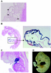
Characterization of the αMyHC-Cre transgenic line during embryogenesis. αMyHC-Cre mice were mated with the ROSA26 reporter line. Embryos at different developmental stages were stained with X-Gal (5-bromo-4-chloro-3-indolyl-β-d-galactopyranoside) solution. (A) Histological section of E8.5 embryo. Arrowheads indicate heart structures. (B) Histological section of blue-stained E9.5 embryo. V, ventricle. (C) Histological section of blue-stained E12.5 embryo. (D) Whole-mount blue-stained E12.5 embryo.
FIG. 3.
FIG. 3.
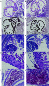
Analysis of sections of normal and Mdm2−/FM αMyHC-Cre mutant embryos. Normal (A, C, E, and G) and mutant (B, D, F, and H to J) embryos are shown at E9.5 (A to D) and E13.5 (E to J). All images are hematoxylin and eosin-stained sections, except for images in panels C and D, which were immunostained with α smooth-muscle actin antibody to label the cardiomyocytes. Yellow bars indicate widths of ventricles (V) and atria (A) in normal and mutant hearts. Arrows in panels E and F indicate the location of the heart, and the arrow in panel H shows abnormal blood deposition. (I) View of Mdm2−/FM αMyHC-Cre mutant atrium. (J) View of Mdm2−/FM αMyHC-Cre mutant liver.
FIG. 4.
FIG. 4.
Analysis of apoptosis and proliferation in heart sections of normal and Mdm2−/FM αMyHC-Cre mutant embryos at E9.5. Normal (A, C, and E) and mutant (B, D, and F) hearts are shown at E9.5. (A and B) TUNEL assay. (C and D) Immunohistochemistry for Ki-67. (E and F) Immunohistochemistry for mouse p53 protein. A, atrium; V, ventricle.
FIG. 5.
FIG. 5.
Heart-specific deletion of Mdm2. (A) PCR of DNA isolated from the tissues of an Mdm2+/FM αMyHC-Cre mouse at 3 weeks of age. Primers A, B, and C are depicted in Fig. 1A. M, 1-kb-plus DNA ladder; ht, heart; lu, lung; li, liver; tl, tail; sk mu, skeletal muscle; wt, wild type. (B) Same PCR as described above, performed on DNA isolated from E9.5 embryos. C, control Mdm2+/FM sample; R, recombined Mdm2+/FM αMyHC-Cre sample.
FIG. 6.
FIG. 6.
αMyHC-Cre showed specific recombination in mouse heart. PCR genotyping was done to detect Mdm4Δ2 and Mdm4FX alleles (see Fig. 1B for locations of primers) by using DNA from different organs of a 2-month-old Mdm4FX/FX αMyHC-Cre mouse. Lane headings are defined in the legend for Fig. 5.
FIG. 7.
FIG. 7.
Deletion of Mdm4 in the heart produced normal morphology at E9.5. Whole-mount β-gal staining was performed on E9.5 mouse embryos. (A) Mdm4+/ αMyHC-Cre ROSA26 mouse. (B) Mdm4−/FX αMyHC-Cre ROSA26 mouse. V, ventricle.

References

    1. Agah, R., P. A. Frenkel, B. A. French, L. H. Michael, P. A. Overbeek, and M. D. Schneider. 1997. Gene recombination in postmitotic cells. Targeted expression of Cre recombinase provokes cardiac-restricted, site-specific rearrangement in adult ventricular muscle in vivo. J. Clin. Investig. 100:169-179. - PMC - PubMed
    1. Bottger, V., A. Bottger, C. Garcia-Echeverria, Y. F. Ramos, A. J. van der Eb, A. G. Jochemsen, and D. P. Lane. 1999. Comparative study of the p53-mdm2 and p53-MDMX interfaces. Oncogene 18:189-199. - PubMed
    1. Chavez-Reyes, A., J. M. Parant, L. L. Amelse, R. M. de Oca Luna, S. J. Korsmeyer, and G. Lozano. 2003. Switching mechanisms of cell death in mdm2- and mdm4-null mice by deletion of p53 downstream targets. Cancer Res. 63:8664-8669. - PubMed
    1. Farley, F. W., P. Soriano, L. S. Steffen, and S. M. Dymecki. 2000. Widespread recombinase expression using FLPeR (Flipper) mice. Genesis 28:106-110. - PubMed
    1. Fernandez, E., Z. Siddiquee, and R. V. Shohet. 2001. Apoptosis and proliferation in the neonatal murine heart. Dev. Dyn. 221:302-310. - PubMed

Publication types

MeSH terms