. 2006 Jan;26(1):230-7.
doi: 10.1128/MCB.26.1.230-237.2006.

Mitogen-activated protein kinase cascade-mediated histone H3 phosphorylation is critical for telomerase reverse transcriptase expression/telomerase activation induced by proliferation

Affiliations

Mitogen-activated protein kinase cascade-mediated histone H3 phosphorylation is critical for telomerase reverse transcriptase expression/telomerase activation induced by proliferation

Zheng Ge et al. Mol Cell Biol. 2006 Jan.

Abstract

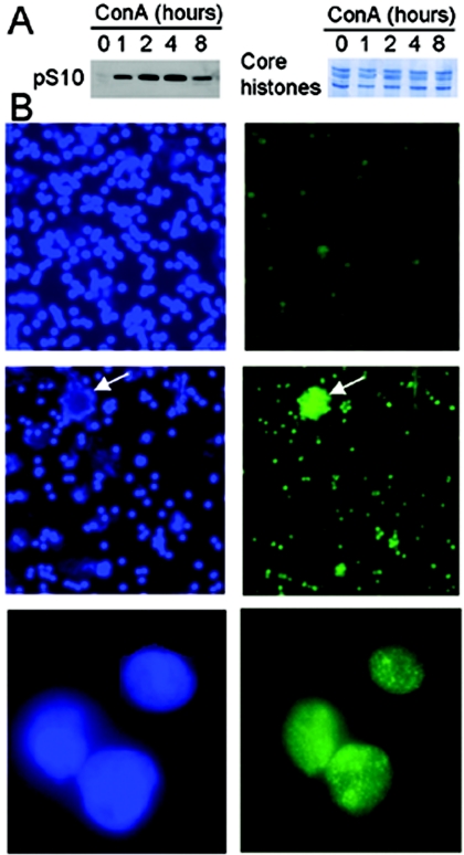
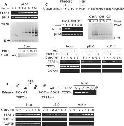
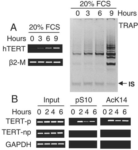
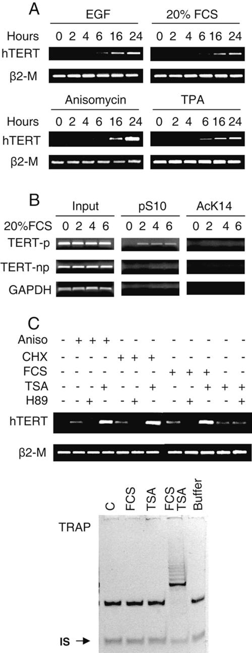
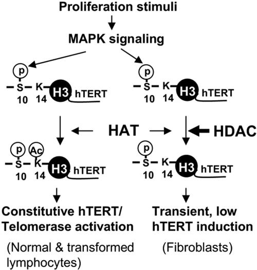
Telomerase activity and telomerase reverse transcriptase (hTERT), the key component of the telomerase complex, are tightly proliferation regulated in normal and malignant cells both in vitro and in vivo; however, underlying mechanisms are unclear. In the present study, we identified mitogen-activated protein kinase (MAPK) cascade-mediated histone H3 ser10 phosphorylation to be a molecular link between proliferation and induction of hTERT/telomerase activity. In normal human T lymphocytes and fibroblasts, growth or stress stimuli known to drive H3 phosphorylation through the MAPK signaling induce hTERT expression and/or telomerase activity that was preceded by phosphorylated histone H3 (ser10) at the hTERT promoter. Blockade of the MAPK-triggered H3 phosphorylation significantly abrogates hTERT induction and ser10 phosphorylation at this promoter. However, H3 ser10 phosphorylation alone resulted in low, transient hTERT induction, as seen in fibroblasts, whereas H3 phosphorylation followed by its acetylation at lys14 robustly trans-activated the hTERT gene accompanying constitutive telomerase activity in normal and malignant T cells. H3 acetylation without phosphorylation similarly exerted weak effects on hTERT expression. These results define H3 phosphorylation as a key to hTERT transactivation induced by proliferation and reveal a fundamental mechanism for telomerase regulation in both normal human cells and transformed T cells.


Figures

FIG. 1.
FIG. 1.
Rapid histone H3 ser10 phosphorylation in normal T cells stimulated with ConA. (A) Western blot analyses of ser10-phosphorylated H3 in the control and ConA-treated T cells (left). The equal loading of histones was verified by Coomassie blue staining of the duplicated gels (right). (B) Immunofluorescence staining of ser10-phosphorylated H3 in the control and ConA-treated T cells. Left and right: 4′,6′-diamidino-2-phenylindole (DAPI) and phosphorylated H3 (ser10) signals, respectively. Top panel, untreated cells (magnification, 20×). Middle (magnification, 20×) and bottom (magnification, 100×) panels, ConA-treated T cells for 1 h. Arrows indicate aggregated T cells with bright signals of phosphorylated H3 after activation.
FIG. 2.
FIG. 2.
Association of H3 ser10 phosphorylation at the hTERT promoter with hTERT induction/telomerase activation in ConA-treated T cells. (A) Up-regulation of the hTERT expression and activation of telomerase in ConA-treated T cells. Upper panel, RT-PCR for hTERT mRNA analysis and TRAP assay for telomerase activity in the control and ConA-treated T cells. IS, internal standard. Lower panel, Western blot (WB) for hTERT protein detection. The specificity of the antibody against hTERT protein was demonstrated by introducing a Flag-hTERT expression vector into telomerase-negative human cells and obtaining appropriate signals with both hTERT and flag antibodies (data not shown). (B) Left upper panel, schematic presentation of the hTERT locus and PCR primer locations for the ChIP assay; right upper panel, validation of the semiquantitative PCR for the hTERT promoter sequence. The input DNA (corresponding to 2% of a chromatin sample) was diluted 0-, 4-, 10-, 25-, and 100-fold as indicated and was then subjected to PCR analyses using TERT-p primers. Lower panel, specific accumulation of histone H3 with ser10 phosphorylation and lys14 acetylation on the hTERT promoter in activated T cells, as determined using ChIP. (C) Attenuated induction of hTERT/telomerase expression and H3 phosphorylation/acetylation at the hTERT promoter by H89 and PD98095 in T cells treated with ConA. Top panel, the inhibitory mechanism of the MAPK signaling-mediated H3 phosphorylation by H89 (10 μM) and PD98095 (40 μM). Middle panel, diminished hTERT mRNA expression (left) and telomerase activity (right) induced by ConA treatment of T cells in the presence of H89 and PD98095. The cells with different treatments for various periods were analyzed for hTERT mRNA and telomerase activity by using RT-PCR and TRAP, respectively. C/H, ConA plus H89; C/P, ConA plus PD98095. IS, internal standard. Bottom panel, the ChIP assay for the occupancy of phosphorylated and acetylated histone H3 at the hTERT promoter in the ConA-treated T cells with and without H89 or PD98095.
FIG. 3.
FIG. 3.
Concomitant up-regulation of hTERT mRNA/telomerase expression and histone H3 phosphorylation/acetylation at the hTERT promoter in quiescent Jurkat cells upon reentry into cell cycle. (A) hTERT transcript (left) and telomerase activity (right) in Jurkat cells during transition from quiescence to G1 phase. Jurkat cells were first incubated in 0.5% FCS-containing medium for 72 h to induce quiescence and then re-fed with 20% of FCS. hTERT mRNA and telomerase activities were determined with the use of RT-PCR and TRAP assay, respectively. IS, internal standard. (B) Histone H3 phosphorylation/acetylation at the hTERT promoter in quiescent and FCS-treated Jurkat cells as determined using ChIP.
FIG. 4.
FIG. 4.
Induction of hTERT mRNA and H3 phosphorylation at the hTERT promoter in normal human fibroblasts by growth and stress stimuli. (A) hTERT mRNA expression in fibroblasts treated with growth and stress stimuli. The fibroblasts were induced to quiescence by incubating them in 0.5% FCS-containing medium for 48 to 72 h and were then treated with EGF (200 ng/ml), 20% FCS, TPA (250 ng/ml), and anisomycin (Aniso; 5 μg/ml) for various periods as indicated. The cells were then analyzed for hTERT mRNA using RT-PCR. (B) Histone H3 phosphorylation on the hTERT promoter preceding hTERT mRNA expression in fibroblasts fed with FCS. (C) Synergistic effects of growth or stress stimuli and HDAC inhibition on induction of hTERT mRNA expression and telomerase activity in fibroblasts. Upper panel, quiescence-induced fibroblasts were treated with EGF, 20% FCS, or anisomycin or CHX (50 μg/ml) in the presence or absence of TSA (1 μM) or H89 (10 μM) and then were analyzed for hTERT mRNA expression. Lower panel, TRAP results of the fibroblasts treated with FCS or TSA and FCS plus TSA. IS, internal standard.
FIG. 5.
FIG. 5.
Working model for histone H3 phosphorylation and acetylation in controlling hTERT transcription and telomerase activation. The model is based on the present finding that both phosphorylation and acetylation of histone H3 are required to fully trans-activate the hTERT gene. First, growth stimuli trigger H3 ser10 phosphorylation at the hTERT promoter via the MAPK cascade in most, if not all, human cells with proliferation capacities. H3 ser10 phosphorylation then promotes HATs to acetylate lys14 in the same histone tail. Depending on the status of HDACs on the hTERT promoter, local H3 lys14 acetylation may occur, and synergistic effects of phosphorylation and acetylation lead to constitutively high hTERT expression and telomerase activation, as seen in normal and malignant T cells. On the other hand, if the role of HDACs on the hTERT promoter is predominant, no further lys14 is acetylated, and consequently ser10 phosphorylation alone induces transient, low levels of hTERT expression, as seen in fibroblasts. Thick arrow, the predominant role for HDACs at the hTERT promoter.

References

    1. Belair, C. D., T. R. Yeager, P. M. Lopez, and C. A. Reznikoff. 1997. Telomerase activity: a biomarker of cell proliferation, not malignant transformation. Proc. Natl. Acad. Sci. USA 94:13677-13682. - PMC - PubMed
    1. Blasco, M. A. 2003. Telomeres and cancer: a tale with many endings. Curr. Opin. Genet. Dev. 13:70-76. - PubMed
    1. Buchkovich, K. J., and C. W. Greider. 1996. Telomerase regulation during entry into the cell cycle in normal human T cells. Mol. Biol. Cell 7:1443-1454. - PMC - PubMed
    1. Chadee, D. N., M. J. Hendzel, C. P. Tylipski, C. D. Allis, D. P. Bazett-Jones, J. A. Wright, and J. R. Davie. 1999. Increased Ser-10 phosphorylation of histone H3 in mitogen-stimulated and oncogene-transformed mouse fibroblasts. J. Biol. Chem. 274:24914-24920. - PubMed
    1. Chadeneau, C., P. Siegel, C. B. Harley, W. J. Muller, and S. Bacchetti. 1995. Telomerase activity in normal and malignant murine tissues. Oncogene 11:893-898. - PubMed

Publication types

MeSH terms

LinkOut - more resources