. 2006 Jan;26(1):362-70.
doi: 10.1128/MCB.26.1.362-370.2006.

Regulation of CD44 alternative splicing by SRm160 and its potential role in tumor cell invasion

Affiliations

Regulation of CD44 alternative splicing by SRm160 and its potential role in tumor cell invasion

Chonghui Cheng et al. Mol Cell Biol. 2006 Jan.

Abstract

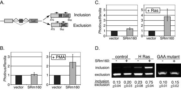
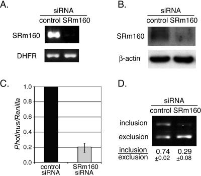
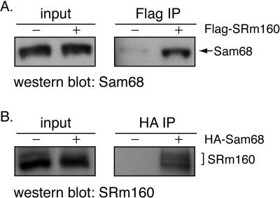
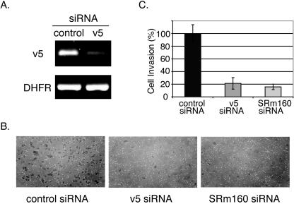
The multiple isoforms of the transmembrane glycoprotein CD44 are produced by alternative RNA splicing. Expression of CD44 isoforms containing variable 5 exon (v5) correlates with enhanced malignancy and invasiveness of some tumors. Here we demonstrate that SRm160, a splicing coactivator, regulates CD44 alternative splicing in a Ras-dependent manner. Overexpression of SRm160 stimulates inclusion of CD44 v5 when Ras is activated. Conversely, small interfering RNA (siRNA)-mediated silencing of SRm160 significantly reduces v5 inclusion. Immunoprecipitation shows association of SRm160 with Sam68, a protein that also stimulates v5 inclusion in a Ras-dependent manner, suggesting that these two proteins interact to regulate CD44 splicing. Importantly, siRNA-mediated depletion of CD44 v5 decreases tumor cell invasion. Reduction of SRm160 by siRNA transfection downregulates the endogenous levels of CD44 isoforms, including v5, and correlates with a decrease in tumor cell invasiveness.


Figures

FIG. 1.
FIG. 1.
SRm160 stimulates CD44 v5 inclusion. (A) The luciferase-based v5 minigene schematic. Exons are shown as boxes and introns are denoted as straight lines. Lines above and below each intron illustrate different joining of exons by alternative splicing. Positions of the translation start codon (ATG) and stop codon are shown. Inclusion of v5 results in expression of an in-frame luciferase protein, whereas exclusion of v5 yields a stop codon upstream of the luciferase gene. The relative locations of the primers used in the PCRs to detect the included and excluded forms are shown as arrows at the top of these forms. (B) Dual luciferase assay of 293T cells. Cells were transiently transfected with the v5 minigene (Photinus luciferase), a Renilla luciferase plasmid (used as an internal transfection control), and either an empty vector (black bars) or SRm160 expression plasmid (gray bars). About 18 h after transfection, cells were treated without PMA (left panel) or with PMA (right panel) for 6 h. After this treatment, cells were harvested and lysed. Luciferase activities were assayed using a Dual-Glo luciferase kit (Promega). The ratios of Photinus and Renilla luciferase activities were normalized to the empty vector transfections (black bars). Error bars denote the range of experimental variations. (C) Relative luciferase activity of cells transiently transfected with the v5 minigene plus either empty vector (black bars) or SRm160 plasmid (gray bars) and cotransfected without or with activated H-Ras V12 plasmid. The ratios of Photinus and Renilla luciferase activities were normalized to the non-SRm160 (empty vector) transfections. (D) RT-PCR analysis of harvested RNA from cells transfected with the constructs described in panel B (without or with SRm160 and without or with activated H-Ras V12) or with a GAA-mutant v5 construct (in the presence of activated H-Ras V12, without or with SRm160). Relative locations of the PCR primers are shown in panel A. PCR products were analyzed in 1.5% agarose gels. The inclusion and exclusion forms of v5 were detected in one PCR. Positions of these two forms are depicted on the left of the gel. Calculated ratios between these forms are shown at the bottom of the gel.
FIG. 2.
FIG. 2.
SRm160 knockdown inhibits v5 inclusion. (A) Quantitative RT-PCR analysis of the SRm160 mRNA harvested from HeLa cells transiently transfected with either control siRNA or SRm160 siRNA. Primers specific to SRm160 were used. Amplification of DHFR mRNA was used as an internal control. (B) Western blot analysis of SRm160 protein levels in siRNA-transfected cells described in panel A. The β-actin protein levels were used as a loading control. (C) Relative luciferase activities of HeLa cells transiently transfected with the v5-Luc minigene, H-Ras V12 plasmid, and a control siRNA or SRm160 siRNA. Ratios of the Photinus and Renilla luciferase activities were normalized to the control siRNA transfections. (D) RT-PCR analysis of RNA from siRNA-transfected cells described in panel C. PCR primers are shown in Fig. 1A. PCR products were analyzed in 1.5% agarose gels. Inclusion and exclusion forms of v5 were detected in one PCR. Positions of these two forms are indicated on the left of the gel. Calculated ratios between these forms are shown at the bottom of the gel.
FIG. 3.
FIG. 3.
SRm160 and Sam68 coimmunoprecipitate. 293T cells were transfected with plasmids containing either Flag-tagged SRm160 (A) or HA-tagged Sam68 (B) or corresponding empty vectors. Cell lysates were immunoprecipitated using anti-Flag or anti-HA antibodies, respectively (indicated at the top of the gels). Total cell lysates (input) as well as immunoprecipitants (IPs) were analyzed by SDS-polyacrylamide gel electrophoresis. Western blot analyses were performed using antibodies of Sam68 (A, Flag IP) or SRm160 (B, HA IP). The positions of Sam68 and SRm160 are indicated on the right of the gels.
FIG. 4.
FIG. 4.
SRm160 is required for splicing of endogenous CD44 variants. (A) Expression pattern of CD44 variants in HeLa cells. The relative locations of PCR primers are depicted at the top of the gels. To analyze different variants of CD44, PCRs were performed using primers that contain different specific 5′ primers pairing to individual variable exons and one 3′ primer pairing to a constitutive exon (filled arrows and indicated as v2 to v10 on top of the gel). PCR amplifications of v2, v3, v6, and v7 variants were also performed using a 5′ primer pairing to a constitutive exon and specific 3′ primers pairing to individual variable exons (open arrows and indicated as v2r, v3r, v6r and v7r). Primers that pair to 5′ and 3′ constitutive exons were used for detection of the CD44 standard form. An agarose gel of PCR products is shown with a 100-bp marker in the left lane. Specific primers used in each PCR are indicated on the top of the gel. (B) RT-PCR analysis of CD44 variant exon inclusion. RNAs were harvested from HeLa cells cotransfected with H-Ras V12 and siRNAs to control, SRm160, and Sam68. Primers for analysis of CD44 variants and the standard form of CD44 are indicated on the left of gels. Amplification of the DHFR mRNA was used as an internal loading control. Agarose gels of PCR analysis of CD44 variants are shown. The relative amounts of inclusion in cells treated with SRm160 and Sam68 siRNAs are normalized to cells treated with control siRNA and plotted as a bar graph. (C) siRNA knockdown of Sam68. RNA analysis of Sam68 from cells treated with control and Sam68 siRNAs is shown in the left panel using Sam68-specific primers. Amplification of the DHFR gene was used as an internal loading control. Western blot analysis of Sam68 knockdown is shown in the right panel. The β-actin protein level was used as a loading control.
FIG. 5.
FIG. 5.
Knockdown of SRm160 reduces tumor cell invasiveness. (A) Quantitative RT-PCR analysis of the v5 mRNA from HeLa cells transiently transfected with either control siRNA or v5 siRNA. Primers specific to v5 were used. Amplification of DHFR mRNA was used as an internal control. (B) Invasion assays. HeLa cells were transfected with control, CD44 v5, or SRm160 siRNA for 48 h. These cells were then suspended in serum-free medium and placed in an invasion chamber and induced to migrate toward serum-containing medium, requiring invasion through a matrigel-coated membrane. After 22 to 24 h, cells that invaded through the membrane were fixed, stained, and counted. Representative photographs of these cells are shown. (C) Quantitation of tumor cell invasion described in panel B. The percentage of cell invasion was normalized to that of cells transfected with control siRNA.

Similar articles

Cited by

References

    1. Babic, I., A. Jakymiw, and D. J. Fujita. 2004. The RNA binding protein Sam68 is acetylated in tumor cell lines, and its acetylation correlates with enhanced RNA binding activity. Oncogene 23:3781-3789. - PubMed
    1. Bennett, K. L., D. G. Jackson, J. C. Simon, E. Tanczos, R. Peach, B. Modrell, I. Stamenkovic, G. Plowman, and A. Aruffo. 1995. CD44 isoforms containing exon V3 are responsible for the presentation of heparin-binding growth factor. J. Cell Biol. 128:687-698. - PMC - PubMed
    1. Black, D. L. 2003. Mechanisms of alternative pre-messenger RNA splicing. Annu. Rev. Biochem. 72:291-336. - PubMed
    1. Blencowe, B. J., R. Issner, J. A. Nickerson, and P. A. Sharp. 1998. A coactivator of pre-mRNA splicing. Genes Dev. 12:996-1009. - PMC - PubMed
    1. Choi, S. H., K. Takahashi, H. Eto, S. S. Yoon, and K. K. Tanabe. 2000. CD44s expression in human colon carcinomas influences growth of liver metastases. Int. J. Cancer 85:523-526. - PubMed

Publication types

MeSH terms