. 2005 Dec 27;102(52):18813-8.
doi: 10.1073/pnas.0507417102. Epub 2005 Dec 14.

TGF-beta regulates the mechanical properties and composition of bone matrix

Affiliations

TGF-beta regulates the mechanical properties and composition of bone matrix

Guive Balooch et al. Proc Natl Acad Sci U S A. .

Abstract

The characteristic toughness and strength of bone result from the nature of bone matrix, the mineralized extracellular matrix produced by osteoblasts. The mechanical properties and composition of bone matrix, along with bone mass and architecture, are critical determinants of a bone's ability to resist fracture. Several regulators of bone mass and architecture have been identified, but factors that regulate the mechanical properties and composition of bone matrix are largely unknown. We used a combination of high-resolution approaches, including atomic-force microscopy, x-ray tomography, and Raman microspectroscopy, to assess the properties of bone matrix independently of bone mass and architecture. Properties were evaluated in genetically modified mice with differing levels of TGF-beta signaling. Bone matrix properties correlated with the level of TGF-beta signaling. Smad3+/- mice had increased bone mass and matrix properties, suggesting that the osteopenic Smad3-/- phenotype may be, in part, secondary to systemic effects of Smad3 deletion. Thus, a reduction in TGF-beta signaling, through its effector Smad3, enhanced the mechanical properties and mineral concentration of the bone matrix, as well as the bone mass, enabling the bone to better resist fracture. Our results provide evidence that bone matrix properties are controlled by growth factor signaling.


Figures

Fig. 1.
Fig. 1.
TGF-β signaling regulates the mechanical properties of bone matrix. (A) AFM topography shows a line of nine nanoindentations (arrow) near an osteocyte lacuna. (B) Force displacement curves were used to calculate the elastic modulus, E (derived from the stiffness, S), and hardness, H. (C) Individual elastic modulus values are for one line of consecutive nanoindentations. Tibia and calvaria from 2-month-old animals with elevated TGF-β signaling (D4 and D5 mice) had decreased elastic modulus and hardness (D). Two-month (D) and neonatal (E) bones with impaired TGF-β signaling (DNTβRII, Smad3+/–, and Smad3–/– mice) had increased elastic modulus and hardness. Error bars show standard deviation. Asterisks indicate a significant difference from wild-type values (P < 0.001).
Fig. 2.
Fig. 2.
Mapping of the mechanical properties and composition of bone matrix reveals local variation in D4 bone matrix. (A) EMM of mid-tibial cortical bone shows the elastic modulus in a color gradient for the 30-μm2 area. A graph shows the values corresponding to the black line in the map. Values drop at the site of an osteocyte lacuna. Average values are indicated. (B) Only D4 matrix exhibited heterogeneity of elastic modulus (blue arrow) that did not correspond to topography. “Placemarker” indents allowed assessment of the same region using Raman microspectroscopy. Modulus variability correlated with heterogeneity of the collagen (C-H stretch band) or hydroxyapatite (PO3–4 band) composition. Lighter colors represent higher Raman peaks. Areas with high mineral and low collagen content (black arrow) had increased elastic modulus, whereas elastic modulus was reduced in areas with low mineral and normal collagen content (white arrow).
Fig. 3.
Fig. 3.
Effect of TGF-β on bone mineral concentration. Color scales indicate the bone matrix mineral concentration in representative XTM cross-sections through 2-month-old tibia (A and B). Quantitative analysis of XTM data from 2-month-old (C) and neonatal (D) bones showed regulation of mineral concentration by TGF-β (*, P < 0.05).
Fig. 4.
Fig. 4.
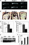
Increased bone mass in Smad3+/– mice with reduced osteoblast TGF-β responsiveness. X-ray analysis of femurs (A) and histomorphometry of von Kossa stained tibiae (B) revealed increased radiodensity, trabecular bone volume, and cortical bone thickness in Smad3+/– bone, relative to wild-type and Smad3–/– bone. (C) Relative bone volume over total volume, as determined by histomorphometry analysis. (D) Differences in cortical bone thickness, as determined by XTM (P < 0.001). (E) Western analysis shows Smad2 and Smad3 expression and phospho-Smad3 (P-Smad3) in calvarial osteoblast cultures. Cells were treated with TGF-β after 6 h of serum starvation. (F) Real-time PCR analysis of Runx2 mRNA expression in calvarial osteoblasts in the absence or presence of 1 ng/ml TGF-β. Values are normalized to RPL19 expression and are shown relative to untreated cells of the same genotype.
Fig. 5.
Fig. 5.
Fracture toughness of femurs from mice with different levels of TGF-β signaling. A three-point bending test of femurs, where fracture was initiated from a sharpened notch (A), was used to measure the fracture toughness of bone, Kc (B). Elevated TGF-β signaling (D4 and D5) decreased, whereas reduced TGF-β signaling (DNTβRII and Smad3+/–) increased Kc compared with wild-type bone (*, P < 0.05). (C) As shown by scanning electron microscopy, fracture-resistant DNTβRII and Smad3+/– bones exhibited extensive crack deflection (white arrows), whereas perpendicular fractures were observed in D4, D5, and Smad3–/– bones. The location of the initiating notch is indicated.

Similar articles

Cited by

References

    1. Currey, J. D. (1999) J. Exp. Biol. 202, 2495–2503. - PubMed
    1. Lane, N. E., Yao, W., Kinney, J. H., Modin, G., Balooch, M. & Wronski, T. J. (2003) J. Bone Miner. Res. 18, 2105–2115. - PubMed
    1. Poli, V., Balena, R., Fattori, E., Markatos, A., Yamamoto, M., Tanaka, H., Ciliberto, G., Rodan, G. A. & Costantini, F. (1994) EMBO J. 13, 1189–1196. - PMC - PubMed
    1. Jamsa, T., Rho, J. Y., Fan, Z., MacKay, C. A., Marks, S. C., Jr., & Tuukkanen, J. (2002) J. Biomech. 35, 161–165. - PubMed
    1. Misof, K., Landis, W. J., Klaushofer, K. & Fratzl, P. (1997) J. Clin. Invest. 100, 40–45. - PMC - PubMed

Publication types

MeSH terms