Comparative Study
. 2005 Dec 14;25(50):11605-13.
doi: 10.1523/JNEUROSCI.4173-05.2005.

Neurosteroid access to the GABAA receptor

Affiliations
Comparative Study

Neurosteroid access to the GABAA receptor

Gustav Akk et al. J Neurosci. .

Abstract

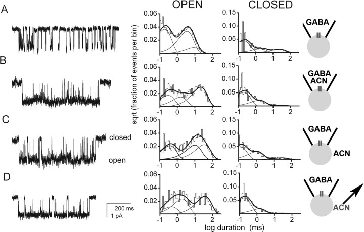
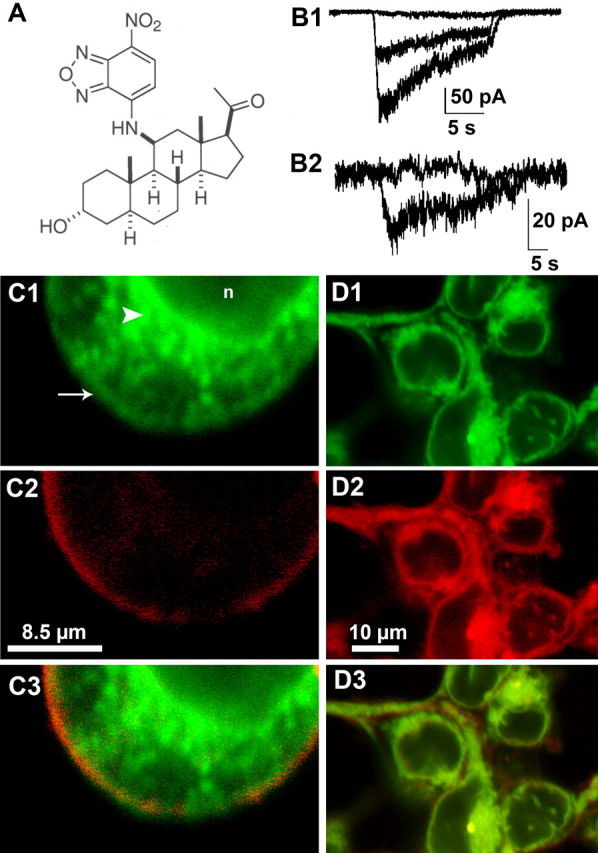
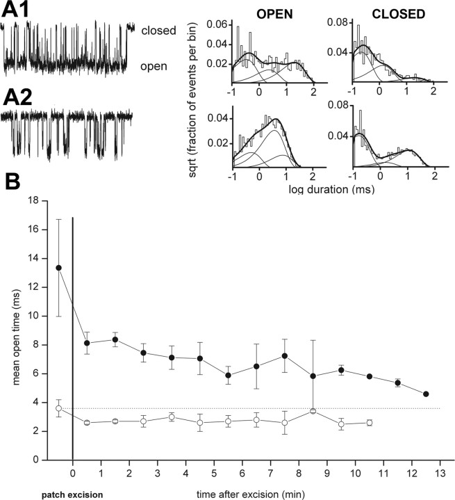
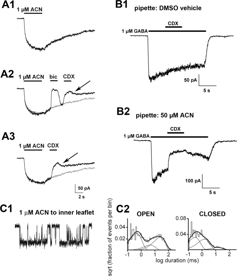
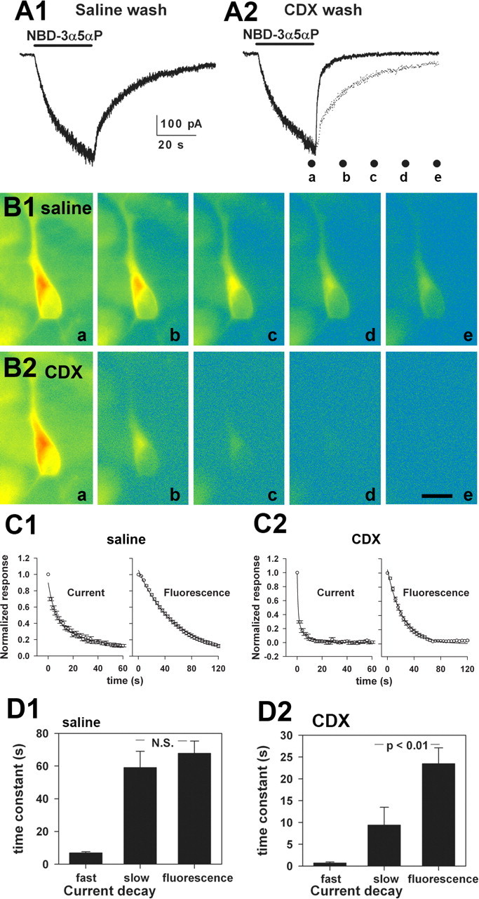
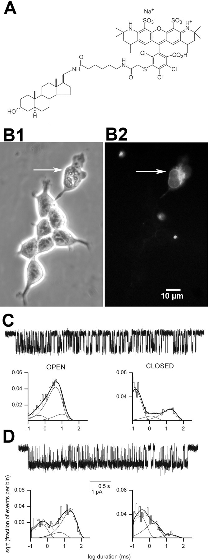
GABAA receptors are a pivotal inhibitory influence in the nervous system, and modulators of the GABAA receptor are important anesthetics, sedatives, anticonvulsants, and anxiolytics. Current views of receptor modulation suggest that many exogenous drugs access and bind to an extracellular receptor domain. Using novel synthetic steroid analogs, we examined the access route for neuroactive steroids, potent GABAA receptor modulators also produced endogenously. Tight-seal recordings, in which direct aqueous drug access to receptor was prevented, demonstrated that steroids can reach the receptor either through plasma membrane lateral diffusion or through intracellular routes. A fluorescent neuroactive steroid accumulated intracellularly, but recordings from excised patches indicated that the intracellular reservoir is not necessary for receptor modulation, although it can apparently equilibrate with the plasma membrane within seconds. A membrane impermeant neuroactive steroid modulated receptor activity only when applied to the inner membrane leaflet, demonstrating that the steroid does not access an extracellular modulatory site. Thus, neuroactive steroids do not require direct aqueous access to the receptor, and membrane accumulation is required for receptor modulation.


Figures

Figure 1.
Figure 1.
Steroid access from extracellular solution is not necessary for robust GABAA receptor modulation. Cell-attached recordings from transfected HEK cells were performed under the conditions schematized to the right of the panels. Channel openings are shown as downward deflections. The graphs represent log-binned open and closed time histograms from the patch as indicated. A, Channel activity in a patch exposed to 50μm GABA in the recording pipette. The open times were 0.16 ms (49%), 3.1 ms (37%), and 7.8 ms (14%). The closed times were 0.19 ms (61%), 1.7 ms (20%), and 21.7 ms (18%). B, Channel activity in a patch exposed to the combination of GABA and 1 μm ACN in the patch pipette. The open times were 0.27 ms (29%), 1.4 ms (24%), and 19.6 ms (47%). The closed times were 0.19 ms (68%), 1.5 ms (28%), and 25.6 ms (5%). Note the increased duration and amplitude of the third component of the open time distribution and the decreased relative weight of the third component of the closed time distribution, as described previously (Akk et al., 2004). C, Potentiation of receptor function was qualitatively similar when the steroid was applied not via the patch pipette but rather to the bath solution after seal formation. The open times were 0.34 ms (33%), 9.2 ms (34%), and 21.3 ms (33%). The closed times were 0.11 ms (57%), 1.0 ms (35%), and 25.0 ms (8%). D, Channel activity from a cell pretreated with ACN. After removal of bath ACN and exposure of the channels to GABA in the pipette, potentiation of channel activity remained. The open times were 0.42 ms (19%), 1.7 ms (38%), and 34.5 ms (43%). The closed times were 0.17 ms (73%), 1.2 ms (23%), and 22.7 ms (4%).
Figure 2.
Figure 2.
A fluorescent GABA-active stero id suggests plasma membrane and intracellular steroid accumulation. A, Structure of NBD-3α5αP. B1, Potentiation of whole-cell GABA responses in HEK cells. The superimposed traces represent the response of a transfected HEK cell to 2μm GABA alone (middle trace), GABA plus 3 μm NBD-3α5αP (largest trace), and GABA, NBD-3α5αP, and 50 μm bicuculline(flat trace). B2, Direct response of another transfected HEK cell to 3μm NBD-3α5αPalone and coapplied with 50μm bicuculline (flat trace). C, NBD-3α5αP (green fluorescence, C1, C3) colocalization with DiI (red fluorescence, C2, C3). Note some colocalization with plasma membrane (arrow) but the prominent intracellular staining (arrowhead) that is excluded from the nucleus(n). C3 is a merged image of C1 and C2. D, NBD-3α5αP (green, D1,D3) colocalization with Nile Red (red, D2, D3), a cell-permeant lipophilic marker. D3 is a merged image of D1 and D2.
Figure 3.
Figure 3.
Patch excision after steroid preincubation and removal does not affect potentiation, suggesting that membrane-accumulated steroid is sufficient for receptor potentiation. A, Channel activity in an inside-out patch from a cell to which ACN was preapplied and then removed before seal formation. The receptors were exposed to 50 μm GABA in the patch pipette. A1, Channel record from 1 min after excision. Histograms pertain to data from 0–2 min after excision. The open times were 0.31 ms (42%), 2.1 ms (23%), and 18.5 ms (35%). The closed times were 0.20 ms (58%), 1.2 ms (32%), and 18.5 ms (9%). A2, The addition of 5 mm methyl-β-cyclodextrin to the bath to remove steroid resulted in an immediate loss of steroid-mediated potentiation. The open times were 0.46 ms (24%), 3.4 ms (57%), and 6.9 ms (19%). The closed times were 0.15 ms (60%), 1.5 ms (9%), and 10.4 ms (31%). B, Time course of the effect of patch excision on channel activity from cells pretreated with ACN(filled circles) or in the presence of GABA alone (open circles). Mean open time was averaged over 1 min blocks. Note the prolonged potentiation of channel activity after ACN pre-exposure and removal. Data show mean ± SD from three to six patches.
Figure 4.
Figure 4.
Intracellular loading can result in potentiation. A, Resurgent steroid effects after cyclodextrin wash. A1, Slow direct gating of GABAA receptors in response to 1 μm ACN in a transfected HEK cell. The cell was rapidly perfused with drug-free saline after ACN removal but nevertheless had a slowly decaying off response, similar to that previously described for neurons and the natural neurosteroid 3α5αP (Shu et al., 2004). A2, In the same cell on another application of ACN, the offset current was reversibly blocked by bicuculline (bic), which is a noncompetitive antagonist with respect to steroid (Ueno et al., 1997). Therefore, in the case of bicuculline, a large part of the resurgence results from bicuculline unbinding before steroid departure. A2, A3, The offset current was partly inhibited by a brief wash with 500 μm γ-cyclodextrin (CDX), which facilitates the removal of steroid (Shu et al., 2004). The resurgent current (arrows) indicates that the replenishment of receptor-accessible steroid, possibly from the intracellular pools, which are inaccessible to cyclodextrin. To facilitate comparison of offset, the dotted trace represents a replot of the control trace in A1, scaled to account for slight rundown between sweeps. Note that the resurgence is less complete than for bicuculline and less than expected from baseline decay (dotted traces). B, Effects of intracellular steroid loading. The currents presented show representative data from a total of four cells under control conditions and five cells loaded with steroid. B1, Response to 1μm GABA in a control transfected HEK cell. Note that the GABA response is insensitive to 500 μm cyclodextrin. B2, In a cell loaded with 50 μm ACN through the whole-cell pipette, 1μm GABA gated a larger response, which was partly sensitive to 500μm γ-cyclodextrin. C1, Channel activity in an inside-out membrane patch (50μm GABA in the pipette), to which 1 μm ACN was applied by bath exchange to the intracellular face. C2, Summary of channel effects. Channel openings are shown downward. The open times were 0.42 ms (48%), 4.2 ms (15%), and 23.3 ms (37%). The closed times were 0.28 ms (48%), 1.4 ms (42%), and 14.5 ms (10%).
Figure 5.
Figure 5.
The bulk intracellular steroid pool equilibrates with the receptor-accessible pool in <1 min. A1, Directly gated current in a hippocampal neuron by 3 μm NBD-3α5αP. After 30 s of application, the cell was washed with saline. A2, The same neuron was rechallenged, but the wash included 500 μm γ-cyclodextrin (CDX). The dotted trace is a replot of A1 for comparison. The dots below the trace indicate time points at which images were taken of cells for B. B1, Pseudocolor fluorescence images (warm colors, high-fluorescence intensity; cool colors, low-fluorescence intensity) of a cell subjected to the protocol in A1. Photographs were from the time points indicated by the labeled dots in A2 (a– e). B2, The same cell was subjected to the protocol indicated in A2. Again, the photographs are from the time points indicated by the dots in A2. Scale bar, 20 μm. C1, C2, The data points show raw normalized decays of NBD-3α5αP-gated currents (left graphs; n = 8 saline-washed and 7 CDX-washed neurons) and fluorescence (right graphs; n = 8 saline-washed and 6 CDX-washed neurons). For current traces, only every 100th data point is represented for clarity. The lines represent superimposed biexponential fits (current decays) and single-exponential fits (fluorescence). Parameters are summarized in D. D1, D2, Summary of exponential fits to the decay phase of NBD-3α5αP-generated currents and decay of intracellular fluorescence from the individual cells represented in C. Current decays were well described by a biexponential fit in all cases. The fast component accounted for 57 ± 5% of the decay in saline and 59 ± 5% in γ-cyclodextrin. Fluorescence decays were fit to fluorescence intensities taken from an intracellular region near the nucleus. To diminish bleaching, images were obtained beginning at the end of the 30 s application of NBD-3α5αP, continuing every 4 s for 60–120 s. Fluorescence decays were well described by a single exponential. Both the fast and slow time constants of current decay were significantly speeded by cyclodextrin wash, as was the time constant of fluorescence wash(p<0.01; note the change in the y-axis between D1 and D2). Data show mean ± SEM from seven to eight cells.
Figure 6.
Figure 6.
Effects of a membrane impermeant steroid. A, Structure of Alexa-3α5αP. B, Phase-contrast (left) and fluorescence (right) images of cells incubated in Alexa-3α5αP for 10 min. Incubation times >60 min failed to label healthy cells. The arrow indicates a cell with a membrane that was purposely ruptured with a sharp patch pipette before Alexa-3α5αP application. Note that only this cell and not intact cells stain with the Alexa-conjugated steroid. Several other pieces of cellular debris also fluorescently stained.C, Cell-attached patch data with GABA (50 μm) plus Alexa-3α5αP(1 μm) in the recording pipette. Channel openings are shown downward. The open times were 0.42 ms (12%), 3.9 ms (73%), and 9.3 ms (15%). The closed times were 0.13 ms (65%), 1.2 ms (10%), and 15.4 ms(24%). D, Inside-outpatch with GABA(50 μm) in the pipette and Alexa-3α5αP(1 μm) applied to the inner membrane face. The open times were 0.41 ms (31%), 4.9 ms (13%), and 20.8 ms (56%). The closed times were 0.27 ms (54%), 1.5 ms (37%), and 16.7 ms (9%). Alexa-3α5αP potentiated receptor function when applied to the intracellular but not the extracellular side of the membrane.

References

    1. Akk G, Steinbach JH (2003) Low doses of ethanol and a neuroactive steroid positively interact to modulate rat GABAA receptor function. J Physiol (Lond) 546: 641–646. - PMC - PubMed
    1. Akk G, Bracamontes J, Steinbach JH (2001) Pregnenolone sulfate block of GABAA receptors: mechanism and involvement of a residue in the M2 region of the α subunit. J Physiol (Lond) 532: 673–684. - PMC - PubMed
    1. Akk G, Bracamontes JR, Covey DF, Evers A, Dao T, Steinbach JH (2004) Neuroactive steroids have multiple actions to potentiate GABAA receptors. J Physiol (Lond) 558: 59–74. - PMC - PubMed
    1. Amin J, Weiss DS (1993) GABAA receptor needs two homologous domains of the β-subunit for activation by GABA but not by pentobarbital. Nature 366: 565–569. - PubMed
    1. Barry PH, Diamond JM (1984) Effects of unstirred layers on membrane phenomena. Physiol Rev 64: 763–872. - PubMed

Publication types

LinkOut - more resources