Comparative Study
. 2005 Dec 14;25(50):11637-44.
doi: 10.1523/JNEUROSCI.3604-05.2005.

Functional magnetic resonance imaging shows oxytocin activates brain regions associated with mother-pup bonding during suckling

Affiliations
Comparative Study

Functional magnetic resonance imaging shows oxytocin activates brain regions associated with mother-pup bonding during suckling

Marcelo Febo et al. J Neurosci. .

Abstract

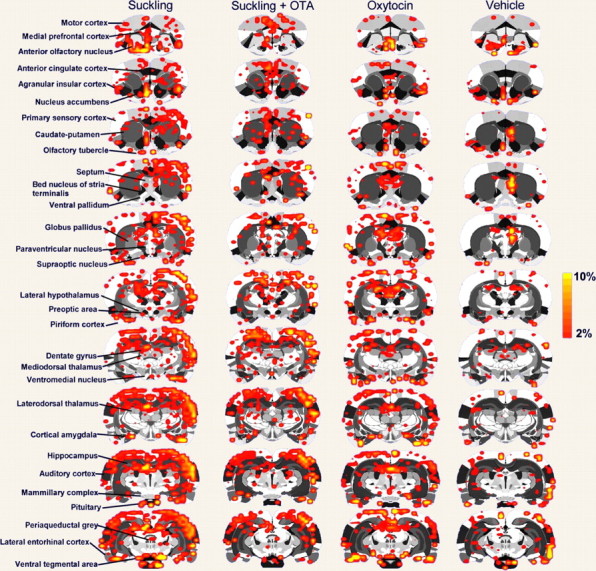
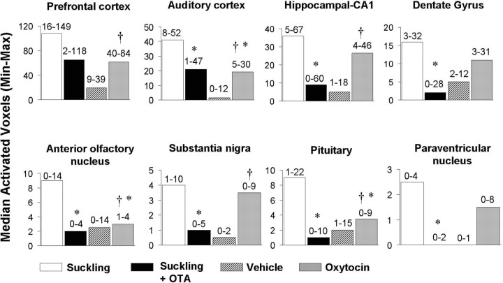
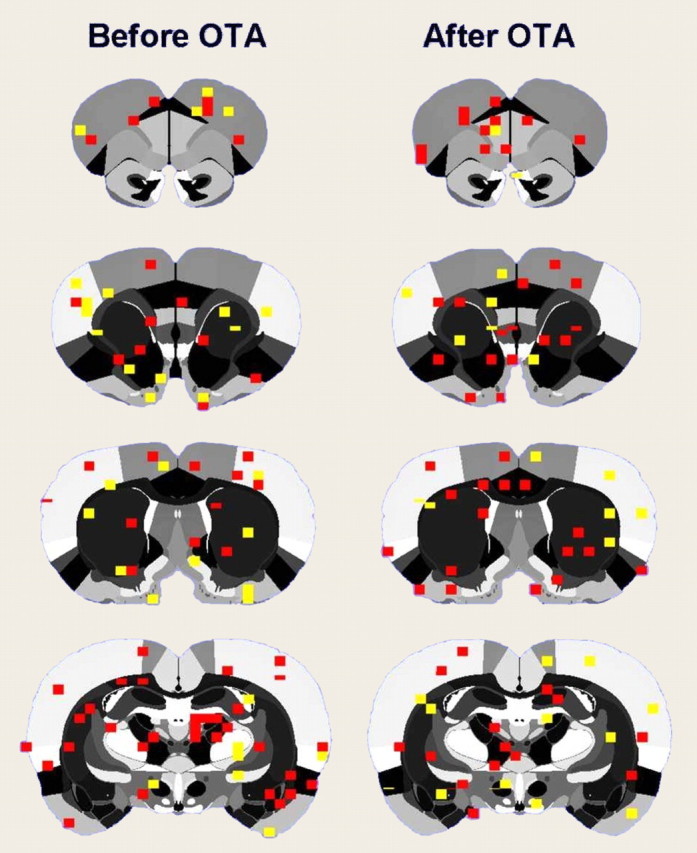
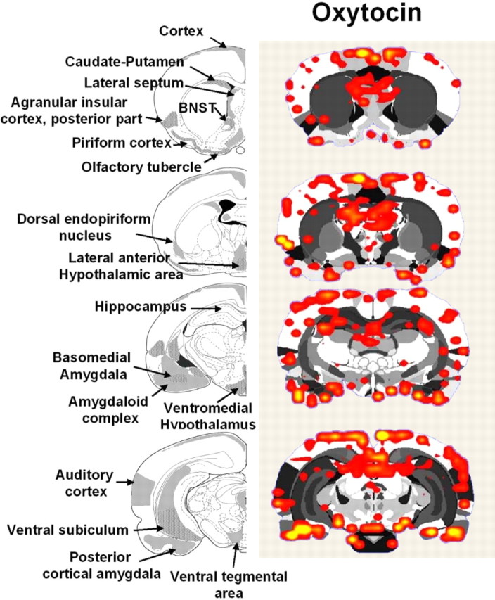
Oxytocin is released in the maternal brain during breastfeeding and may help strengthen the mother-infant relationship. Here, we used functional magnetic resonance imaging to determine whether oxytocin modulates brain activity in postpartum day 4-8 dams receiving suckling stimulation. During imaging sessions, dams were exposed to pup suckling before and after administration of an oxytocin receptor antagonist. Another group of dams received oxytocin alone. Changes in brain activation in response to suckling closely matched that elicited by oxytocin administration. The overlapping brain areas included the olfactory system, nucleus accumbens, insular cortex, prefrontal cortex, ventral tegmental area, cortical amygdala, and several cortical and hypothalamic nuclei. Blockade of oxytocin receptors largely attenuated activation in these regions. The data suggest that oxytocin may strengthen mother-infant bond formation partly by acting through brain areas involved in regulating olfactory discrimination, emotions, and reward.


Figures

Figure 1.
Figure 1.
Experimental setup for imaging awake postpartum dams during suckling. a, A body tube opening allows pups to gain access to two pairs of teats on either side of the ventrum. Pups are placed under the chassis at a close distance. b, Contact with the ventrum and teat is established within seconds during a functional scan. c, Side view showing a suckling pup.
Figure 2.
Figure 2.
Brain regions used for validating the suckling–stimulation paradigm during functional imaging. Atlas coronal sections and representative functional images highlight the somatosensory cortex (a), paraventricular nucleus (c), and pituitary (d) activated with suckling. Scale bar hue indicates the percentage-change value. b, BOLD signal change over time during suckling stimulation (expressed as percentage change from baseline).
Figure 3.
Figure 3.
Two-dimensional statistical maps of brain activation in response to suckling or oxytocin. Activation maps are composites of P4–P8 dams that received suckling stimulation before and after oxytocin receptor blockade or oxytocin and vehicle administration. Colored pixels represent brain areas that showed signal intensity values significantly increased above baseline. Scale bar hue indicates the percentage-change value. Statistical significance was determined using a voxel-wise t test analysis with false-positive detection filtering, comparing baseline and stimulation periods (α = 0.05; Bonferroni corrected). Various regions of interest are indicated to the left.
Figure 4.
Figure 4.
Median number of voxels activated by suckling, oxytocin, or vehicle administration. Data are shown for P4–P8 dams that received suckling stimulation before (empty bars) and after (dark bars) oxytocin receptor blockade, or with oxytocin (gray bars) and vehicle (patterned bars) alone. Numbers on top of the bars indicate the data range across individual animals (minimum and maximum). Symbols indicate statistical differences at p < 0.05 using a nonparametric Kruskal–Wallis test. *Significantly different from suckling alone. Significantly different from vehicle.
Figure 5.
Figure 5.
Effect of oxytocin or an oxytocin antagonist on evoked cortical activity. Compositive BOLD activation maps in rats given electrical (1 mA) stimulation of the hindpaw both before and after blockade of oxytocin receptors with an antagonist are shown. The colored pixels represent statistically significant increases in BOLD signal (p < 0.05; t test).
Figure 6.
Figure 6.
Brain areas showing positive BOLD responses to oxytocin in postpartum dams. Atlas coronal sections on the left highlight brain areas containing moderate-to-high levels of oxytocin receptors, as reported in the literature (moderate, gray colored areas; high, gray/dotted areas) (Tribollet et al., 1988; Veinante and Freund-Mercier, 1997; Vaccari et al., 1998). The adjacent activation maps (n = 6) show regions of positive BOLD signal changes after administration of oxytocin. Colored pixels represent brain areas that showed signal intensity values significantly increased above baseline. Statistical significance was determined using a voxel-wise t test analysis with false-positive detection filtering, comparing baseline and stimulation periods (α = 0.05; Bonferroni corrected).

References

    1. Byrnes EM, Rigero BA, Bridges RS (2002) Dopamine antagonists during parturition disrupt maternal care and the retention of maternal behavior in rats. Pharmacol Biochem Behav 73: 869–875. - PubMed
    1. Champagne F, Diorio J, Sharma S, Meaney MJ (2001) Naturally occurring variations in maternal behavior in the rat are associated with differences in estrogen-inducible central oxytocin receptors. Proc Natl Acad Sci USA 98: 12736–12741. - PMC - PubMed
    1. Febo M, Segarra AC, Tenney JR, Brevard ME, Duong TQ, Ferris CF (2004) Imaging cocaine-induced changes in the mesocorticolimbic dopaminergic system of conscious rats. J Neurosci Methods 139: 167–176. - PMC - PubMed
    1. Ferris CF, Kulkarni P, Sullivan Jr JM, Harder JA, Messenger TL, Febo M (2005) Pup suckling is more rewarding than cocaine: evidence from functional magnetic resonance imaging and three-dimensional computational analysis. J Neurosci 25: 149–156. - PMC - PubMed
    1. Fleming A, Vaccarino F, Tambosso L, Chee P (1979) Vomeronasal and olfactory system modulation of maternal behavior in the rat. Science 203: 372–374. - PubMed

Publication types

LinkOut - more resources